

What are lymph nodes?
Lymph nodes are small, bean-shaped organs spread along the lymphatic system. Each is surrounded by a tough capsule of connective tissue. Lymph enters a node through afferent lymphatic vessels and leaves through efferent vessels, and on the way the node removes debris and pathogens from the lymph. (OpenStax Anatomy and Physiology 2e, 21.1: Anatomy of the Lymphatic and Immune Systems.)
Nodes are found throughout the body but are concentrated in certain places: near the groin, armpits, neck, chest and abdomen. Their swelling during an infection is one sign of the close link between the lymphatic and immune systems. (OpenStax Anatomy and Physiology 2e, 21.1: Anatomy of the Lymphatic and Immune Systems.)
In everyday speech, enlarged neck nodes are often called "swollen glands", although they are lymph nodes, not glands. The American Academy of Family Physicians' review lists the head and neck node groups that can be felt as submental, submandibular, anterior and posterior cervical, pre-auricular and supraclavicular. (Gaddey HL, Riegel AM. Unexplained Lymphadenopathy: Evaluation and Differential Diagnosis. Am Fam Physician 2016.)
Where are the lymph nodes in the neck?
The head and neck nodes are grouped as superficial and deep. The superficial groups form a ring around the junction of the head and neck: occipital, mastoid (post-auricular), pre-auricular, parotid, submandibular and submental nodes, plus facial nodes on the cheek and superficial cervical nodes in the neck. (TeachMeAnatomy: Lymphatic Drainage of the Head and Neck.)
The deep cervical nodes receive all the lymph of the head and neck, either directly or after it has passed through the superficial nodes. They form a vertical chain along the internal jugular vein inside the carotid sheath. (TeachMeAnatomy: Lymphatic Drainage of the Head and Neck.)
TeachMeAnatomy lists the deep groups as prelaryngeal, pretracheal, paratracheal, retropharyngeal, infrahyoid, jugulodigastric (tonsillar), jugulo-omohyoid and supraclavicular nodes, divided into superior and inferior sets. (TeachMeAnatomy: Lymphatic Drainage of the Head and Neck.)
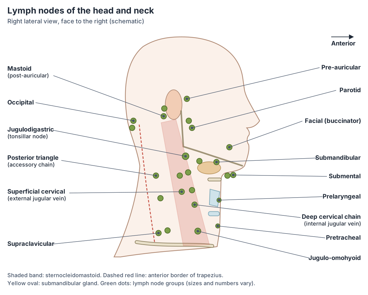
Occipital, mastoid, pre-auricular, parotid and facial nodes
Occipital nodes, usually one to three, lie at the back of the head near the lateral border of trapezius and drain the occipital scalp. Mastoid (post-auricular) nodes, usually two, lie behind the ear and drain the back of the neck, the upper ear and the back of the ear canal. (TeachMeAnatomy: Lymphatic Drainage of the Head and Neck.)
Pre-auricular nodes, one to three in number, lie in front of the auricle and drain the superficial face and temple. Parotid nodes lie on the parotid gland, with a deeper set inside it; between them they drain the nose and nasal cavity, the external acoustic meatus, the middle ear and the lateral edge of the orbit, and the deep group also drains the nasopharynx. (TeachMeAnatomy: Lymphatic Drainage of the Head and Neck.)
Facial nodes (infraorbital, buccinator and supramandibular) are scattered along the cheek and drain the lining of the nose and cheek, the eyelids and the conjunctiva. (TeachMeAnatomy: Lymphatic Drainage of the Head and Neck.)
Head and neck lymph node groups and what they drain
| Node group | Where it is | Main areas drained |
|---|---|---|
| Occipital (1 to 3 nodes) | Back of the head, near the lateral border of trapezius | Occipital part of the scalp |
| Mastoid / post-auricular (usually 2) | Behind the ear | Posterior neck, upper ear, back of the external auditory meatus |
| Pre-auricular (1 to 3) | In front of the auricle | Superficial face and temporal region |
| Parotid (superficial and deep) | On and in the parotid gland | Nose, nasal cavity, external acoustic meatus, middle ear, lateral orbit; deep nodes also nasopharynx |
| Facial (buccinator, infraorbital, supramandibular) | On the face along the cheek | Nasal and cheek mucosa, eyelids, conjunctiva |
| Submental | Under the chin, superficial to mylohyoid | Central lower lip, floor of the mouth, tip of the tongue |
| Submandibular (3 to 6) | In the submandibular triangle | Cheeks, side of the nose, upper lip, lateral lower lip, gums, front of the tongue; also submental and facial nodes |
| Superficial cervical | Along the anterior and external jugular veins | Skin of the front and side of the neck |
| Deep cervical (including jugulodigastric and jugulo-omohyoid) | Along the internal jugular vein in the carotid sheath | All lymph of the head and neck, directly or via the superficial groups |
Locations, numbers and drainage areas from TeachMeAnatomy.
Submental and submandibular lymph nodes
The submental nodes lie under the chin, superficial to the mylohyoid muscle, in the area often called the submental triangle. They drain the central part of the lower lip, the floor of the mouth and the tip of the tongue. (TeachMeAnatomy: Lymphatic Drainage of the Head and Neck.)
The submandibular nodes, usually three to six, lie in the submandibular triangle under the body of the mandible. They drain the cheeks, the side of the nose, the upper lip, the outer parts of the lower lip, the gums and the front of the tongue, and they also receive lymph from the submental and facial nodes. (TeachMeAnatomy: Lymphatic Drainage of the Head and Neck.)
In the level system, submental nodes are level Ia, lying between the anterior bellies of the two digastric muscles, and submandibular nodes are level Ib, behind and lateral to the anterior belly of digastric. (Radiopaedia: Cervical lymph node levels.)
Deep cervical, jugulodigastric and supraclavicular nodes
The jugulodigastric node, one of the upper deep cervical nodes, is also called the tonsillar node. The palatine tonsils drain to it and to the other upper deep cervical nodes, so it is often enlarged in tonsillitis. (TeachMeAnatomy: The Tonsils and Adenoids (Waldeyer's Ring).)
Superficial cervical nodes lie along the anterior jugular vein, draining the front of the neck, and along the external jugular vein, draining the superficial side of the neck. (TeachMeAnatomy: Lymphatic Drainage of the Head and Neck.)
The left supraclavicular node, known as Virchow's node, receives lymph from the abdominal cavity. Its enlargement (Troisier's sign) can be a sign that a stomach cancer has spread. (TeachMeAnatomy: Lymphatic Drainage of the Head and Neck.)
Neck lymph node levels I to VI
Clinicians describe neck nodes by levels, which use landmarks that can be seen on scans and in surgery: the mandible, hyoid bone, cricoid cartilage, clavicle, sternocleidomastoid and trapezius. Radiopaedia notes that its scheme is a synthesis of radiologically useful boundaries and that definitions differ between specialties. (Radiopaedia: Cervical lymph node levels.)
Level I lies above the hyoid and below the mandible and mylohyoid (Ia submental, Ib submandibular). Levels II, III and IV follow the internal jugular chain under the sternocleidomastoid: level II from the skull base to the hyoid, level III from the hyoid to the cricoid cartilage, and level IV from the cricoid to the clavicle. (Radiopaedia: Cervical lymph node levels.)
Level V is the posterior triangle, between the back edge of the sternocleidomastoid and the front edge of the trapezius. It is split at the level of the cricoid into Va, which includes nodes along the spinal accessory nerve, and Vb, which includes the lateral supraclavicular nodes. Level IV includes the medial supraclavicular nodes, including Virchow's node. (Radiopaedia: Cervical lymph node levels.)
Level VI is the central compartment, from the hyoid to the suprasternal notch and between the two common carotid arteries. It contains the prelaryngeal (Delphian), pretracheal, paratracheal and perithyroid nodes. A level VII is used in different ways by different specialties and is not standardised. (Radiopaedia: Cervical lymph node levels.)
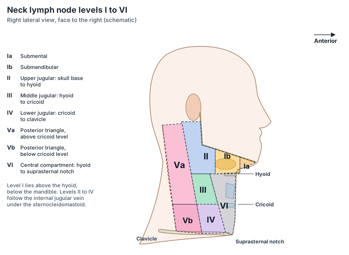
Neck lymph node levels I to VI
| Level | Name | Boundaries (simplified) |
|---|---|---|
| Ia | Submental | Between the anterior bellies of the two digastric muscles, above the hyoid |
| Ib | Submandibular | Below the mandible and mylohyoid, behind the anterior belly of digastric, in front of the back of the submandibular gland |
| II (IIa, IIb) | Upper internal jugular | Skull base to the lower border of the hyoid, from the back of the submandibular gland to the back of the sternocleidomastoid; IIa in front of or touching the internal jugular vein, IIb behind it |
| III | Middle internal jugular | Lower border of the hyoid to the lower border of the cricoid cartilage, under the sternocleidomastoid |
| IV | Lower internal jugular | Lower border of the cricoid to the clavicle; includes the medial supraclavicular nodes (Virchow's node) |
| V (Va, Vb) | Posterior triangle | Behind the sternocleidomastoid and in front of the trapezius, skull base to clavicle; Va above and Vb below the cricoid level |
| VI | Central (anterior) compartment | Hyoid to the suprasternal notch, between the two common carotid arteries; prelaryngeal, pretracheal, paratracheal and perithyroid nodes |
Simplified from Radiopaedia, which notes that definitions differ between specialties. A level VII is used inconsistently and is not standardised.
Waldeyer's ring (the tonsillar ring)
Waldeyer's ring is a ring of lymphoid tissue around the upper part of the pharynx. Its top is the pharyngeal tonsil in the roof of the nasopharynx; when enlarged, this is called the adenoids. On each side are the tubal tonsils around the openings of the Eustachian tubes and the palatine tonsils between the palatoglossal and palatopharyngeal arches. The lingual tonsil on the back third of the tongue completes the ring below. (TeachMeAnatomy: The Tonsils and Adenoids (Waldeyer's Ring).)
The tonsils are mucosa-associated lymphoid tissue (MALT) containing T cells, B cells and macrophages. They act as a first line of defence against pathogens entering through the nose and mouth. (TeachMeAnatomy: The Tonsils and Adenoids (Waldeyer's Ring).)
OpenStax describes tonsils as lymphoid nodules along the inner surface of the pharynx, with the pharyngeal tonsil at the back of the throat and the palatine tonsils on either side. (OpenStax Anatomy and Physiology 2e, 21.1: Anatomy of the Lymphatic and Immune Systems.)
Tonsillitis, inflammation of the palatine tonsils, is usually viral; bacteria cause about a third of cases. A peritonsillar abscess (quinsy) is a collection of pus beside the tonsil that needs drainage. (TeachMeAnatomy: The Tonsils and Adenoids (Waldeyer's Ring).)
Where does lymph from the head and neck go?
Vessels leaving the deep cervical nodes join to form a jugular lymphatic trunk on each side. The left jugular trunk joins the thoracic duct at the root of the neck, which empties into the venous system at the left subclavian vein. The right jugular trunk joins the right lymphatic duct, which empties at the right subclavian vein. (TeachMeAnatomy: Lymphatic Drainage of the Head and Neck.)
The right lymphatic duct receives lymph only from the upper right side of the body; all other regions drain through the thoracic duct. (OpenStax Anatomy and Physiology 2e, 21.1: Anatomy of the Lymphatic and Immune Systems.)
What is normal when you feel a lymph node?
Being able to feel a small lymph node is common. A family-medicine review notes that about half of otherwise healthy children have palpable lymph nodes at any one time, and that many healthy adults have inguinal (groin) nodes up to 2 cm across. (Gaddey HL, Riegel AM. Unexplained Lymphadenopathy: Evaluation and Differential Diagnosis. Am Fam Physician 2016.)
The same review defines lymphadenopathy as nodes that are abnormal in size, for example larger than 1 cm, or in consistency, while also noting that no particular size proves a node is malignant. Multiple small nodes that feel like shot pellets under the skin ("shotty" nodes) usually reflect a reaction to a viral infection. (Gaddey HL, Riegel AM. Unexplained Lymphadenopathy: Evaluation and Differential Diagnosis. Am Fam Physician 2016.)
Features that clinicians take more seriously include nodes that are hard, matted together or fixed to surrounding tissue, and nodes in certain places: palpable supraclavicular nodes are always considered abnormal. Tenderness alone is non-specific and may reflect inflammation from infection. This is educational information only; anyone worried about a lump should see a health professional. (Gaddey HL, Riegel AM. Unexplained Lymphadenopathy: Evaluation and Differential Diagnosis. Am Fam Physician 2016.)
Abdominal lymph nodes
The abdomen has its own large node groups. The pre-aortic nodes sit around the origins of the arteries to the gut and are divided into coeliac, superior mesenteric and inferior mesenteric groups. The superior and inferior mesenteric nodes collect lymph from mesenteric and colonic nodes and drain into the coeliac nodes, which give rise to the intestinal lymph trunks. (Kenhub: Lymph nodes of the thorax and abdomen.)
Together with the lateral aortic and retro-aortic nodes, the pre-aortic nodes make up the para-aortic lymph nodes. The lateral aortic group drains into the right and left lumbar lymph trunks, and lymph from the abdominal organs drains almost entirely through the thoracic duct. (Kenhub: Lymph nodes of the thorax and abdomen.)
Frequently asked questions
Where are the lymph nodes in the neck?
Neck lymph nodes lie in a ring where the head meets the neck (occipital, behind the ear, in front of the ear, over the parotid gland, under the jaw and under the chin) and in a deep chain along the internal jugular vein under the sternocleidomastoid muscle, from below the angle of the jaw down to just above the clavicle. Further groups lie in front of the larynx and trachea.
What are the lymph node levels of the neck?
Neck nodes are grouped into levels: I submental and submandibular, II upper jugular (skull base to hyoid), III middle jugular (hyoid to cricoid), IV lower jugular (cricoid to clavicle), V posterior triangle between sternocleidomastoid and trapezius, and VI central compartment in front of the larynx and trachea from the hyoid to the suprasternal notch.
What is the submental lymph node?
The submental nodes lie under the chin, superficial to the mylohyoid muscle, between the anterior bellies of the two digastric muscles (level Ia). They drain the central lower lip, the floor of the mouth and the tip of the tongue, and they send lymph on to the submandibular and deep cervical nodes.
What is Waldeyer's ring?
Waldeyer's ring is the ring of lymphoid tissue around the upper pharynx. It is made of the pharyngeal tonsil (adenoids when enlarged) above, the tubal tonsils and palatine tonsils on each side, and the lingual tonsil on the back of the tongue below. These tonsils are a first line of defence against pathogens entering through the nose and mouth.
Is it normal to feel a lymph node in your neck?
Often, yes. About half of healthy children have nodes that can be felt at any time, and small nodes commonly enlarge briefly during infections. Medical reviews define enlargement as nodes over about 1 cm or with an unusual consistency, and treat hard, fixed or supraclavicular nodes more seriously. This is general information; a doctor should assess any lump that worries you.
What are "glands" in the neck?
When people talk about swollen glands in the neck they usually mean enlarged lymph nodes, such as the submandibular nodes under the jaw or the upper deep cervical (jugulodigastric) nodes. True glands in the area include the salivary glands (parotid, submandibular and sublingual) and the thyroid, which are different structures.
References
- OpenStax. Anatomy and Physiology 2e, §21.1 Anatomy of the Lymphatic and Immune Systems. Rice University, 2022.
- TeachMeAnatomy. Lymphatic Drainage of the Head and Neck.
- TeachMeAnatomy. The Tonsils and Adenoids.
- Radiopaedia. Cervical lymph node levels.
- Gaddey HL, Riegel AM. Unexplained Lymphadenopathy: Evaluation and Differential Diagnosis. American Family Physician 2016;94(11):896-903.
- Kenhub. Lymph nodes of the thorax and abdomen.
Related anatomy
- Neck musclesNeck muscles explained: sternocleidomastoid, suprahyoid, infrahyoid, scalene, prevertebral and suboccipital groups…
- Internal jugular veinInternal jugular vein (IJ vein) anatomy: jugular foramen to brachiocephalic vein, carotid sheath relations…
- Carotid arteriesCarotid arteries explained: common carotid, bifurcation, internal carotid, the 8 external carotid branches with a…
- TongueTongue anatomy explained: parts and papillae of the dorsum, the underside with the frenulum and sublingual folds…
- SpleenSpleen location explained: left upper quadrant under ribs 9 to 11, behind the stomach. Relations, ligaments, blood…
Explore the whole body in the interactive 3D human anatomy model, or browse the A–Z list of human structures.