

Tongue at a glance
- What it is
- The tongue is a muscular organ of the mouth involved in taste, chewing, swallowing and speech.
- Location
- It lies in the floor of the oral cavity and extends back into the oropharynx, anchored to the hyoid and mandible.
- Function
- It manipulates food, enables taste and speech, and starts swallowing.
- Body system
- Digestive · single, midline structure
- FMA ID
- FMA54640 (Foundational Model of Anatomy)
Key facts
- Has intrinsic and extrinsic muscles; the hypoglossal nerve (CN XII) supplies all except palatoglossus (vagus nerve).
- Taste: facial nerve via chorda tympani (anterior two-thirds), glossopharyngeal nerve (posterior third).
- Papillae: filiform, fungiform, vallate and foliate.
Clinical relevance
Hypoglossal nerve injury causes the tongue to deviate toward the affected side on protrusion.
Parts of the tongue and the dorsum
The tongue has three parts: the apex (tip), the most mobile part at the front; the body, which forms most of the tongue; and the root at the back. Its upper surface is called the dorsum and its lower surface the inferior (ventral) surface. (Kenhub: Tongue.)
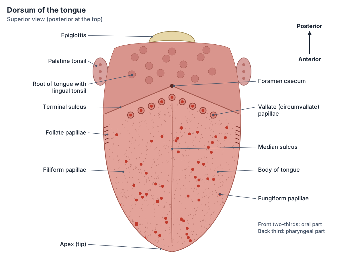
A shallow median sulcus runs back along the middle of the dorsum. It marks where the two halves of the tongue fused in the embryo and ends at a small pit, the foramen caecum. From the foramen caecum the V-shaped terminal sulcus (sulcus terminalis) runs forwards and outwards on each side. It divides the dorsum into an oral part in front (the front two-thirds) and a pharyngeal part behind (the back third). (Kenhub: Tongue.)
The pharyngeal part, at the back, carries the lingual tonsils, collections of lymphoid tissue at the base of the tongue. Lingual glands in the lamina propria of the tongue secrete mucus and a watery serous fluid containing the enzyme lingual lipase, which plays a minor role in breaking down fats. (OpenStax Anatomy and Physiology 2e, 23.3: The Mouth, Pharynx, and Esophagus.)
Papillae and taste buds on the dorsum
The oral part of the dorsum is covered with papillae, raised bumps that contain the structures for taste. OpenStax lists four types: circumvallate (vallate), foliate, filiform and fungiform. Taste buds containing gustatory receptor cells sit on the papillae. (OpenStax Anatomy and Physiology 2e, 14.1: Sensory Perception.)
Filiform papillae are the most numerous; they are keratinised, give the tongue its rough feel and carry no taste buds. Fungiform papillae are mushroom-shaped and scattered over the dorsum, with a few taste buds on top. Foliate papillae are a row of slits on the back of each side of the tongue near the terminal sulcus. The 8 to 12 large vallate papillae form a V just in front of the terminal sulcus, each surrounded by a trench into which the glands of von Ebner drain. OpenStax adds that filiform papillae carry touch receptors rather than taste buds. (Kenhub: Tongue.)
Taste buds contain gustatory receptor cells that detect five basic tastes: sweet, salty, sour, bitter and umami (savoury, linked to glutamate). OpenStax also mentions research suggesting a possible sixth taste for fats. (OpenStax Anatomy and Physiology 2e, 14.1: Sensory Perception.)
Underside of the tongue: frenulum, veins and sublingual folds
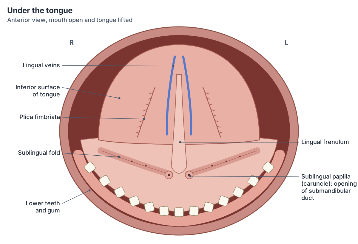
The inferior surface of the tongue is smooth and thin. In the midline a fold of mucous membrane, the lingual frenulum, tethers the tongue to the floor of the mouth. (OpenStax Anatomy and Physiology 2e, 23.3: The Mouth, Pharynx, and Esophagus.)
On each side of the frenulum the lingual veins show through the thin mucosa, and lateral to them is a fringed fold called the plica fimbriata. The deep lingual vein runs along the underside of the tongue towards the root, where it joins the sublingual vein. (Kenhub: Tongue.)
On the floor of the mouth, on either side of the base of the frenulum, are two small bumps called the sublingual papillae (sublingual caruncles). The submandibular duct opens at each papilla. Running backwards and outwards from them is the sublingual fold (plica sublingualis), a ridge of mucosa raised by the sublingual gland underneath; 8 to 20 small sublingual ducts open along its crest. The lingual nerve and the submandibular duct run along the inner side of the gland. (TeachMeAnatomy: The Sublingual Gland.)
OpenStax describes the submandibular glands secreting saliva into the mouth through the submandibular ducts, and the sublingual glands through the smaller sublingual ducts, so the floor of the mouth under the tongue is where much of the saliva enters the mouth. (OpenStax Anatomy and Physiology 2e, 23.3: The Mouth, Pharynx, and Esophagus.)
Intrinsic muscles: changing the shape of the tongue
The intrinsic muscles lie entirely within the tongue and have no attachment to bone. There are four paired intrinsic muscles, named after the direction of their fibres: superior longitudinal, inferior longitudinal, transverse and vertical. They change the shape of the tongue rather than its position. (TeachMeAnatomy: The Tongue.)
The superior longitudinal muscle shortens the tongue and curls the tip upwards; the inferior longitudinal muscle shortens it and turns the tip down. The transverse muscle narrows and lengthens the tongue, and the vertical muscle flattens and widens it. A fibrous median septum divides the tongue into right and left halves. (Kenhub: Tongue.)
Extrinsic muscles: genioglossus, hyoglossus, styloglossus and palatoglossus
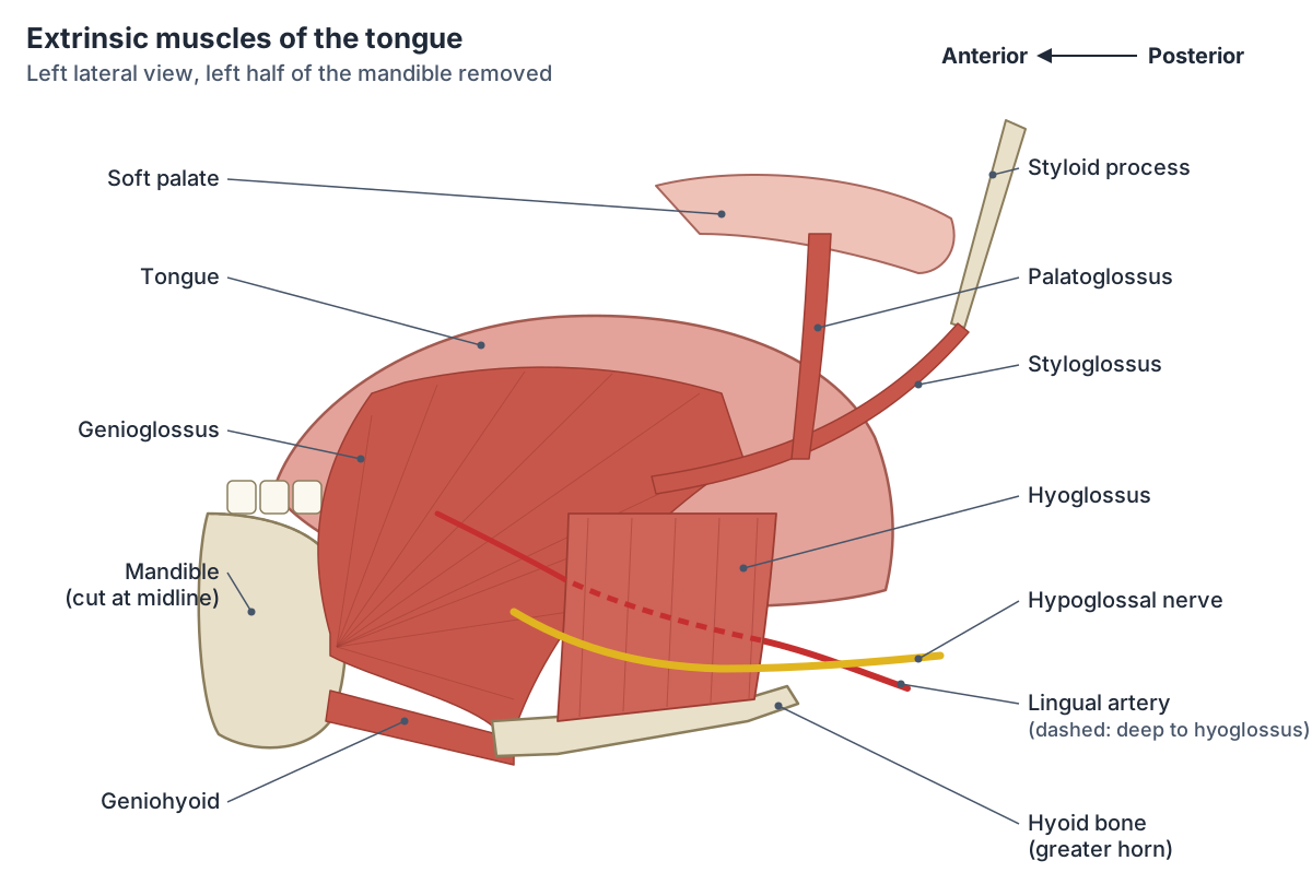
The four extrinsic muscles arise outside the tongue and move it as a whole: forwards, backwards, up and down. OpenStax lists them as genioglossus, which moves the tongue down and forwards; styloglossus, which pulls it up and back; palatoglossus, which raises the back of the tongue; and hyoglossus, which moves it down and flattens it. (OpenStax Anatomy and Physiology 2e, 11.3: Axial Muscles of the Head, Neck, and Back.)
Genioglossus arises from the genial tubercle (mental spine) on the inner surface of the mandible near the midline and spreads back into the tongue, inserting into the dorsum and lingual aponeurosis and, by its lowest fibres, the body of the hyoid. It protrudes the tongue (sticks it out) and depresses it. (Kenhub: Tongue.)
Hyoglossus is a thin, flat sheet of muscle on the side of the tongue. It arises from the whole length of the greater horn and the front of the body of the hyoid bone and runs upwards to insert into the side of the tongue, between styloglossus on the outside and the inferior longitudinal muscle on the inside. It depresses and retracts the tongue and is supplied by the hypoglossal nerve. The submandibular ganglion, which hangs from the lingual nerve, lies on its outer surface. (Radiopaedia: Hyoglossus muscle.)
Hyoglossus is an important landmark in the floor of the mouth. The lingual artery runs forwards deep to it, between hyoglossus and genioglossus, on its way to the tip of the tongue, while the hypoglossal nerve runs forwards on the muscle to reach the tongue. (Kenhub: Nerve and blood supply of the tongue.)
Styloglossus runs from the styloid process of the temporal bone to the side of the tongue and retracts and elevates it. Palatoglossus runs from the palatine aponeurosis of the soft palate to the tongue and raises the back of the tongue. (TeachMeAnatomy: The Tongue.)
Kenhub adds that palatoglossus closes the oropharyngeal isthmus, the opening between the mouth and the oropharynx, and that it is the only tongue muscle not supplied by the hypoglossal nerve: its nerve supply comes through the pharyngeal plexus. (Kenhub: Tongue.)
Muscles of the tongue
| Muscle | Attachments | Main action | Nerve |
|---|---|---|---|
| Superior longitudinal (intrinsic) | Submucosa and septum to the margins and apex | Shortens the tongue and curls the tip up | Hypoglossal (CN XII) |
| Inferior longitudinal (intrinsic) | Root of the tongue and hyoid to the apex | Shortens the tongue and curls the tip down | Hypoglossal (CN XII) |
| Transverse (intrinsic) | Median septum to the lateral margins | Narrows and lengthens the tongue | Hypoglossal (CN XII) |
| Vertical (intrinsic) | Dorsum to the inferior surface (lingual aponeurosis) | Flattens and widens the tongue | Hypoglossal (CN XII) |
| Genioglossus (extrinsic) | Superior mental spine (genial tubercle) of the mandible to the dorsum and the body of the hyoid | Protrudes and depresses the tongue | Hypoglossal (CN XII) |
| Hyoglossus (extrinsic) | Body and greater horn of the hyoid to the side of the tongue | Depresses and retracts the tongue | Hypoglossal (CN XII) |
| Styloglossus (extrinsic) | Styloid process of the temporal bone to the side of the tongue | Retracts and elevates the tongue | Hypoglossal (CN XII) |
| Palatoglossus (extrinsic) | Palatine aponeurosis of the soft palate to the side of the tongue | Elevates the back of the tongue; narrows the opening into the pharynx | Vagus (CN X), via the pharyngeal plexus |
Attachments and actions from TeachMeAnatomy and Kenhub.
Nerve supply: movement, touch and taste
All the intrinsic and extrinsic muscles of the tongue are supplied by the hypoglossal nerve (cranial nerve XII), except palatoglossus, which is supplied by the vagus nerve (cranial nerve X). The hypoglossal nerve leaves the skull through the hypoglossal canal of the occipital bone, passes down near the angle of the mandible, crosses the internal and external carotid arteries and then turns forwards into the tongue. (TeachMeAnatomy: The Hypoglossal Nerve (CN XII).)
General sensation (touch, temperature and pain) from the front two-thirds of the tongue travels in the lingual nerve, a branch of the mandibular division of the trigeminal nerve. Taste from the same area travels in the chorda tympani, a branch of the facial nerve that joins the lingual nerve. The back third sends both touch and taste through the glossopharyngeal nerve (cranial nerve IX). (TeachMeAnatomy: The Tongue.)
A small area at the very back of the tongue sends taste through the vagus nerve. OpenStax gives the same three-nerve pattern for taste: facial nerve for the anterior two-thirds, glossopharyngeal for the posterior third and vagus for the extreme back of the tongue. (OpenStax Anatomy and Physiology 2e, 14.1: Sensory Perception.)
Blood supply and lymphatic drainage
The main artery of the tongue is the lingual artery, a branch of the external carotid artery that arises at the level of the greater horn of the hyoid. It gives dorsal lingual branches to the root, the deep lingual artery to the body, and the sublingual artery to the floor of the mouth and the sublingual gland. (Kenhub: Nerve and blood supply of the tongue.)
Veins from the tongue drain into the lingual vein and then the internal jugular vein. Lymph from the front two-thirds drains to the submental and submandibular lymph nodes and then to the deep cervical nodes; lymph from the back third drains directly to the deep cervical nodes. (TeachMeAnatomy: The Tongue.)
How the tongue develops
The tongue begins to form at about day 23 from swellings in the floor of the embryonic pharynx. The front, oral part comes from paired lateral lingual swellings and a median swelling of the first pharyngeal arch; the back part comes from the second, third and fourth arches. The tongue muscles develop separately from occipital myotomes, which explains why they are supplied by the hypoglossal nerve. (Kenhub: Tongue.)
The foramen caecum is the remnant of the thyroglossal duct. If part of the duct does not close, a midline thyroglossal cyst or fistula can form. (TeachMeAnatomy: The Tongue.)
Clinical notes: hypoglossal nerve damage and tongue-tie
To test the hypoglossal nerve, the patient is asked to stick out the tongue. With a lower motor neuron lesion of the nerve the tongue deviates towards the damaged side, and that side may show wasting and twitching (fasciculations). (TeachMeAnatomy: The Hypoglossal Nerve (CN XII).)
In tongue-tie (ankyloglossia) the lingual frenulum is unusually short, limiting movement of the tongue. In babies it can cause difficulty breastfeeding, and it can be treated by a minor operation to release the frenulum. (TeachMeAnatomy: The Tongue.)
In Pierre Robin sequence, a defect of pharyngeal arch development, the tongue falls backwards (glossoptosis) and can obstruct the airway. (Kenhub: Tongue.)
Structures next to the tongue
The nearest named structures in the 3D model, measured from the centre of each part:
- Genioglossus · muscular
- Gingiva of lower jaw · digestive
- Mylohyoid · muscular
- Hyoglossus · muscular
- Mandible · skeletal
- Sublingual gland · digestive
- Geniohyoid · muscular
- Lower first secondary molar tooth · digestive
- Submandibular gland · digestive
- Lower second secondary premolar tooth · digestive
Frequently asked questions
What is the dorsum of the tongue?
The dorsum is the upper surface of the tongue. It is divided by the V-shaped terminal sulcus into an oral part (the front two-thirds), covered with filiform, fungiform, foliate and vallate papillae, and a pharyngeal part (the back third), which carries the lingual tonsil. A median sulcus runs along its midline to the foramen caecum at the point of the V.
What is under the tongue?
Under the tongue are the lingual frenulum in the midline, the lingual veins and fringed plica fimbriata on each side of it, and, on the floor of the mouth, the sublingual papillae where the submandibular ducts open. Behind them the sublingual folds overlie the sublingual glands, whose small ducts open along the folds.
What does the hyoglossus muscle do?
Hyoglossus depresses and retracts the tongue, pulling it down and back. It is a flat extrinsic muscle that runs from the body and greater horn of the hyoid bone up to the side of the tongue and is supplied by the hypoglossal nerve. The lingual artery runs deep to it, which makes it a key landmark in the floor of the mouth.
How many muscles are in the tongue?
Each half of the tongue has eight muscles: four intrinsic muscles (superior longitudinal, inferior longitudinal, transverse and vertical) that change its shape, and four extrinsic muscles (genioglossus, hyoglossus, styloglossus and palatoglossus) that change its position. All are supplied by the hypoglossal nerve except palatoglossus, which is supplied by the vagus nerve.
What nerves carry taste from the tongue?
Taste from the front two-thirds of the tongue is carried by the chorda tympani branch of the facial nerve (CN VII). Taste from the back third, including the vallate papillae, is carried by the glossopharyngeal nerve (CN IX). A small area near the epiglottis sends taste through the vagus nerve (CN X). Ordinary touch from the front two-thirds travels in the lingual nerve.
References
- OpenStax. Anatomy and Physiology 2e, §23.3 The Mouth, Pharynx, and Esophagus. Rice University, 2022.
- OpenStax. Anatomy and Physiology 2e, §11.3 Axial Muscles of the Head, Neck, and Back. Rice University, 2022.
- OpenStax. Anatomy and Physiology 2e, §14.1 Sensory Perception. Rice University, 2022.
- TeachMeAnatomy. The Tongue.
- TeachMeAnatomy. The Hypoglossal Nerve (CN XII).
- TeachMeAnatomy. The Sublingual Gland.
- Kenhub. Tongue.
- Kenhub. Nerve and blood supply of the tongue.
- Radiopaedia. Hyoglossus muscle.
- Foundational Model of Anatomy: Tongue (FMA54640). EMBL-EBI Ontology Lookup Service.
- Mitsuhashi N. et al. (2009). BodyParts3D: 3D structure database for anatomical concepts. Nucleic Acids Research 37, D782–D785. Data © The Database Center for Life Science, CC BY 4.0.
Related anatomy
- HyoglossusHyoglossus: A flat extrinsic tongue muscle rising from the hyoid bone. See where it is in an interactive 3D human…
- GenioglossusGenioglossus: The largest extrinsic tongue muscle, forming most of the tongue bulk. See where it is in an interactive…
- Hyoid boneHyoid bone: A U-shaped bone in the neck. See where it is in an interactive 3D human body, what it does and which…
- Sublingual glandSublingual gland: The sublingual gland is the smallest of the three major salivary glands. See where it is in an…
- Muscles of masticationThe muscles of mastication explained: masseter, temporalis, medial and lateral pterygoid, with labelled diagrams, the…
- TeethHuman teeth explained: incisors, canine (cuspid) teeth, premolars and molars, Universal and FDI numbering chart, tooth…
Explore the whole body in the interactive 3D human anatomy model, or browse the A–Z list of human structures.