

How are the neck muscles grouped?
The muscles of the front of the neck are divided by the hyoid bone: the suprahyoid muscles lie above it and the infrahyoid muscles below it. Sternocleidomastoid, the main muscle that bends and turns the head, runs across the side of the neck, and the three scalene muscles (anterior, middle and posterior) lie deeper at the side. (OpenStax Anatomy and Physiology 2e, 11.3: Axial Muscles of the Head, Neck, and Back.)
Deeper still, in front of the vertebral column, are the prevertebral muscles: longus capitis, longus colli and the rectus capitis anterior and lateralis. The neck fascia is described in three layers: the investing layer around trapezius and sternocleidomastoid, the visceral (pretracheal) layer around the thyroid, infrahyoid muscles and the air and food passages, and the prevertebral layer over the prevertebral muscles, the scalenes and the phrenic nerve. (O'Rahilly R et al. Basic Human Anatomy, Chapter 50: The Neck (Dartmouth Medical School).)
At the back of the neck, under the skull, lie the four small suboccipital muscles. They are covered by semispinalis capitis, one of the deep back muscles, which forms the roof of the suboccipital triangle. (TeachMeAnatomy: The Suboccipital Muscles.)
Trapezius, splenius, erector spinae and the deep back muscles, layer by layer. Back muscles.
Sternocleidomastoid and platysma
Platysma is a thin, four-sided sheet of muscle in the skin of the front of the neck. It arises from the skin over the deltoid and pectoralis major and inserts into the lower border of the mandible and the skin around the mouth. It is supplied by the cervical branch of the facial nerve (cranial nerve VII). (O'Rahilly R et al. Basic Human Anatomy, Chapter 50: The Neck (Dartmouth Medical School).)
Sternocleidomastoid arises by two heads, from the front of the manubrium of the sternum and from the upper surface of the medial third of the clavicle. It inserts on the lateral surface of the mastoid process and the lateral part of the superior nuchal line. It is supplied mainly by the accessory nerve (cranial nerve XI), with contributions from cervical nerves 2 to 4. (O'Rahilly R et al. Basic Human Anatomy, Chapter 50: The Neck (Dartmouth Medical School).)
One sternocleidomastoid bends the neck to its own side and turns the head to the opposite side. Acting together, the two muscles bend the lower neck forward while tilting the head back at the upper cervical joints. The muscle lies superficially, so it is easy to see and feel, and the carotid pulse can be felt along its front edge. Beneath it run the common carotid artery, the internal jugular vein, the vagus nerve and the ansa cervicalis. Spasm of the muscle causes wryneck (torticollis), with the head tilted towards the affected side and turned away from it. (Kenhub: Sternocleidomastoid muscle.)
Anterior and posterior triangles of the neck
Sternocleidomastoid divides each side of the neck into an anterior triangle in front of it and a posterior triangle behind it. (OpenStax Anatomy and Physiology 2e, 11.3: Axial Muscles of the Head, Neck, and Back.)
The anterior triangle is bounded by the lower border of the mandible above, the anterior border of sternocleidomastoid laterally and the midline of the neck medially. Its roof is the investing fascia. It is divided into four smaller triangles: submental, submandibular, carotid and muscular. (TeachMeAnatomy: The Anterior Triangle of the Neck.)
The posterior triangle is bounded by the posterior border of sternocleidomastoid in front, the anterior border of trapezius behind and the middle third of the clavicle below. Its roof is the investing fascia and its floor is the prevertebral fascia over splenius capitis, levator scapulae and the three scalene muscles. The inferior belly of omohyoid crosses it, dividing it into an occipital triangle above and a subclavian triangle below. (TeachMeAnatomy: The Posterior Triangle of the Neck.)
The posterior triangle contains the accessory nerve (cranial nerve XI), which runs quite superficially, branches of the cervical plexus including the phrenic nerve (C3 to C5), the external jugular vein, the last part of the subclavian artery, the trunks of the brachial plexus, and the transverse cervical and suprascapular vessels. (TeachMeAnatomy: The Posterior Triangle of the Neck.)
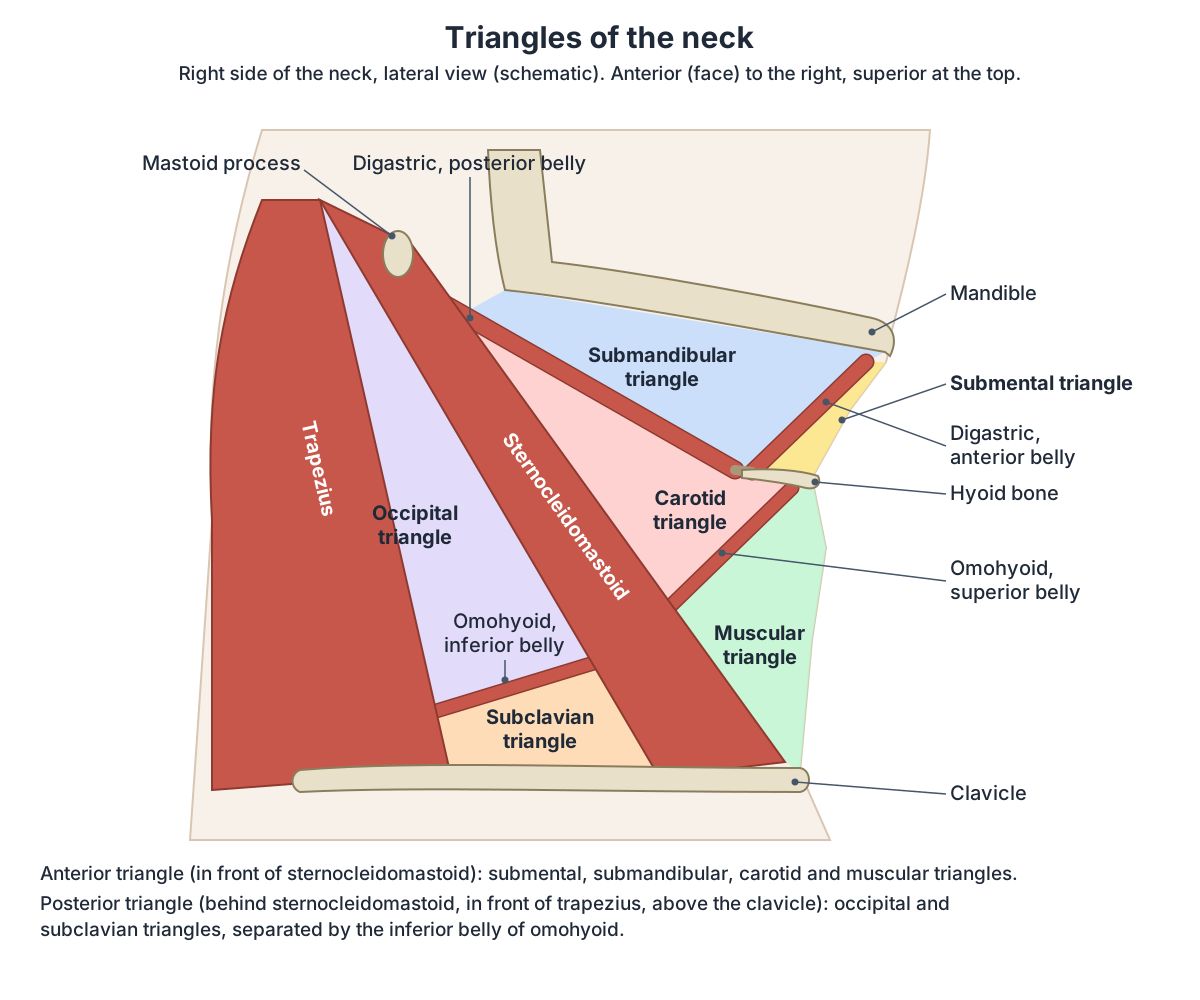
Submental, submandibular, carotid and muscular triangles
The submental triangle lies under the chin. It is bounded by the hyoid bone below, the midline medially and the anterior belly of digastric laterally. Its floor is the mylohyoid muscle and it contains the submental lymph nodes. The submandibular triangle lies between the body of the mandible and the anterior and posterior bellies of digastric and contains the submandibular gland, lymph nodes and the facial artery and vein. (TeachMeAnatomy: The Anterior Triangle of the Neck.)
The carotid triangle is bounded by the posterior belly of digastric above, the anterior border of sternocleidomastoid laterally and the superior belly of omohyoid below. It contains the common carotid artery, which divides into the internal and external carotid arteries here, the internal jugular vein and the hypoglossal and vagus nerves. The muscular triangle lies below the hyoid between the midline, the superior belly of omohyoid and the lower sternocleidomastoid; it contains the infrahyoid muscles, the pharynx and the thyroid and parathyroid glands. (TeachMeAnatomy: The Anterior Triangle of the Neck.)
The carotid sheath is a sleeve of deep cervical fascia on each side of the neck. It contains the common and internal carotid arteries, the internal jugular vein and the vagus nerve. (O'Rahilly R et al. Basic Human Anatomy, Chapter 50: The Neck (Dartmouth Medical School).)
Suprahyoid muscles: digastric, stylohyoid, mylohyoid and geniohyoid
The suprahyoid muscles raise the hyoid bone, the floor of the mouth and the larynx during swallowing (deglutition). Digastric has anterior and posterior bellies; stylohyoid moves the hyoid backwards and lifts the larynx; mylohyoid lifts the hyoid and helps press the tongue up against the roof of the mouth; geniohyoid pulls the hyoid forwards and can also lower (depress) the mandible. (OpenStax Anatomy and Physiology 2e, 11.3: Axial Muscles of the Head, Neck, and Back.)
Digastric has two bellies joined by an intermediate tendon, held to the hyoid by a fibrous sling. The anterior belly arises from the digastric fossa of the mandible and is supplied by a branch of the mandibular nerve (from the trigeminal nerve, cranial nerve V). The posterior belly arises from the mastoid process and is supplied by the facial nerve (cranial nerve VII). Digastric lowers the mandible and raises the hyoid. (TeachMeAnatomy: The Suprahyoid Muscles.)
Stylohyoid runs from the styloid process of the temporal bone to the side of the hyoid and is supplied by the facial nerve. Mylohyoid runs from the mylohyoid line of the mandible to the hyoid and is supplied by the mandibular branch of the trigeminal nerve. Geniohyoid runs from the inferior mental spine of the mandible to the hyoid and is supplied by fibres from C1 that travel with the hypoglossal nerve. (TeachMeAnatomy: The Suprahyoid Muscles.)
Infrahyoid (strap) muscles
The four infrahyoid muscles, sternohyoid, omohyoid, sternothyroid and thyrohyoid, are flat strap-like muscles. Sternohyoid and omohyoid lie in a superficial plane, sternothyroid and thyrohyoid in a deeper one. Together they anchor the hyoid bone and control the position of the larynx. (O'Rahilly R et al. Basic Human Anatomy, Chapter 50: The Neck (Dartmouth Medical School).)
Sternohyoid runs from the manubrium and the back of the medial end of the clavicle to the body of the hyoid. Sternothyroid runs from the back of the manubrium and the first costal cartilage to the oblique line of the thyroid cartilage, and pulls the larynx down. Thyrohyoid runs from the oblique line of the thyroid cartilage up to the hyoid; it can raise the larynx when the hyoid is fixed. Omohyoid has an inferior belly from the upper border of the scapula and a superior belly to the hyoid, joined by an intermediate tendon connected to the carotid sheath. (Kenhub: Infrahyoid muscles.)
Sternohyoid, omohyoid and sternothyroid are supplied by the anterior rami of C1 to C3 through the ansa cervicalis, a nerve loop in the front of the neck. Thyrohyoid is the exception: it is supplied by fibres from C1 carried within the hypoglossal nerve. (TeachMeAnatomy: The Infrahyoid Muscles.)
Suprahyoid and infrahyoid muscles
| Muscle | Attachments | Innervation | Main action |
|---|---|---|---|
| Digastric | Anterior belly: digastric fossa of mandible. Posterior belly: mastoid process. Intermediate tendon slung to hyoid | Anterior belly: mandibular nerve (CN V3). Posterior belly: facial nerve (CN VII) | Depresses mandible, elevates hyoid |
| Stylohyoid | Styloid process to side of hyoid | Facial nerve (CN VII) | Pulls hyoid up and back at the start of swallowing |
| Mylohyoid | Mylohyoid line of mandible to hyoid | Mandibular nerve (CN V3) | Elevates hyoid and floor of mouth |
| Geniohyoid | Inferior mental spine of mandible to hyoid | C1 fibres via hypoglossal nerve | Elevates hyoid, depresses mandible |
| Sternohyoid | Manubrium and medial clavicle to body of hyoid | Ansa cervicalis (C1–C3) | Depresses hyoid |
| Omohyoid | Upper border of scapula to hyoid, via intermediate tendon | Ansa cervicalis (C1–C3) | Depresses hyoid |
| Sternothyroid | Back of manubrium and first costal cartilage to oblique line of thyroid cartilage | Ansa cervicalis (C1–C3) | Depresses larynx |
| Thyrohyoid | Oblique line of thyroid cartilage to hyoid | C1 fibres via hypoglossal nerve | Depresses hyoid; elevates larynx if hyoid fixed |
Sources: TeachMeAnatomy (suprahyoid and infrahyoid muscles); Kenhub (infrahyoid muscles).
Scalene muscles
There are three scalene muscles on each side. Anterior scalene arises from the anterior tubercles of the transverse processes of C3 to C6 and attaches to the scalene tubercle of the first rib. Middle scalene, the largest and longest, arises from the posterior tubercles of C2 to C7 and attaches to the upper surface of the first rib. Posterior scalene, the smallest and deepest, arises from the posterior tubercles of C5 to C7 and attaches to the second rib. All are supplied by anterior rami of cervical nerves. (TeachMeAnatomy: The Scalene Muscles.)
The scalenes lift the first and second ribs and bend the neck to the same side. They act as accessory muscles of breathing; their visible use in a person at rest is a sign of respiratory distress. The brachial plexus and the subclavian artery pass between the anterior and middle scalenes, while the subclavian vein crosses in front of the anterior scalene and the phrenic nerve runs down its front surface. (TeachMeAnatomy: The Scalene Muscles.)
Not all textbooks agree on whether the scalenes work in quiet breathing. One classic text states that the scalenes may act as muscles of inspiration even during quiet breathing. (O'Rahilly R et al. Basic Human Anatomy, Chapter 50: The Neck (Dartmouth Medical School).)
OpenStax describes the scalenes as working together to flex, laterally flex and rotate the head, and notes that they contribute to deep inhalation. Of the three, the middle scalene is the longest and the posterior scalene the smallest. (OpenStax Anatomy and Physiology 2e, 11.3: Axial Muscles of the Head, Neck, and Back.)
Prevertebral muscles: longus colli, longus capitis and rectus capitis anterior
Longus colli lies directly on the front of the cervical and upper thoracic vertebrae and has three parts. The superior oblique part runs from the anterior tubercles of C3 to C5 to the anterior tubercle of the atlas (C1). The vertical intermediate part runs from the bodies of C5 to T3 to the bodies of C2 to C4. The inferior oblique part runs from the bodies of T1 to T3 to the anterior tubercles of C5 and C6. It is supplied by the anterior rami of C2 to C6; both muscles together flex the neck. (Kenhub: Prevertebral muscles.)
Longus capitis arises from the anterior tubercles of C3 to C6 and inserts on the basilar part of the occipital bone; it flexes the head and is supplied by C1 to C3. Rectus capitis anterior runs from the lateral mass of the atlas to the base of the occipital bone and flexes the head. Rectus capitis lateralis runs from the transverse process of the atlas to the jugular process of the occipital bone, bends the head to the side and helps stabilise the atlanto-occipital joint. (Kenhub: Prevertebral muscles.)
Longus colli is active in talking, coughing and swallowing. (O'Rahilly R et al. Basic Human Anatomy, Chapter 50: The Neck (Dartmouth Medical School).)
Suboccipital muscles and the suboccipital triangle
The suboccipital muscles are four small muscles under the back of the skull, all supplied by the suboccipital nerve (the posterior ramus of C1). Rectus capitis posterior major runs from the spinous process of C2 to the lateral part of the inferior nuchal line. Rectus capitis posterior minor runs from the posterior tubercle of C1 to the medial part of the inferior nuchal line. Both extend the head, and the major also rotates it. (TeachMeAnatomy: The Suboccipital Muscles.)
Obliquus capitis inferior runs from the spinous process of C2 to the transverse process of C1; it is the only suboccipital muscle with no attachment to the skull. It extends and rotates the head. Obliquus capitis superior runs from the transverse process of C1 to the occipital bone between the superior and inferior nuchal lines and extends the head. (TeachMeAnatomy: The Suboccipital Muscles.)
Three of these muscles outline the suboccipital triangle: rectus capitis posterior major above and medially, obliquus capitis superior above and laterally, and obliquus capitis inferior below. Its floor is the posterior atlanto-occipital membrane and the posterior arch of C1, and its roof is semispinalis capitis. It contains the vertebral artery, the suboccipital venous plexus and the suboccipital nerve. (TeachMeAnatomy: The Suboccipital Muscles.)
Scalene, prevertebral and suboccipital muscles
| Muscle | Attachments | Innervation | Main action |
|---|---|---|---|
| Anterior scalene | Anterior tubercles C3–C6 to scalene tubercle of rib 1 | Anterior rami C5–C6 | Elevates rib 1; lateral flexion of neck |
| Middle scalene | Posterior tubercles C2–C7 to upper surface of rib 1 | Anterior rami C3–C8 | Elevates rib 1; lateral flexion of neck |
| Posterior scalene | Posterior tubercles C5–C7 to rib 2 | Anterior rami C6–C8 | Elevates rib 2; lateral flexion of neck |
| Longus colli | Vertebral bodies and anterior tubercles C1–T3 (three parts) | Anterior rami C2–C6 | Flexes neck; one side rotates neck to opposite side |
| Longus capitis | Anterior tubercles C3–C6 to basilar occipital bone | Anterior rami C1–C3 | Flexes head |
| Rectus capitis anterior | Lateral mass of atlas to basilar occipital bone | Anterior rami C1–C2 | Flexes head |
| Rectus capitis lateralis | Transverse process of atlas to jugular process of occipital bone | Anterior ramus C1 | Lateral flexion of head |
| Rectus capitis posterior major | Spinous process C2 to lateral inferior nuchal line | Suboccipital nerve (posterior ramus C1) | Extends and rotates head |
| Rectus capitis posterior minor | Posterior tubercle C1 to medial inferior nuchal line | Suboccipital nerve | Extends head |
| Obliquus capitis superior | Transverse process C1 to occipital bone between nuchal lines | Suboccipital nerve | Extends head |
| Obliquus capitis inferior | Spinous process C2 to transverse process C1 | Suboccipital nerve | Extends and rotates head |
Sources: TeachMeAnatomy (scalene and suboccipital muscles); Kenhub (prevertebral muscles).
Neck muscles in the 3D model
- Digastric
- A two-bellied muscle above the hyoid bone linking the mandible and the mastoid region.
- Geniohyoid
- A narrow muscle above the mylohyoid in the floor of the mouth.
- Inferior oblique part of longus colli
- The lower oblique fibres of longus colli on the front of the cervical spine.
- Longus capitis
- A prevertebral muscle in front of the upper cervical spine.
- Mylohyoid
- A flat muscle forming the muscular floor of the mouth.
- Obliquus capitis inferior
- A suboccipital muscle below the atlas.
- Obliquus capitis superior
- A small suboccipital muscle forming the superolateral border of the suboccipital triangle.
- Omohyoid
- A thin infrahyoid strap muscle with two bellies joined by an intermediate tendon.
- Rectus capitis anterior
- A small deep prevertebral muscle at the base of the skull.
- Rectus capitis lateralis
- A small deep muscle lateral to the atlanto-occipital joint.
- Rectus capitis posterior major
- A suboccipital muscle forming the superomedial border of the suboccipital triangle.
- Rectus capitis posterior minor
- A small suboccipital muscle lying medial to rectus capitis posterior major.
- Scalenus anterior
- A deep neck muscle and an important landmark in the root of the neck.
- Scalenus medius
- The largest and longest of the scalene muscles.
- Scalenus posterior
- The smallest and deepest of the scalene muscles.
- Sternocleidomastoid
- A thick superficial neck muscle forming the main landmark dividing the anterior and posterior triangles.
- Sternohyoid
- A thin infrahyoid strap muscle at the front of the neck.
- Sternothyroid
- An infrahyoid strap muscle lying deep to sternohyoid.
- Stylohyoid
- A slender suprahyoid muscle running from the styloid process to the hyoid bone.
- Superior oblique part of longus colli
- The upper oblique part of longus colli, a prevertebral neck muscle.
- Thyrohyoid
- A short infrahyoid muscle continuing upward from sternothyroid.
- Vertical intermediate part of longus colli
- The central vertical part of longus colli, a prevertebral neck muscle.
Frequently asked questions
What are the main muscles of the neck?
The main neck muscle groups are the superficial muscles (platysma and sternocleidomastoid), the suprahyoid muscles (digastric, stylohyoid, mylohyoid, geniohyoid), the infrahyoid muscles (sternohyoid, omohyoid, sternothyroid, thyrohyoid), the scalenes, the prevertebral muscles (longus colli, longus capitis, rectus capitis anterior and lateralis) and the suboccipital muscles at the back.
What are the borders of the anterior triangle of the neck?
The anterior triangle is bounded by the lower border of the mandible above, the anterior border of sternocleidomastoid laterally and the midline of the neck medially. It is subdivided by the digastric and omohyoid muscles into the submental, submandibular, carotid and muscular triangles. The carotid triangle contains the carotid bifurcation, internal jugular vein and vagus and hypoglossal nerves.
What are the borders of the posterior triangle of the neck?
The posterior triangle lies between the posterior border of sternocleidomastoid, the anterior border of trapezius and the middle third of the clavicle. Its floor is formed by splenius capitis, levator scapulae and the scalenes, covered by prevertebral fascia. The accessory nerve crosses it superficially, and the inferior belly of omohyoid divides it into occipital and subclavian triangles.
What are the four suboccipital muscles?
The suboccipital muscles are rectus capitis posterior major, rectus capitis posterior minor, obliquus capitis superior and obliquus capitis inferior. They lie under the back of the skull between the occipital bone, atlas and axis, and are all supplied by the suboccipital nerve (posterior ramus of C1). They extend the head, and some rotate it.
What passes between the scalene muscles?
The brachial plexus and the subclavian artery pass between the anterior and middle scalene muscles. The subclavian vein runs in front of the anterior scalene, not between the muscles, and the phrenic nerve runs down the front of the anterior scalene. This gap is where local anaesthetic is injected for an interscalene nerve block.
What does the longus colli muscle do?
Longus colli lies on the front of the cervical and upper thoracic vertebrae in three parts. Acting on both sides it flexes the neck, and acting on one side it rotates the neck to the opposite side. It is also active during talking, coughing and swallowing. It is supplied by the anterior rami of C2 to C6.
What is in the carotid sheath?
The carotid sheath is a tube of deep cervical fascia on each side of the neck. It contains the common carotid artery and, higher up, the internal carotid artery, the internal jugular vein and the vagus nerve. It lies under the anterior border of sternocleidomastoid, which is why the carotid pulse can be felt there.
References
- OpenStax. Anatomy and Physiology 2e, §11.3 Axial Muscles of the Head, Neck, and Back. Rice University, 2022.
- O'Rahilly R, Müller F, Carpenter S, Swenson R. Basic Human Anatomy, Chapter 50: The Neck. Dartmouth Medical School (online edition).
- Kenhub. Sternocleidomastoid muscle.
- TeachMeAnatomy. The Anterior Triangle of the Neck.
- TeachMeAnatomy. The Posterior Triangle of the Neck.
- TeachMeAnatomy. The Suprahyoid Muscles.
- TeachMeAnatomy. The Infrahyoid Muscles.
- Kenhub. Infrahyoid muscles.
- TeachMeAnatomy. The Scalene Muscles.
- Kenhub. Prevertebral muscles.
- TeachMeAnatomy. The Suboccipital Muscles.
- Mitsuhashi N. et al. (2009). BodyParts3D: 3D structure database for anatomical concepts. Nucleic Acids Research 37, D782–D785. Data © The Database Center for Life Science, CC BY 4.0.
Related anatomy
- Back musclesBack muscles explained layer by layer: trapezius, rhomboids, serratus posterior, erector spinae, multifidus and…
- Splenius capitisSplenius capitis anatomy: origin, insertion, nerve and blood supply, actions, relations and how it differs from…
- Levator scapulaeLevator scapulae muscle anatomy: origin on C1 to C4, insertion on the scapula, actions on the shoulder blade and neck…
- Cervical vertebraeCervical vertebrae explained: typical C3 to C6 features, the atlas (C1), axis and dens (C2), C7 vertebra prominens, AO…
- Carotid arteriesCarotid arteries explained: common carotid, bifurcation, internal carotid, the 8 external carotid branches with a…
- LarynxLarynx anatomy with 3D model and diagrams: thyroid, cricoid, arytenoid and epiglottis cartilages, vocal folds…
Explore the whole body in the interactive 3D human anatomy model, or browse the A–Z list of human structures.