

Female reproductive organs: what the diagram shows
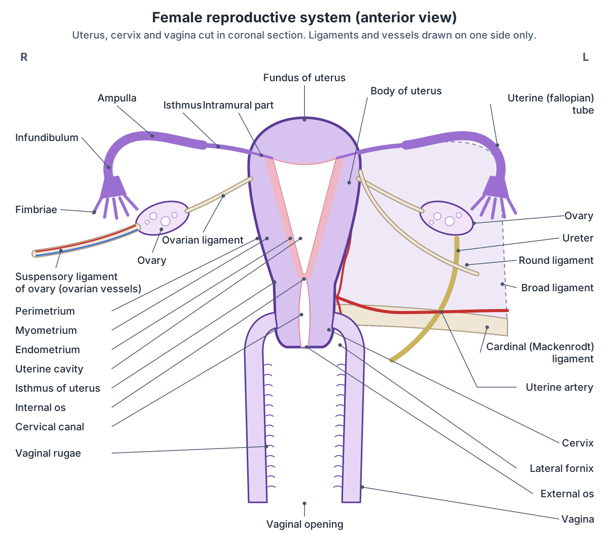
The ovaries are paired, almond-sized organs about 2 to 3 cm long in the pelvic cavity. Each uterine tube carries the oocyte from the ovary to the uterus; the tube lies close to its ovary but is not directly attached to it. The uterus is a muscular organ about 7 cm long and 5 cm wide outside pregnancy, and its narrow lower part, the cervix, projects into the vagina. (OpenStax Anatomy and Physiology 2e, 27.2: Anatomy and Physiology of the Ovarian Reproductive System.)
The vagina is a muscular canal about 10 cm long. It is the entrance to the reproductive tract and the exit for menstrual flow and for the baby at birth. (OpenStax Anatomy and Physiology 2e, 27.2: Anatomy and Physiology of the Ovarian Reproductive System.)
The uterus, tubes and ovaries are held in place by folds of peritoneum and bands of connective tissue called ligaments. TeachMeAnatomy groups their support into three levels: the broad and round ligaments above, the cardinal, pubocervical and uterosacral ligaments in the middle, and the pelvic floor (levator ani, perineal membrane and perineal body) below. (TeachMeAnatomy: Ligaments of the Female Reproductive Tract.)
Where are the ovaries?
The ovaries lie in the pelvis, one on each side of the uterus. Each is attached to the back of the broad ligament by a fold of peritoneum called the mesovarium, and its vessels and nerves enter at the hilum. Like the testes, the ovaries develop higher up, in the mesonephric ridge, and descend, but unlike the testes they stop in the pelvis. (TeachMeAnatomy: The Ovaries.)
The ovaries produce oocytes and secrete oestrogen and progesterone in response to luteinising hormone (LH) and follicle-stimulating hormone (FSH) from the pituitary gland. The outer cortex contains the follicles, each holding an oocyte; the inner medulla is loose connective tissue with the vessels and nerves. (TeachMeAnatomy: The Ovaries.)
Each ovarian artery arises from the abdominal aorta below the renal arteries, with a contribution from the uterine artery. The left ovarian vein drains into the left renal vein and the right into the inferior vena cava. Lymph drains to the para-aortic nodes beside the aorta. (TeachMeAnatomy: The Ovaries.)
Uterine (fallopian) tubes: parts and function
The uterine tubes, also called fallopian tubes, oviducts or salpinges, are about 10 to 12 cm long and 1 to 4 mm wide. From lateral to medial, each has five parts: the fimbriae, the infundibulum, the ampulla, the isthmus and the interstitial (intramural) part inside the wall of the uterus. (Radiopaedia: Fallopian tube.)
The fimbriae are about 25 finger-like projections that drape over the ovary. The infundibulum is the funnel-shaped end that opens into the peritoneal cavity. The ampulla is the widest and longest part, more than half the length of the tube, and the usual site of fertilisation. The isthmus, next to the uterus, is the narrowest part. (Radiopaedia: Fallopian tube.)
The tubes run in the upper border of the broad ligament. Their lining has ciliated cells and non-ciliated secretory peg cells that move the egg towards the uterus and nourish it, and the smooth muscle wall contracts in waves (peristalsis), most strongly when oestrogen levels are high. (TeachMeAnatomy: The Fallopian Tubes.)
The ovarian artery supplies the lateral third of the tube and the uterine artery the medial two-thirds. (Radiopaedia: Fallopian tube.)
Parts of the uterine (fallopian) tube
| Part | Position | Key features |
|---|---|---|
| Fimbriae | Lateral end, over the ovary | About 25 finger-like projections that catch the egg |
| Infundibulum | Funnel-shaped lateral end | Opens into the peritoneal cavity (abdominal ostium) |
| Ampulla | Middle, more than half the length | Widest and longest part; usual site of fertilisation |
| Isthmus | Next to the uterus | Narrowest part |
| Intramural (interstitial) part | Inside the uterine wall | Opens into the uterine cavity at the cornu |
Based on Radiopaedia (fallopian tube). The whole tube is about 10 to 12 cm long.
Where is the uterus? Location and position
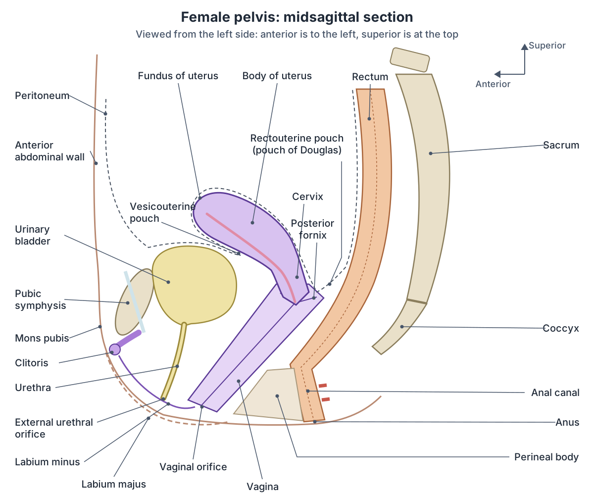
The uterus lies in the centre of the pelvis, behind and above the urinary bladder and in front of the rectum. In most adults it is anteverted (tilted forwards relative to the vagina) and anteflexed (the body bent forwards on the cervix). Its exact position changes as the bladder fills. (TeachMeAnatomy: The Uterus.)
Peritoneum covers the uterus and dips down in front of and behind it. The uterovesical (vesicouterine) pouch lies between the uterus and the bladder; the rectouterine pouch, or pouch of Douglas, lies between the uterus and the rectum. The broad and round ligaments and the uterine vessels lie at its sides. (Radiopaedia: Uterus.)
The rectouterine pouch is the lowest part of the peritoneal cavity when a woman lies on her back, so fluid such as blood, pus or ascites collects there. Its front wall is formed by the uterus and the posterior fornix of the vagina. (Radiopaedia: Rectouterine pouch.)
Radiopaedia defines version as the angle between the axes of the cervix and the vagina (anteversion is under 90 degrees) and flexion as the angle between the axes of the body and the cervix (anteflexion is under 180 degrees). Both change with filling of the bladder and rectum. (Radiopaedia: Uterus.)
A retroverted uterus tilts backwards and sits directly above the vagina. An abnormal position does not cause problems in itself, but a retroverted uterus is more likely to prolapse when abdominal pressure rises, especially after damage to the pelvic floor. (TeachMeAnatomy: The Uterus.)
Uterus anatomy: fundus, body, isthmus and cervix
The adult uterus is shaped like an upside-down pear, about 7.5 cm long, 5 cm wide at the top and 2.5 cm thick, and weighs about 30 to 40 g. It has two main divisions, the body and the cervix, joined at a slight narrowing called the isthmus. (Radiopaedia: Uterus.)
The fundus of the uterus is the rounded top of the body, above the level where the uterine tubes enter. The body narrows downwards from the fundus to the isthmus. Its cavity is a flattened triangle, with its base at the fundus between the openings of the tubes and its apex at the internal os. (Radiopaedia: Uterus.)
The body is the usual site where the embryo (blastocyst) implants. The uterine artery, a branch of the internal iliac artery, supplies the uterus. It crosses above the ureter about 1 cm lateral to the internal os, remembered as 'water under the bridge', and surgeons protect the ureter here during hysterectomy. (TeachMeAnatomy: The Uterus.)
Layers of the uterus wall
The uterine wall has three layers. The perimetrium is the outer serous layer. The myometrium is a thick layer of smooth muscle that produces the contractions of labour. The endometrium is the inner lining. (OpenStax Anatomy and Physiology 2e, 27.2: Anatomy and Physiology of the Ovarian Reproductive System.)
The endometrium has two parts. The deep stratum basalis changes little during the menstrual cycle and is not shed. The superficial stratum functionalis responds to oestrogen and progesterone, thickens, and is shed at menstruation, then regrows from the basalis. (TeachMeAnatomy: The Uterus.)
The perimetrium is a double layer of peritoneum continuous with the peritoneum of the abdomen. In pregnancy the myometrium enlarges both by cells getting bigger (hypertrophy) and by cells multiplying (hyperplasia). (TeachMeAnatomy: The Uterus.)
On MRI, three zones can be seen in the wall: the bright endometrium, a dark inner band of myometrium called the junctional zone, and the intermediate outer myometrium. (Radiopaedia: Uterus.)
The cervix
The cervix is the lower part of the uterus. The ectocervix projects into the vagina and has a central opening, the external os. The endocervical canal runs from the external os up to the internal os, where it opens into the uterine cavity. (TeachMeAnatomy: The Cervix.)
The ectocervix is covered by non-keratinised stratified squamous epithelium, while the endocervical canal is lined by mucus-secreting simple columnar epithelium. The cervix lets sperm enter the uterus, especially around ovulation when its mucus thins, and acts as a barrier that helps keep the uterus sterile. (TeachMeAnatomy: The Cervix.)
Ligaments of the uterus and ovary, including the Mackenrodt ligament
The broad ligament is a flat double sheet of peritoneum that runs from each side of the uterus to the side wall of the pelvis. It has three parts: the mesometrium around the uterus, the mesosalpinx enclosing the uterine tube, and the mesovarium attaching the ovary. It contains the ovarian and uterine arteries. (TeachMeAnatomy: Ligaments of the Female Reproductive Tract.)
The round ligament is a remnant of the embryonic gubernaculum. It runs from the uterine horn through the inguinal canal to the labium majus. The ovarian ligament (ligamentum ovarii proprium, the ligament of the ovary) is a fibrous band inside the broad ligament that joins the ovary to the side of the uterus just below the tube. The suspensory ligament of the ovary (infundibulopelvic ligament) runs from the ovary to the pelvic wall and carries the ovarian artery, vein, nerves and lymphatics. (TeachMeAnatomy: Ligaments of the Female Reproductive Tract.)
The cardinal ligament, also called the transverse cervical ligament or Mackenrodt's ligament, runs from the side of the cervix and the lateral vaginal fornix to the lateral pelvic wall, at the level of the ischial spines, along the base of the broad ligament. It houses the uterine artery and veins. The uterosacral ligaments attach the cervix to the sacrum, and the pubocervical ligaments attach it to the back of the pubic symphysis. (TeachMeAnatomy: Ligaments of the Female Reproductive Tract.)
Radiopaedia describes the cardinal ligament as a condensation of connective tissue, vessels and nerves rather than a discrete ligament. With the uterosacral and pubocervical ligaments it suspends the cervix and upper vagina and helps prevent pelvic organ prolapse. The ureter passes under the uterine vessels as it crosses the ligament, so surgery here can injure either. (Radiopaedia: Cardinal ligament.)
Ligaments of the uterus and ovary
| Ligament | Runs from and to | Contains or does |
|---|---|---|
| Broad ligament | Sides of the uterus to the lateral pelvic walls | Peritoneal fold (mesometrium, mesosalpinx, mesovarium); contains ovarian and uterine arteries |
| Round ligament of uterus | Uterine horn, through the inguinal canal, to the labium majus | Remnant of the gubernaculum; helps keep the uterus anteverted |
| Ovarian ligament (ligamentum ovarii proprium) | Ovary to the side of the uterus below the tube | Fibrous band inside the broad ligament |
| Suspensory ligament of ovary | Ovary to the lateral pelvic wall | Ovarian artery, vein, nerves and lymphatics |
| Cardinal (transverse cervical, Mackenrodt) ligament | Cervix and upper vagina to the lateral pelvic wall | Uterine artery and veins; main support of the cervix |
| Uterosacral ligament | Cervix to the sacrum | Supports the uterus from behind |
| Pubocervical ligament | Cervix to the back of the pubic symphysis | Supports the uterus from the front |
From TeachMeAnatomy (ligaments of the female reproductive tract) and Radiopaedia (cardinal ligament).
Vagina anatomy: parts, layers and vaginal rugae
The vagina is a distensible fibromuscular tube that runs upwards and backwards from the external vaginal orifice to the cervix. Its front and back walls normally lie against each other, so in cross-section it is H-shaped rather than round. (TeachMeAnatomy: The Vagina.)
In front of the vagina are the bladder and urethra; behind it are the rectouterine pouch, the rectum and the anal canal; at the sides are the ureters and levator ani. Its upper end surrounds the cervix, forming the fornices: a shallow anterior fornix and a deeper posterior fornix. (TeachMeAnatomy: The Vagina.)
The vaginal wall has three layers: an outer fibrous adventitia, a middle layer of smooth muscle, and an inner stratified squamous epithelium on a lamina propria. The epithelium is folded into transverse ridges called vaginal rugae, which increase the surface area and allow the vagina to stretch. Lactobacilli in the vagina produce lactic acid, keeping the pH below 4.5. (OpenStax Anatomy and Physiology 2e, 27.2: Anatomy and Physiology of the Ovarian Reproductive System.)
The vagina has no glands of its own; it is lubricated by cervical mucus. It is supplied by the uterine and vaginal arteries. Most of it has autonomic nerves only; just the lowest fifth has somatic sensation, through the deep perineal nerve from the pudendal nerve. Lymph from the upper third drains to the external iliac nodes, the middle third to the internal iliac nodes and the lower third to the superficial inguinal nodes. (TeachMeAnatomy: The Vagina.)
External female genitalia (vulva)
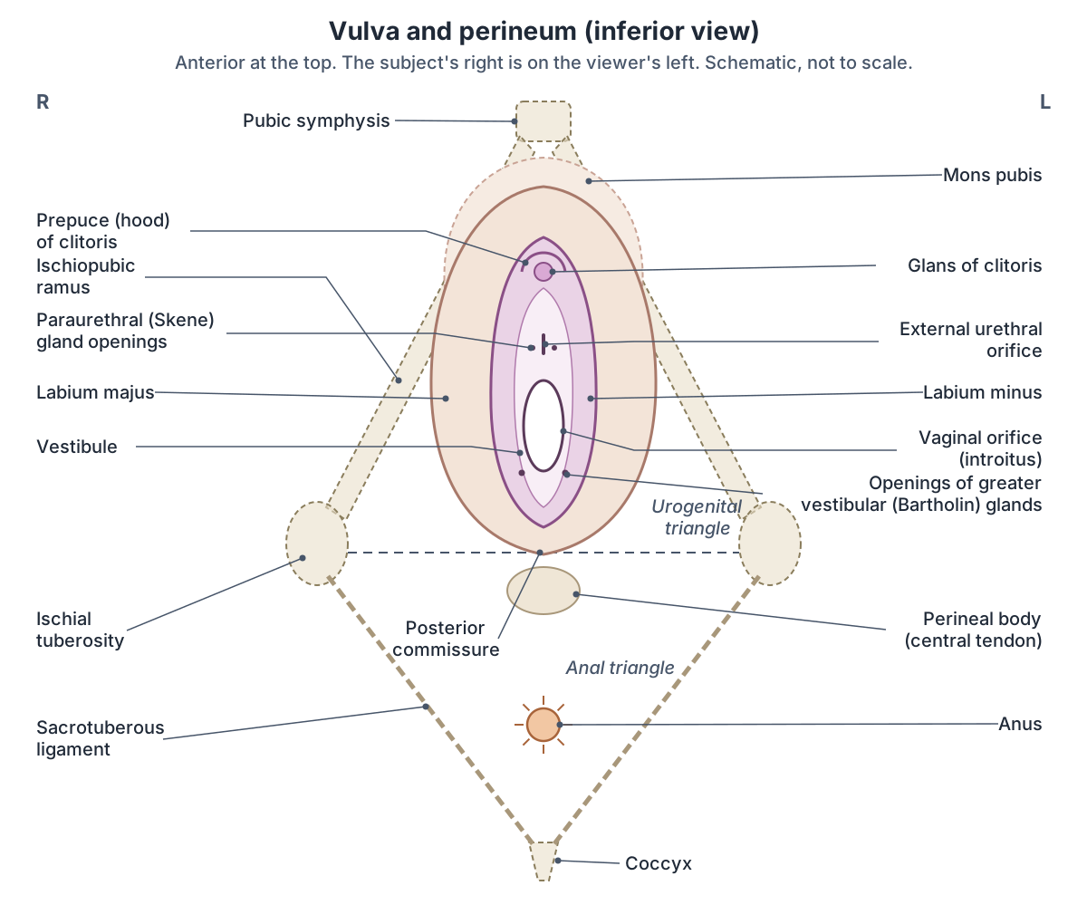
The vulva is the collective name for the external genitalia. The mons pubis is a pad of fat over the pubic symphysis. The labia majora are two hair-bearing skin folds running back from the mons to the posterior commissure over the perineal body. The labia minora are two hairless folds inside them, which join in front to form the hood (prepuce) of the clitoris and behind to form the fourchette. (TeachMeAnatomy: The Vulva.)
The vestibule is the area enclosed by the labia minora. It contains the external urethral orifice and the vaginal orifice (introitus). The greater vestibular (Bartholin) glands lie on each side of the vaginal orifice and release lubricating mucus through small ducts during arousal. (TeachMeAnatomy: The Vulva.)
The external urethral orifice lies in the vestibule in front of the vaginal opening and about 2 to 3 cm behind the clitoris. The paraurethral (Skene) glands lie on either side of the lower urethra and are considered the female equivalent of the prostate. (TeachMeAnatomy: The Urethra.)
The clitoris is the female homologue of the penis. Its body is formed by two corpora cavernosa, which continue backwards as the crura along each inferior pubic ramus; the glans lies at the front under the prepuce. Older texts do not count the bulbs of the vestibule as part of the clitoris, but newer sources suggest that view is outdated. The prepuce formed by the labia minora is the homologue of the penile foreskin. (Radiopaedia: Clitoris.)
The vulva is supplied by the internal and external pudendal arteries. Its front is supplied by the ilioinguinal nerve and the genital branch of the genitofemoral nerve, and its back by the pudendal nerve and the posterior cutaneous nerve of the thigh. Lymph drains to the superficial inguinal nodes. (TeachMeAnatomy: The Vulva.)
Perineum area and the perineal body
The perineum is the region below the pelvic floor, between the thighs. Anatomically it is diamond-shaped, bounded by the pubic symphysis in front, the tip of the coccyx behind, and the ischiopubic rami, ischial tuberosities and sacrotuberous ligaments at the sides. In everyday clinical use, perineum often means just the skin between the vaginal opening and the anus. (TeachMeAnatomy: The Perineum.)
A line between the two ischial tuberosities divides it into an anterior urogenital triangle, containing the external genitalia and urethra, and a posterior anal triangle, containing the anus, external anal sphincter and the fat-filled ischioanal fossae. (TeachMeAnatomy: The Perineum.)
The perineal membrane is a tough sheet of fascia across the urogenital triangle, pierced by the urethra and vagina. Above it, the deep perineal pouch contains part of the urethra, the external urethral sphincter and part of the vagina. Below it, the superficial perineal pouch contains the erectile tissue of the clitoris, the ischiocavernosus, bulbospongiosus and superficial transverse perineal muscles and the greater vestibular glands. (TeachMeAnatomy: The Perineum.)
The perineal body (central tendon of the perineum) is a fibromuscular mass where the two triangles meet, between the vagina and the anal canal. Levator ani, bulbospongiosus, the transverse perineal muscles and the anal and urethral sphincters attach to it. It supports the back wall of the vagina and can be stretched or torn in childbirth; an episiotomy is a controlled cut to avoid uncontrolled tearing. (TeachMeAnatomy: The Perineum.)
In women the bulbospongiosus covers the bulbs of the vestibule and the greater vestibular glands. It arises from the perineal body, narrows the vaginal orifice and is supplied by the pudendal nerve. (Radiopaedia: Bulbospongiosus muscle.)
External urethral sphincter
The female urethra is about 4 cm long. It runs from the bladder neck down through the pelvic floor and perineal membrane and opens into the vestibule. Because it is short, women are more prone to urinary tract infections such as cystitis. (TeachMeAnatomy: The Urethra.)
The external urethral sphincter is skeletal muscle under voluntary control in the deep perineal pouch. In women it has three parts: the sphincter urethrae, a ring around the upper two-thirds of the urethra; the compressor urethrae, paired bands from the ischiopubic rami that loop around the urethra above the perineal membrane; and the urethrovaginal sphincter, a flat band that encircles both the urethra and the vagina. (Kenhub: Urethral sphincters.)
It is supplied by perineal branches of the pudendal nerve (S2 to S4). It keeps the urethra closed between voids and can stop the flow voluntarily, while the smooth muscle internal urethral sphincter at the bladder neck is under autonomic control. In women the external sphincter also pulls the urethra down against the front wall of the vagina, which helps continence. (Kenhub: Urethral sphincters.)
Mammary glands (breast)
The breasts are accessory organs of the reproductive system. They lie on the front of the chest over pectoralis major and serratus anterior, from the side of the sternum to the mid-axillary line and from the 2nd to the 6th costal cartilages, with an axillary tail extending towards the armpit. (TeachMeAnatomy: The Breasts.)
The mammary glands are modified sweat glands that make milk. About 15 to 20 lactiferous ducts open at the nipple; each widens into a lactiferous sinus and leads back to a lobe of milk-secreting alveoli. Suspensory (Cooper's) ligaments attach the breast tissue to the overlying skin. (OpenStax Anatomy and Physiology 2e, 27.2: Anatomy and Physiology of the Ovarian Reproductive System.)
About 75 percent of breast lymph drains to the axillary lymph nodes, which is why these nodes are examined in breast cancer. (TeachMeAnatomy: The Breasts.)
Clinical relevance
Fibroids are benign, oestrogen-dependent tumours of the myometrium; most cause no symptoms, but large ones can cause heavy periods, pain or infertility. Endometriosis is endometrial tissue outside the uterus, most often on the ovaries and uterine ligaments, and is associated with painful periods and infertility. (TeachMeAnatomy: The Uterus.)
In an ectopic pregnancy a fertilised egg implants in the uterine tube, often after earlier inflammation (salpingitis) has partly blocked it. It is an emergency because the tube can rupture and bleed. (TeachMeAnatomy: The Fallopian Tubes.)
Ovarian cysts are fluid-filled sacs that are common before the menopause and usually benign; large ones can cause pain or twisting of the ovary (torsion). Ovarian cancer often causes vague symptoms and is found late. (TeachMeAnatomy: The Ovaries.)
A blocked Bartholin gland duct can form a cyst, which may become infected (bartholinitis). (TeachMeAnatomy: The Perineum.)
Frequently asked questions
Where is the uterus located?
The uterus lies in the middle of the pelvis, behind and above the urinary bladder and in front of the rectum. In most women it is anteverted and anteflexed, tipping forwards over the bladder. Its cervix projects into the top of the vagina, and peritoneal pouches lie in front of it (vesicouterine pouch) and behind it (rectouterine pouch).
What are the layers of the uterus?
The uterine wall has three layers. The outer perimetrium is a serous layer of peritoneum. The thick middle myometrium is smooth muscle that contracts in labour. The inner endometrium has a deep stratum basalis that stays and a superficial stratum functionalis that thickens each cycle and is shed during menstruation.
What are the parts of the uterine tube?
From the ovary towards the uterus, each uterine (fallopian) tube has fimbriae, the funnel-shaped infundibulum, the wide ampulla where fertilisation usually happens, the narrow isthmus, and the intramural (interstitial) part that passes through the uterine wall. The tube is about 10 to 12 cm long and lies in the upper edge of the broad ligament.
What is the Mackenrodt ligament?
The Mackenrodt ligament is another name for the cardinal or transverse cervical ligament. It runs from the side of the cervix and upper vagina to the lateral pelvic wall at the base of the broad ligament and carries the uterine artery and veins. With the uterosacral and pubocervical ligaments, it supports the cervix and helps prevent prolapse.
What are vaginal rugae?
Vaginal rugae are transverse folds of the stratified squamous epithelium that lines the vagina. They increase the surface area of the vaginal wall and allow it to stretch, for example during childbirth. The wall under the rugae has a lamina propria, a smooth muscle layer and an outer fibrous adventitia.
What are the parts of the vulva?
The vulva includes the mons pubis, the labia majora and labia minora, the clitoris with its hood (prepuce), and the vestibule between the labia minora. The vestibule contains the external urethral orifice in front and the vaginal orifice behind it, with the openings of the greater vestibular (Bartholin) glands on each side of the vaginal orifice.
What is the perineum?
Anatomically, the perineum is the diamond-shaped region below the pelvic floor, between the pubic symphysis, ischial tuberosities and coccyx. A line between the ischial tuberosities divides it into the urogenital triangle in front and the anal triangle behind. Clinically, the term often refers to the area of skin between the vaginal opening and the anus, over the perineal body.
References
- OpenStax. Anatomy and Physiology 2e, §27.2 Anatomy and Physiology of the Ovarian Reproductive System. Rice University, 2022.
- Jones O. The Uterus. TeachMeAnatomy.
- Jones O. The Fallopian Tubes. TeachMeAnatomy.
- Jones O. The Ovaries. TeachMeAnatomy.
- Jones O. The Vagina. TeachMeAnatomy.
- Jones O. Ligaments of the Female Reproductive Tract. TeachMeAnatomy.
- Jones O. The Cervix. TeachMeAnatomy.
- Jones O. The Vulva. TeachMeAnatomy.
- Jones O. The Perineum. TeachMeAnatomy.
- Jones O. The Urethra. TeachMeAnatomy.
- Jones O. The Breasts. TeachMeAnatomy.
- Uterus. Radiopaedia reference article.
- Fallopian tube. Radiopaedia reference article.
- Cardinal ligament. Radiopaedia reference article.
- Rectouterine pouch. Radiopaedia reference article.
- Clitoris. Radiopaedia reference article.
- Bulbospongiosus muscle. Radiopaedia reference article.
- Urethral sphincters. Kenhub anatomy library.
Related anatomy
- Male reproductive systemMale reproductive anatomy with labelled diagrams: scrotum, testes, epididymis, ductus deferens, seminal gland…
- PelvisPelvis bones explained: ilium, ischium and pubis, obturator foramen, pelvic ligaments and male vs female pelvis, with…
- SacrumSacrum anatomy explained: promontory, ala, sacral foramina, crests and hiatus, joints and ligaments, sacrum vs coccyx…
- UreterWhat do the ureters do? Ureter function, course, relations, the three narrowings where kidney stones lodge, blood…
- KidneyWhere are your kidneys? Kidney location on the back (T12 to L3), coverings, relations, cortex, medulla, calyces, renal…
Explore the whole body in the interactive 3D human anatomy model, or browse the A–Z list of human structures.