

What are the muscles of mastication?
Mastication is the medical word for chewing. The muscles of mastication are four paired muscles that move the mandible at the temporomandibular joint: masseter, temporalis, medial pterygoid and lateral pterygoid. They develop from the first pharyngeal arch and are therefore supplied by the nerve of that arch, the mandibular nerve, a branch of the trigeminal nerve (cranial nerve V). (TeachMeAnatomy: The Muscles of Mastication.)
OpenStax describes masseter as the main chewing muscle because it elevates the mandible to close the mouth, with temporalis helping to elevate and retract the jaw. The lateral pterygoid depresses and protracts the mandible and moves it from side to side, and the medial pterygoid elevates it and assists side-to-side movement. (OpenStax Anatomy and Physiology 2e, 11.3: Axial Muscles of the Head, Neck, and Back.)
Chewing is the first stage of mechanical digestion. The lips and cheeks keep food between the teeth, and the teeth tear and crush it into smaller particles that can be mixed with saliva and swallowed. The cheek muscle buccinator and the tongue help with this, but they are not muscles of mastication: buccinator is a muscle of facial expression supplied by the facial nerve. (OpenStax Anatomy and Physiology 2e, 23.3: The Mouth, Pharynx, and Esophagus.)
Muscles of mastication: origin, insertion, nerve and action
| Muscle | Origin | Insertion | Nerve (all from V3) | Main actions |
|---|---|---|---|---|
| Masseter (musculus masseter) | Superficial part: maxillary process of the zygomatic bone and lower border of the zygomatic arch. Deep part: zygomatic arch | Lateral surface of the ramus and angle of the mandible (masseteric tuberosity); deep part higher on the ramus | Masseteric nerve | Elevates the mandible (closes the jaw); anterior fibres help protrude it |
| Temporalis (musculus temporalis) | Floor of the temporal fossa below the temporal line, and the temporal fascia | Apex and medial surface of the coronoid process of the mandible | Deep temporal nerves | Anterior fibres elevate the mandible; posterior fibres retract it |
| Medial pterygoid (internal pterygoid, musculus pterygoideus medialis) | Deep head: medial surface of the lateral pterygoid plate. Superficial head: maxillary tuberosity and pyramidal process of the palatine bone | Medial surface of the ramus and angle of the mandible | Nerve to medial pterygoid | Elevates the mandible; helps protrude it and move it side to side |
| Lateral pterygoid (musculus pterygoideus lateralis) | Superior head: infratemporal crest and surface of the greater wing of the sphenoid. Inferior head: lateral surface of the lateral pterygoid plate | Superior head: capsule and articular disc of the TMJ. Inferior head: pterygoid fovea on the neck of the condylar process | Nerve to lateral pterygoid | Both sides: protrude the mandible and open the mouth. One side: moves the jaw to the opposite side |
Attachments from TeachMeAnatomy and Kenhub. All four muscles are supplied by branches of the mandibular division of the trigeminal nerve (V3).
Masseter muscle (musculus masseter)
Masseter is the thick, rectangular muscle you can feel at the back of the cheek when you clench your teeth. Its superficial part arises from the maxillary process of the zygomatic bone and the front two-thirds of the lower border of the zygomatic arch, and inserts on the masseteric tuberosity on the lower lateral surface of the ramus, near the angle of the mandible. Its deep part has more vertical fibres that arise from the zygomatic arch and insert higher on the ramus. (Kenhub: Masseter muscle.)
Masseter elevates the mandible to close the jaw with force, and its anterior fibres help to protrude it. It is supplied by the masseteric nerve from the mandibular nerve and by the masseteric artery from the maxillary artery. The parotid duct crosses its outer surface before turning inwards through buccinator to open inside the cheek. (Kenhub: Masseter muscle.)
TeachMeAnatomy calls masseter the most powerful muscle of mastication. Kenhub, on the other hand, describes temporalis as the strongest muscle acting on the TMJ; the comparison depends on how strength is measured, and both muscles are powerful jaw closers. (TeachMeAnatomy: The Muscles of Mastication.)
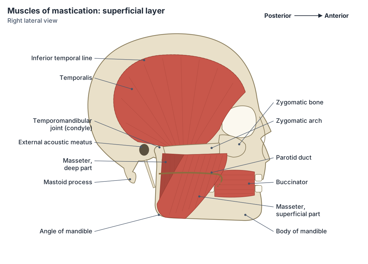
Temporalis muscle (musculus temporalis)
Temporalis is a broad, fan-shaped muscle on the side of the skull. It arises from most of the temporal fossa below the temporal line and from the temporal fascia that covers it. Its fibres converge into a tendon that passes deep to the zygomatic arch and inserts on the apex and medial surface of the coronoid process of the mandible. (Kenhub: Temporalis muscle.)
The anterior, more vertical fibres of temporalis elevate the mandible, and the posterior, almost horizontal fibres retract it (pull it backwards). It is supplied by the anterior, middle and posterior deep temporal nerves from the mandibular nerve, and by the deep temporal branches of the maxillary artery with help from the superficial temporal artery. (Kenhub: Temporalis muscle.)
The temporal fascia over the muscle can be harvested by surgeons as a graft to repair a perforated eardrum, an operation called myringoplasty. (TeachMeAnatomy: The Muscles of Mastication.)
Medial pterygoid muscle (internal pterygoid)
The medial pterygoid, also called the internal pterygoid or musculus pterygoideus internus in older texts, is a thick muscle on the inner side of the ramus. Its deep head arises from the medial surface of the lateral pterygoid plate of the sphenoid bone. Its small superficial head arises from the maxillary tuberosity and the pyramidal process of the palatine bone. (Kenhub: Medial pterygoid muscle.)
Both heads run downwards, backwards and laterally to insert by a tendinous sheet into a rough triangular area on the medial surface of the ramus and angle of the mandible. Masseter inserts on the opposite, lateral surface of the same region, so the two muscles grip the angle of the mandible from both sides. (Kenhub: Medial pterygoid muscle.)
Acting together, the two medial pterygoids elevate the mandible and, with the lateral pterygoids, protrude it. One medial pterygoid acting alone rotates the mandible slightly medially, helping to swing the chin towards the other side. The muscle is supplied by the nerve to medial pterygoid from the mandibular nerve. The lingual and inferior alveolar nerves pass between it and the ramus, which is relevant to dental nerve blocks. (Kenhub: Medial pterygoid muscle.)
Lateral pterygoid muscle (lat pterygoid)
The lateral pterygoid is a short, thick muscle with two heads that runs almost horizontally backwards to the jaw joint. Its superior head arises from the infratemporal crest and surface of the greater wing of the sphenoid. Its much larger inferior head arises from the lateral surface of the lateral pterygoid plate. (Kenhub: Lateral pterygoid muscle.)
The superior head inserts into the front of the capsule and articular disc of the TMJ, and the inferior head inserts into the pterygoid fovea, a small pit on the front of the neck of the mandible's condylar process. Because its fibres are horizontal, the lateral pterygoid is the main muscle that pulls the mandible forwards (protrusion). (TeachMeAnatomy: The Muscles of Mastication.)
When both lateral pterygoids contract, the inferior heads pull the condyles forwards and the superior heads pull the discs forwards with them, so the jaw protrudes and the mouth opens. When one side contracts, the condyle on that side moves forwards and inwards and the chin swings to the opposite side; this works with the medial pterygoid of the same side when we grind food. (Kenhub: Lateral pterygoid muscle.)
The lateral pterygoid is supplied by the nerve to lateral pterygoid from the mandibular nerve and by pterygoid branches of the maxillary artery. The maxillary artery and the buccal nerve pass between its two heads, and the lingual and inferior alveolar nerves appear at its lower border. (Kenhub: Lateral pterygoid muscle.)
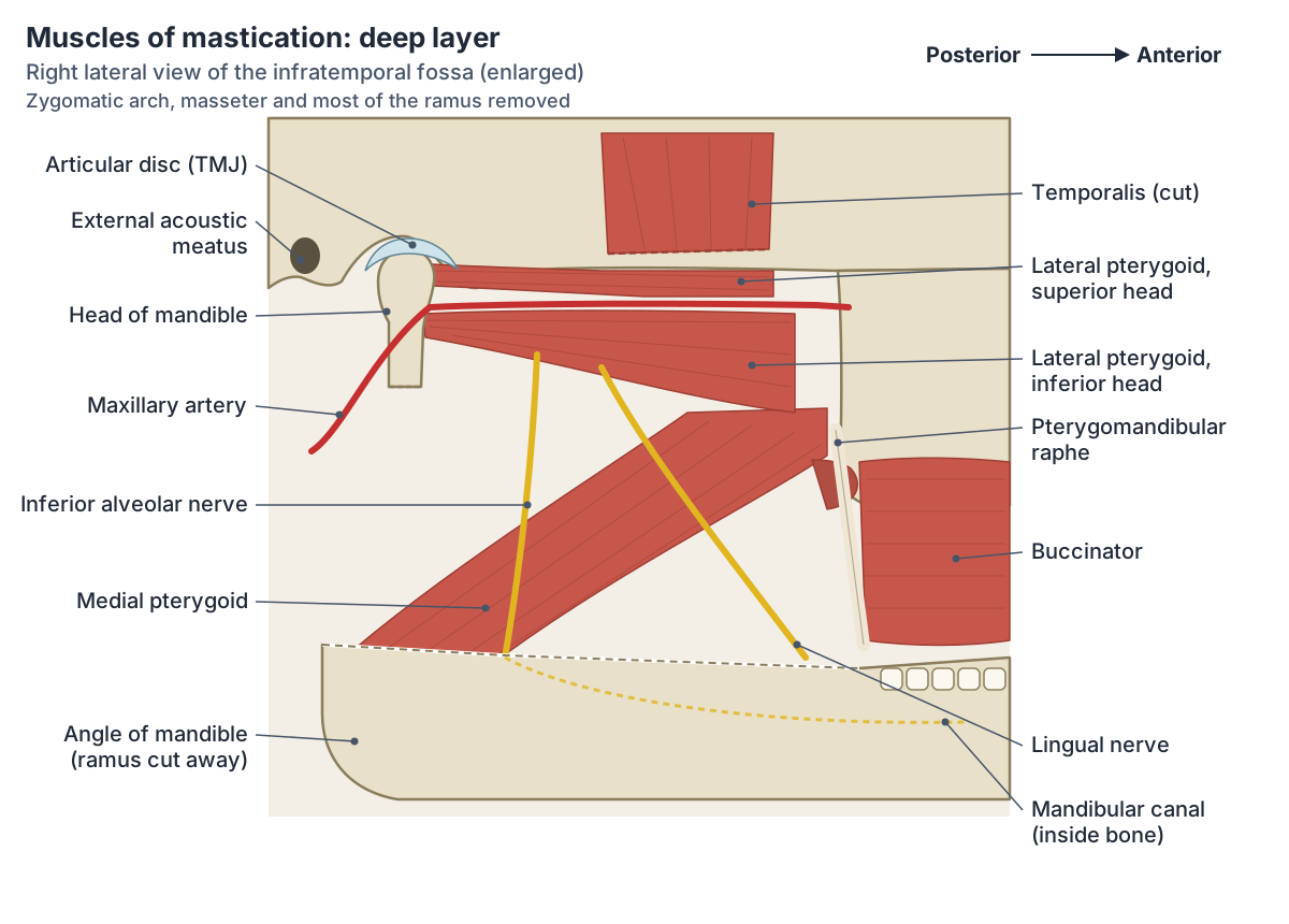
The infratemporal fossa (fossa infratemporalis)
The infratemporal fossa is the space below the skull base, deep to the ramus of the mandible. Its lateral wall is the ramus and condylar process of the mandible; its medial wall is the lateral pterygoid plate; in front is the back wall of the maxillary sinus; behind is the carotid sheath; its roof is the greater wing of the sphenoid; and the medial pterygoid forms its floor. (TeachMeAnatomy: The Infratemporal Fossa.)
The medial and lateral pterygoids lie inside the fossa, while masseter and temporalis attach to its borders. The lateral pterygoid divides the contents: branches of the mandibular nerve lie deep to it and the maxillary artery runs superficial to it. The mandibular nerve enters through the foramen ovale and gives the auriculotemporal, buccal, lingual and inferior alveolar nerves; the chorda tympani joins the lingual nerve here. (TeachMeAnatomy: The Infratemporal Fossa.)
The fossa also contains the pterygoid venous plexus, which connects with the cavernous sinus inside the skull. This connection is a route by which infection from the face can spread into the cranial cavity. Superiorly the fossa opens into the temporal fossa, and it communicates with the pterygopalatine fossa through the pterygomaxillary fissure. (TeachMeAnatomy: The Infratemporal Fossa.)
TMJ anatomy: mandibular fossa, articular eminence and disc
The temporomandibular joint is an atypical synovial joint between the condylar process (head) of the mandible and the mandibular fossa and articular eminence of the temporal bone. The articular eminence, also called the articular tubercle, is the rounded ridge of bone just in front of the mandibular fossa. (Radiopaedia: Temporomandibular joint.)
Unlike most synovial joints, the TMJ's articular surfaces are covered by fibrocartilage instead of hyaline cartilage. A fibrocartilage articular disc separates the bones completely and divides the joint into two compartments, each with its own synovial membrane. (Kenhub: Temporomandibular joint.)
In side view the disc has a bow-tie shape, with thick anterior and posterior bands joined by a thin intermediate zone. Behind the posterior band is the loose, richly innervated retrodiscal tissue (bilaminar zone). The capsule is loose above the disc and tight below it, anchoring the disc to the condylar neck, and the lateral pterygoid inserts into the capsule, the disc and the condylar neck. (Radiopaedia: Temporomandibular joint.)
Gliding (translation) for protrusion, retraction and side-to-side movement happens in the upper (discotemporal) compartment, while hinge-like rotation for opening and closing happens in the lower (discomandibular) compartment. When the mouth opens wide, the condyle and disc translate forwards so that the condyle comes to lie beneath the articular eminence. (Radiopaedia: Temporomandibular joint.)
The joint is reinforced by the lateral (temporomandibular) ligament, a thickening of the capsule from the articular tubercle to the neck of the mandible that prevents posterior dislocation. The sphenomandibular and stylomandibular ligaments lie outside the capsule. The joint is supplied mainly by the auriculotemporal and masseteric branches of the mandibular nerve and by branches of the external carotid artery, especially the superficial temporal artery. (TeachMeAnatomy: The Temporomandibular Joint.)
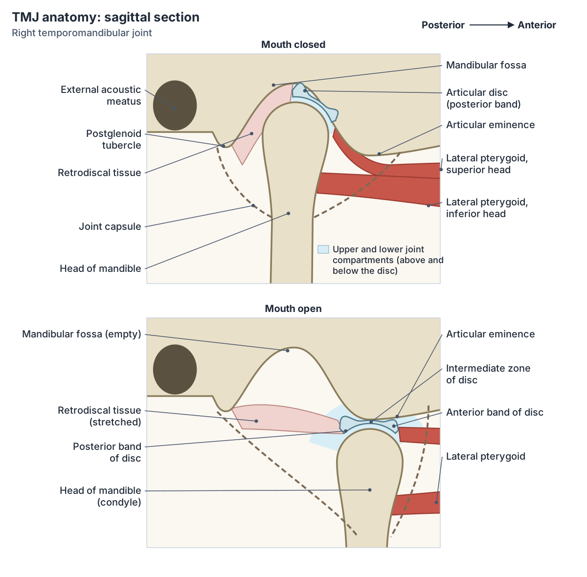
How the muscles of mastication move the jaw
Elevation, the powerful closing movement, is produced by temporalis, masseter and the medial pterygoid. Depression, opening the mouth, is mainly due to gravity, and the digastric, geniohyoid and mylohyoid muscles help when there is resistance. Protrusion is produced by the lateral pterygoid with help from the medial pterygoid, and retraction by the posterior fibres of temporalis. (TeachMeAnatomy: The Temporomandibular Joint.)
Grinding food from side to side is not a separate movement at one joint. It comes from alternating protrusion and retraction on each side, so the mandible swings around one condyle and then the other. (TeachMeAnatomy: The Temporomandibular Joint.)
In practice the two kinds of movement are combined: Kenhub's TMJ article notes that rotation and translation usually happen together during normal opening and closing. (Kenhub: Temporomandibular joint.)
The lateral pterygoid also works while the mouth closes. Its inferior heads contract eccentrically (lengthening while active) to control the backward glide of the condyles into the fossae, and the retrodiscal tissue behind the disc limits how far forward the condyle can travel. (Kenhub: Lateral pterygoid muscle.)
Jaw movements and the muscles that produce them
| Movement at the TMJ | Main muscles |
|---|---|
| Elevation (closing the mouth) | Temporalis, masseter, medial pterygoid |
| Depression (opening the mouth) | Mainly gravity; lateral pterygoid; digastric, geniohyoid and mylohyoid against resistance |
| Protrusion (jaw forwards) | Lateral pterygoid, assisted by medial pterygoid |
| Retraction (jaw backwards) | Posterior fibres of temporalis |
| Side-to-side (grinding) | Lateral and medial pterygoids of one side, then the other (alternating protrusion and retraction) |
The pterygomandibular raphe
The pterygomandibular raphe is a tendinous band that runs from the hamulus of the medial pterygoid plate to the mylohyoid ridge on the inner surface of the mandible. The buccinator attaches to its front edge and the superior pharyngeal constrictor to its back edge. (Radiopaedia: Pterygomandibular raphe.)
Inside the mouth the raphe is covered by a fold of mucosa, the pterygomandibular fold, between the anterior pillar of the tonsil and the retromolar area behind the last molar. Dentists use this fold as a landmark when giving an inferior alveolar nerve block to numb the lower teeth. (Radiopaedia: Pterygomandibular raphe.)
Clinical notes: jaw dislocation, trismus and the jaw jerk
The most common TMJ injury is anterior dislocation, which can happen during a wide yawn, a large bite or a blow to the side of the face. The head of the mandible slides forwards out of the mandibular fossa, in front of the articular eminence, and the patient cannot close the mouth. Posterior dislocation is very rare because the postglenoid tubercle and the strong lateral ligament resist it. (TeachMeAnatomy: The Temporomandibular Joint.)
Trismus is a persistent spasm of the masseter that makes it hard to open the mouth. It is a feature of tetanus and of abscesses in the space beneath masseter, which usually spread from an infected lower third molar. The masseter (jaw jerk) reflex is also tested in clinical examination; an abnormal response may point to a lesion of the trigeminal nerve or brainstem. (Kenhub: Masseter muscle.)
Tension in temporalis is a common cause of pain over the temple, for example with tooth grinding (bruxism) or after long dental treatment with the mouth open. Kenhub notes that temple pain also needs inflammation of the superficial temporal artery (giant cell arteritis) to be ruled out. (Kenhub: Temporalis muscle.)
Muscles of mastication in the 3D model
- Mandible
- The lower jaw bone.
- Maxilla
- The upper jaw bone, paired.
- Pterygomandibular raphe
- A tendinous band between the pterygoid hamulus and the mandible, not a true muscle.
- Sphenoid bone
- A butterfly-shaped bone at the base of the skull.
- Temporal bone
- A paired bone forming part of the side and base of the skull.
- Zygomatic bone
- The cheekbone.
Frequently asked questions
What are the 4 muscles of mastication?
The four muscles of mastication are masseter, temporalis, medial pterygoid and lateral pterygoid, one of each on both sides. Masseter and temporalis lie on the side of the head and close the jaw. The two pterygoids lie deep to the ramus of the mandible in the infratemporal fossa. All four are supplied by the mandibular division of the trigeminal nerve (V3).
What nerve supplies the muscles of mastication?
All four are supplied by the mandibular nerve (V3), the third division of the trigeminal nerve, which leaves the skull through the foramen ovale. Masseter is supplied by the masseteric nerve, temporalis by the deep temporal nerves, and each pterygoid by its own nerve to medial or lateral pterygoid. This follows from their development from the first pharyngeal arch.
Which muscle of mastication opens the mouth?
The lateral pterygoid is the only muscle of mastication that helps open the mouth. When both lateral pterygoids contract they pull the condyles and discs forwards and depress the chin. Most opening is due to gravity, with the digastric, geniohyoid and mylohyoid muscles below the jaw helping against resistance. Masseter, temporalis and medial pterygoid all close the mouth.
What is the difference between the medial and lateral pterygoid?
Both arise from the lateral pterygoid plate of the sphenoid bone. The medial pterygoid runs downwards to the inner surface of the angle of the mandible and closes the jaw. The lateral pterygoid runs horizontally backwards to the neck of the condyle and the TMJ disc and pulls the jaw forwards, opens the mouth and moves the jaw to the opposite side.
What is the articular eminence?
The articular eminence, or articular tubercle, is the rounded ridge of the temporal bone just in front of the mandibular fossa. With the fossa it forms the upper surface of the temporomandibular joint. When the mouth opens wide, the condyle and articular disc glide forwards to sit beneath the eminence; if the condyle slips in front of it, the jaw dislocates.
Is the buccinator a muscle of mastication?
No. Buccinator keeps food between the teeth while chewing, but it is a muscle of facial expression supplied by the facial nerve. It arises in part from the pterygomandibular raphe and runs forward in the cheek to the corner of the mouth. The muscles of mastication proper are the four muscles supplied by the mandibular nerve.
References
- OpenStax. Anatomy and Physiology 2e, §11.3 Axial Muscles of the Head, Neck, and Back. Rice University, 2022.
- OpenStax. Anatomy and Physiology 2e, §23.3 The Mouth, Pharynx, and Esophagus. Rice University, 2022.
- TeachMeAnatomy. The Muscles of Mastication.
- TeachMeAnatomy. The Temporomandibular Joint.
- TeachMeAnatomy. The Infratemporal Fossa.
- Kenhub. Masseter muscle.
- Kenhub. Temporalis muscle.
- Kenhub. Medial pterygoid muscle.
- Kenhub. Lateral pterygoid muscle.
- Kenhub. Temporomandibular joint.
- Radiopaedia. Temporomandibular joint.
- Radiopaedia. Pterygomandibular raphe.
- Mitsuhashi N. et al. (2009). BodyParts3D: 3D structure database for anatomical concepts. Nucleic Acids Research 37, D782–D785. Data © The Database Center for Life Science, CC BY 4.0.
Related anatomy
- MandibleMandible anatomy with a labelled diagram: body, ramus, angle, condylar and coronoid processes, foramina, muscle…
- Temporal boneThe temporal bone (os temporale) explained: petrous part and inner ear, mastoid process, zygomatic and styloid…
- Sphenoid boneThe sphenoid bone (os sphenoidale): sella turcica and hypophyseal fossa, greater and lesser wings, pterygoid process…
- Facial musclesFace muscles labeled: labelled diagrams and a table of every muscle of facial expression, from procerus and…
- TongueTongue anatomy explained: parts and papillae of the dorsum, the underside with the frenulum and sublingual folds…
- Pterygomandibular raphePterygomandibular raphe: A tendinous band between the pterygoid hamulus and the mandible, not a true muscle. See where…
Explore the whole body in the interactive 3D human anatomy model, or browse the A–Z list of human structures.