

Sacrum at a glance
- What it is
- A triangular bone of five fused sacral vertebrae.
- Location
- Lies at the back of the pelvis between the hip bones (sacroiliac joints), below L5 and above the coccyx.
- Function
- Transfers weight from the spine to the pelvis.
- Body system
- Skeletal · single, midline structure
- FMA ID
- FMA16202 (Foundational Model of Anatomy)
Key facts
- Features the sacral promontory, four pairs of anterior and four pairs of posterior sacral foramina, and the sacral hiatus.
- Its auricular surfaces form the sacroiliac joints with the ilia, and the anterior and posterior sacral foramina transmit the rami of S1–S4.
- The sacral canal continues the vertebral canal and opens at the sacral hiatus, usually at S4–S5.
Clinical relevance
Sacral hiatus is used for caudal epidural anaesthesia.
What is the sacrum?
The sacrum is the second-lowest part of the vertebral column and forms the back wall of the bony pelvis. It is shaped like an upside-down triangle: the wide base faces upwards and the narrow apex points down. It is also curved, hollow (concave) at the front and rounded (convex) at the back. (Radiopaedia: Sacrum.)
The sacrum begins as five separate sacral vertebrae. They start to fuse after about age 20 and the process is not complete until middle age. The coccyx below it is formed from fused coccygeal vertebrae. The wide upper end carries body weight; the narrow lower tip does not. (OpenStax Anatomy and Physiology 2e, 7.3: The Vertebral Column.)
Kenhub calls the sacrum the keystone of the human body: it links the spine to the two iliac bones and contributes to the stability of the hips. (Kenhub: Sacrum.)
When you stand, the front (pelvic) surface of the sacrum faces forwards and downwards and its back (dorsal) surface faces backwards and upwards. The front and back surfaces give attachment to the ligaments and muscles of the pelvis. (TeachMeAnatomy: The Sacrum.)
In the 3D model on this page the sacrum is shown with the fifth lumbar vertebra and the hip bones. The model comes from BodyParts3D, an adult male data set, and has no coccyx, so the coccyx is covered in the text and diagram instead.
Parts of the sacrum: base, promontory, ala and apex
The base is the upper surface of the first sacral vertebra (S1). It meets the fifth lumbar vertebra (L5) and the L5/S1 intervertebral disc. The front edge of the S1 body projects forwards as the sacral promontory, which forms the back edge of the pelvic inlet (pelvic brim). (TeachMeAnatomy: The Sacrum.)
On each side of the S1 body is a wing-like ala (plural alae). The alae grow out sideways from S1. The parts of the sacrum around and lateral to the anterior foramina come from costal (rib) elements fused to the vertebrae, while the posterior foramina are formed by the vertebral parts. (Radiopaedia: Sacrum.)
Behind the base, S1 keeps two superior articular processes. Their concave facets face backwards and inwards and form the facet (zygapophyseal) joints with the inferior articular processes of L5. S1 is the largest sacral vertebra and still shows recognisable pedicles and transverse processes. (Kenhub: Sacrum.)
The apex is the narrow lower end, the body of S5. It carries an oval facet that joins the coccyx. The lateral surfaces carry the ear-shaped auricular surfaces, which articulate with the ilium of each hip bone. Just behind each auricular surface is a rough area with depressions for the strong sacroiliac ligaments. (Kenhub: Sacrum.)
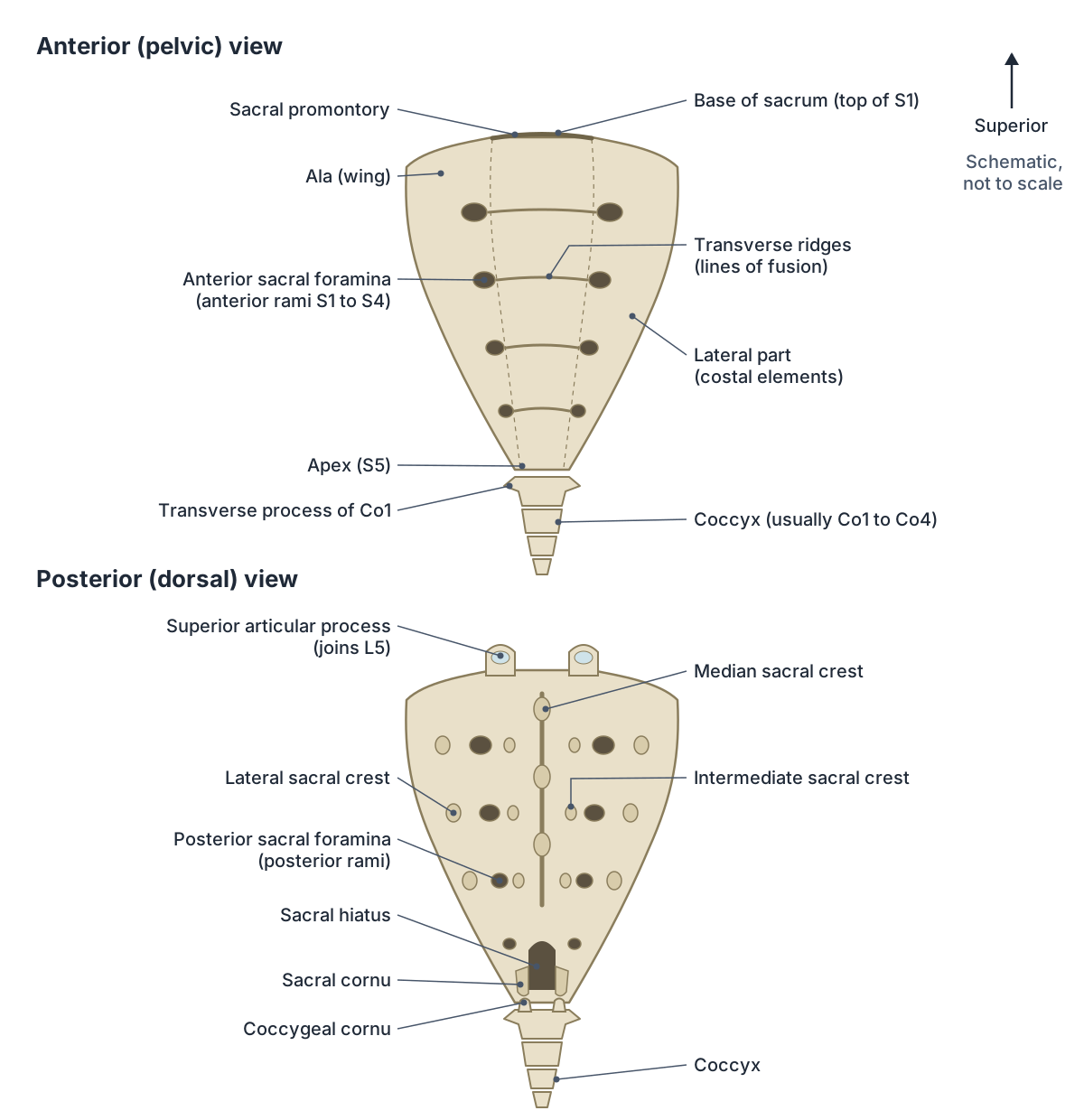
Anterior (pelvic) surface of the sacrum
The pelvic surface is smoother than the back. Four transverse ridges cross it: they mark where the bodies of the five sacral vertebrae, and the discs between them, have fused. At each end of a ridge is an anterior sacral foramen, giving four pairs in all. (TeachMeAnatomy: The Sacrum.)
The four pairs of anterior sacral foramina transmit the anterior (ventral) rami of S1 to S4. The fifth sacral nerve leaves lower down, through the sacral hiatus. The ventral rami of S1 to S4 join the lumbosacral trunk (L4 and L5) to form the sacral plexus on the front of the piriformis muscle. (Radiopaedia: Sacrum.)
The sacral plexus gives rise to the sciatic nerve (L4 to S3), the pudendal nerve (S2 to S4) and the superior (L4 to S1) and inferior (L5 to S2) gluteal nerves. Medial to the anterior sacral foramina lie the sympathetic trunks, which run down the pelvic surface of the sacrum. (Radiopaedia: Sacral plexus.)
Posterior (dorsal) surface and the sacral crests
The dorsal surface is rough because the fused parts of the vertebrae form three kinds of vertical ridge, called crests. The median sacral crest runs down the middle and is formed by the fused spinous processes. TeachMeAnatomy describes it from S1 to S3, while Kenhub gives S1 to S4, so the number of spines in the crest varies. (TeachMeAnatomy: The Sacrum.)
Either side of the median crest is an intermediate sacral crest, formed by the fused articular processes. Further out is the lateral sacral crest, formed by the fused transverse processes. The posterior sacroiliac ligaments attach to the intermediate and lateral crests, and the sacrotuberous ligament attaches to the lateral crest. (TeachMeAnatomy: The Sacrum.)
Between the intermediate and lateral crests are four pairs of posterior sacral foramina. They carry the smaller posterior (dorsal) rami of the sacral nerves. At the lower end, the inferior articular processes of S5 form two small projections, the sacral cornua (horns), which articulate with the coccygeal cornua. (Radiopaedia: Sacrum.)
The sacral canal and sacral hiatus
The sacral canal is the triangular tunnel through the sacrum. It continues the vertebral canal of the lumbar spine and ends at the sacral hiatus. It holds the sacral and coccygeal nerve roots, the spinal meninges as far down as S2, and the filum terminale. The epidural space continues all the way to the hiatus. (Radiopaedia: Sacrum.)
The spinal cord itself does not reach the sacrum. Kenhub places its end around L2, so the sacral canal contains the cauda equina and the filum terminale rather than cord. The dural sac usually ends at about S2, and the filum terminale continues down to attach to the coccyx. (Kenhub: Sacrum.)
The sacral hiatus is an opening on the back of the sacrum, usually at the level of S5, where the S5 spinous process (and sometimes the one above) has not formed. It is flanked by the sacral cornua and covered by the sacrococcygeal ligament, fat and skin. The fifth sacral nerve exits through it. (Radiopaedia: Sacral hiatus.)
Because the hiatus can be felt in the upper part of the natal cleft and leads straight into the epidural space, it is the route for caudal epidural injections, used for sacral and lower lumbar nerve root pain. (Radiopaedia: Sacral hiatus.)
Joints of the sacrum
The sacrum takes part in three sets of joints. Above, the lumbosacral joint links it with L5 through the L5/S1 disc and the paired facet joints. Below, the sacrococcygeal joint links its apex with the first coccygeal segment. On each side, the sacroiliac joint links the sacrum with the ilium. (Radiopaedia: Sacrum.)
The sacroiliac joint is part synovial and part fibrous. Its L-shaped auricular surfaces are irregular and interlock; the sacral side is covered by hyaline cartilage and the iliac side by fibrocartilage. Movement is only a few degrees of gliding and rotation, and stability comes mainly from strong ligaments. Movement increases in pregnancy because these ligaments become laxer. (Radiopaedia: Sacroiliac joint.)
The main ligaments are the anterior, interosseous and posterior sacroiliac ligaments, the iliolumbar ligament, and the sacrospinous and sacrotuberous ligaments. The interosseous sacroiliac ligament is the main bond between the sacrum and ilium. (Kenhub: Sacrum.)
The sacrospinous ligament runs from the sacrum to the ischial spine and the sacrotuberous ligament from the sacrum to the ischial tuberosity. Together they hold the sacrum steady as it carries the weight of the body, and they turn the sciatic notches of the hip bone into the greater and lesser sciatic foramina. (OpenStax Anatomy and Physiology 2e, 8.3: The Pelvic Girdle and Pelvis.)
Muscles and ligaments attached to the sacrum
| Surface | Muscles | Ligaments |
|---|---|---|
| Pelvic (anterior) surface | Piriformis (about S2 to S4), coccygeus (lower sacrum) | Anterior sacroiliac, anterior sacrococcygeal (continuation of the anterior longitudinal ligament) |
| Dorsal (posterior) surface | Multifidus, erector spinae | Supraspinous (median crest), posterior sacroiliac (intermediate and lateral crests), posterior sacrococcygeal |
| Lateral borders | Gluteus maximus | Sacrotuberous, sacrospinous, interosseous sacroiliac |
| Base and ala | Iliacus (some fibres from the ala) | Iliolumbar and lumbosacral ligaments reinforce the L5/S1 joint |
Compiled from TeachMeAnatomy, Kenhub and Radiopaedia (see references).
Blood supply and relations
Arteries reach the sacrum from the iliolumbar and the superior and inferior lateral sacral arteries, which come from the posterior division of the internal iliac artery, and from the median sacral artery, which comes from the abdominal aorta. Veins drain into the vertebral venous plexus, the median sacral vein (to the common iliac vein or inferior vena cava) and the lateral sacral veins (to the internal iliac vein). (Radiopaedia: Sacrum.)
In front of the sacrum lie the presacral fascia, the peritoneum at S1 and S2 and the rectum at S3 to S5. In the midline run the median sacral vessels and the superior rectal artery. On each side lie the sympathetic chain, lateral sacral vessels, piriformis, sacral plexus, coccygeus and levator ani. (Radiopaedia: Sacrum.)
Male vs female sacrum
The female sacrum is wider, shorter and less curved than the male sacrum, and its promontory projects less into the pelvic cavity. This helps give the female pelvic inlet its rounder, oval shape, compared with the heart-shaped male inlet. (OpenStax Anatomy and Physiology 2e, 8.3: The Pelvic Girdle and Pelvis.)
Kenhub adds that the female sacrum is tilted more obliquely backwards, which enlarges the pelvic cavity. Together these differences widen the front-to-back diameters of the female pelvis, which matters for childbirth. (Kenhub: Sacrum.)
Sacrum vs coccyx: what is the difference?
The sacrum and coccyx are both made of fused vertebrae at the lower end of the spine, but they differ in size and job. The sacrum is five fused vertebrae that carry the weight of the trunk to the hip bones. The coccyx (tailbone) is a small, tapering bone below it that carries no weight when standing, though it can take some weight when sitting. (OpenStax Anatomy and Physiology 2e, 7.3: The Vertebral Column.)
The coccyx usually has four rudimentary segments (Co1 to Co4) that fuse into one bone, but some people have three or five, and Co1 often stays separate. Unlike the sacrum, the coccyx has no vertebral canal, pedicles, laminae or spinous processes. (TeachMeAnatomy: The Coccyx.)
The largest coccygeal features are the coccygeal cornua on the back of Co1, which project upwards to meet the sacral cornua. Together the two pairs of cornua close a small gap for the posterior division of S5. Each coccygeal segment ossifies from its own centre, the first at about 1 to 4 years and the last at about 14 to 20 years. (Radiopaedia: Coccyx.)
The sacrococcygeal joint is a fibrocartilaginous joint (symphysis). It allows only slight passive bending forwards and backwards, for example during defecation and childbirth. Five ligaments support it: the anterior, deep posterior, superficial posterior and lateral sacrococcygeal ligaments, and the interarticular ligaments between the cornua. (TeachMeAnatomy: The Coccyx.)
The coccyx anchors the gluteus maximus, the levator ani of the pelvic floor and the anococcygeal ligament, which helps hold the anus in place. The coccygeus muscle and the sacrospinous and sacrotuberous ligaments attach to its sides. (Radiopaedia: Coccyx.)
Sacrum vs coccyx at a glance
| Feature | Sacrum | Coccyx |
|---|---|---|
| Vertebrae fused | 5 (S1 to S5) | Usually 4 (range 3 to 5) |
| Shape | Large inverted triangle, concave in front | Small, tapering 'tail' |
| Weight bearing | Yes, carries the trunk to the hip bones | No when standing; some when sitting |
| Canal and foramina | Sacral canal, 4 pairs of anterior and 4 pairs of posterior foramina | No vertebral canal |
| Joints | L5 above, ilium on each side, coccyx below | Sacrum above (sacrococcygeal joint) |
| In the 3D model | Yes | Not included |
Common variants: sacralisation, lumbarisation and spina bifida occulta
Lumbosacral transitional vertebrae are found in about a quarter of people. In sacralisation, L5 is partly or fully absorbed into the sacrum (about 17%); in lumbarisation, S1 behaves like an extra lumbar vertebra (about 2%). If they are not recognised, a procedure can be performed at the wrong spinal level. (Radiopaedia: Lumbosacral transitional vertebra.)
Spina bifida occulta is the mildest form of spina bifida. The bony arch at the back of a vertebra does not fully close, usually in the lower lumbosacral spine. It is estimated to affect 10 to 20% of people in most Western countries and usually causes no symptoms. (Radiopaedia: Spina bifida occulta.)
Clinical relevance: sacrum and tailbone pain
A sacral insufficiency fracture is a stress fracture in weakened bone under normal loads. It mainly affects older women, and osteoporosis is the commonest risk factor. People present with low back pain without a major injury. MRI is more sensitive than CT, and a bone scan may show an H-shaped (Honda sign) pattern. (Radiopaedia: Sacral insufficiency fracture.)
Coccydynia is pain in and around the coccyx, usually brought on by pressure such as sitting. It is about five times more common in women. Many cases have no clear cause; others follow a fall onto the buttocks or a difficult childbirth. (Radiopaedia: Coccydynia.)
A coccyx fracture usually follows a sudden fall onto the buttocks and is normally managed without surgery. Sacrococcygeal teratoma is a tumour at the base of the spine in newborns, seen in about 1 in 35,000 live births; most are benign. (TeachMeAnatomy: The Coccyx.)
At the lumbosacral junction, spondylolisthesis (one vertebra slipping forwards on the one below) most often affects L5 on S1, and the sacroiliac joint is a common source of low back pain. (Kenhub: Sacrum.)
Structures next to the sacrum
The nearest named structures in the 3D model, measured from the centre of each part:
- Internal iliac vein · cardiovascular
- Median sacral vein · cardiovascular
- Superior rectal vein · cardiovascular
- Superior rectal artery · cardiovascular
- Superior gluteal vein · cardiovascular
- Rectum · digestive
- Internal iliac artery · cardiovascular
- Obturator vein · cardiovascular
- Coccygeus · muscular
- Seminal vesicle · reproductive
Frequently asked questions
What is the sacrum?
The sacrum is a triangular bone at the base of the spine made of five fused sacral vertebrae. It sits between the two hip bones at the back of the pelvis, joins L5 above and the coccyx below, and passes the weight of the upper body to the pelvis. Fusion of its vertebrae starts after about age 20.
Where is the sacrum located?
The sacrum is at the bottom of the spine, below the fifth lumbar vertebra and above the tailbone. It forms the back wall of the pelvis and is wedged between the right and left hip bones, which it meets at the two sacroiliac joints. Its lower part lies just above the cleft between the buttocks.
What is the difference between the sacrum and the coccyx?
The sacrum is the larger bone, made of five fused vertebrae, and it carries the weight of the trunk to the hip bones. The coccyx, or tailbone, sits below it and usually has four small fused segments. The coccyx bears no weight when standing, has no vertebral canal and mainly anchors muscles and ligaments of the pelvic floor and buttock.
How many holes are in the sacrum?
The sacrum has four pairs of anterior sacral foramina on its front and four pairs of posterior sacral foramina on its back, sixteen in all. The front foramina carry the anterior rami of S1 to S4 and the back foramina carry the smaller posterior rami. The sacral canal also opens at the top and at the sacral hiatus at the bottom.
What is the sacral promontory?
The sacral promontory is the forward-projecting front edge of the upper surface of the first sacral vertebra. It forms the back of the pelvic brim, the boundary between the greater and lesser pelvis. It projects less in females, which helps give the female pelvic inlet its oval shape.
What is the ala of the sacrum?
The ala (wing) of the sacrum is the broad, flat part on each side of the body of S1. The two alae extend sideways towards the hip bones and form part of the pelvic brim. Fibres of the iliacus muscle arise from the ala, and the sacroiliac joint lies along the lateral side of the upper sacrum.
What is the sacral hiatus and why does it matter?
The sacral hiatus is the opening at the lower end of the sacral canal on the back of the sacrum, usually at S5, between the two sacral cornua. It is covered by the sacrococcygeal ligament and can be felt at the top of the natal cleft. Doctors pass a needle through it to give a caudal epidural injection.
References
- OpenStax. Anatomy and Physiology 2e, §7.3 The Vertebral Column. Rice University, 2022.
- OpenStax. Anatomy and Physiology 2e, §8.3 The Pelvic Girdle and Pelvis. Rice University, 2022.
- Jones O. The Sacrum. TeachMeAnatomy.
- Jones O. The Coccyx. TeachMeAnatomy.
- Sacrum. Radiopaedia reference article.
- Coccyx. Radiopaedia reference article.
- Sacral hiatus. Radiopaedia reference article.
- Sacroiliac joint. Radiopaedia reference article.
- Sacral plexus. Radiopaedia reference article.
- Lumbosacral transitional vertebra. Radiopaedia reference article.
- Spina bifida occulta. Radiopaedia reference article.
- Sacral insufficiency fracture. Radiopaedia reference article.
- Coccydynia. Radiopaedia reference article.
- Sacrum. Kenhub anatomy library.
- Sacrum and coccyx. Kenhub study unit.
- Foundational Model of Anatomy: Sacrum (FMA16202). EMBL-EBI Ontology Lookup Service.
- Mitsuhashi N. et al. (2009). BodyParts3D: 3D structure database for anatomical concepts. Nucleic Acids Research 37, D782–D785. Data © The Database Center for Life Science, CC BY 4.0.
Related anatomy
- PelvisPelvis bones explained: ilium, ischium and pubis, obturator foramen, pelvic ligaments and male vs female pelvis, with…
- Skeletal systemExplore a realistic 3D human skeleton: the 206 bones, axial vs appendicular skeleton, bone types, bony landmarks and…
- Vertebral columnHuman spine anatomy: cervical, thoracic and lumbar vertebrae, curvatures, discs, facet joints and spinal ligaments…
- Hip boneHip bone: The large irregular bone of the pelvis formed by the fusion of the ilium, ischium and pubis. See where it is…
- Fifth lumbar vertebraFifth lumbar vertebra: The fifth of the five lumbar vertebrae, labelled L5, in the lower back. See where it is in an…
- Spinal cordSpinal cord anatomy with labelled diagrams: cross-section, grey matter horns, spinothalamic and dorsal column tracts…
- Lower limb nervesLower limb nerves explained: lumbar and sacral plexus diagram, femoral, saphenous, sciatic, tibial, common peroneal…
Explore the whole body in the interactive 3D human anatomy model, or browse the A–Z list of human structures.