

What are teeth and how are they arranged?
Teeth are hard organs, similar to bones, used to tear, grind and otherwise break down food mechanically before it is swallowed. Each tooth sits in a socket (alveolus) in the maxilla or mandible. The gums (gingivae) line the sockets and wrap around the neck of each tooth, and the periodontal ligament holds the tooth in its socket. (OpenStax Anatomy and Physiology 2e, 23.3: The Mouth, Pharynx, and Esophagus.)
The teeth form two arches, each split into a left and a right quadrant. In the permanent dentition there are 16 teeth in each arch and 8 in each quadrant. The upper (maxillary) arch is fixed to the skull, while the lower (mandibular) arch moves at the temporomandibular joint. (Dentalcare: Types of Teeth, Structures, Location and Functions.)
A tooth is named by dentition, arch, quadrant and type, from the general to the specific. For example, "permanent mandibular right lateral incisor" means a permanent tooth, in the lower jaw, on the patient's right, and it is the second tooth from the midline. (TeachMeAnatomy: Child and Adult Dentition.)
Types of teeth and their functions
Each quadrant of the adult mouth holds two incisors, one canine, two premolars and three molars, from front to back. (TeachMeAnatomy: Child and Adult Dentition.)
The shape of each type matches its job. Incisors have sharp edges for biting into food. Canines (cuspids) have a pointed cusp to tear food. Premolars (bicuspids) have two rounded cusps for mashing food, and molars, the largest and most posterior teeth, have several cusps that crush food so it is ready to swallow. (OpenStax Anatomy and Physiology 2e, 23.3: The Mouth, Pharynx, and Esophagus.)
Premolars are found only in the permanent dentition. They usually have two cusps, which is why they are also called bicuspids, although they can have two or three; the lower second premolar often has three. The upper first premolar usually has two roots and the other premolars one. (Dentalcare: Types of Teeth and their Functions.)
Types of teeth: number, shape and function
| Tooth type | Permanent teeth | Deciduous teeth | Shape | Main job |
|---|---|---|---|---|
| Incisors (central and lateral) | 8 (4 upper, 4 lower) | 8 | Straight, sharp cutting edge | Biting into and cutting food |
| Canines (cuspids) | 4 (2 upper, 2 lower) | 4 | One pointed cusp, one long root | Piercing, holding and tearing food |
| Premolars (bicuspids) | 8 (4 upper, 4 lower) | 0 | Usually 2 cusps (2 to 3); upper first premolar has 2 roots | Mashing and crushing food |
| Molars | 12 (6 upper, 6 lower), including 4 wisdom teeth | 8 | Broad crown with several cusps; upper molars usually 3 roots, lower molars 2 | Crushing and grinding food before swallowing |
Counts from OpenStax and TeachMeAnatomy; cusp and root details from Dentalcare. Sources differ slightly on molar cusps (3 to 5 in TeachMeAnatomy, 4 to 5 in Dentalcare).
Incisor teeth: central vs lateral incisors
There are 8 incisors in both the deciduous and the permanent dentition, 4 in the upper jaw and 4 in the lower jaw. Their straight edges are built for cutting food. The central incisors sit either side of the midline, and the lateral incisors sit between the central incisors and the canines. (TeachMeAnatomy: Child and Adult Dentition.)
The maxillary lateral incisor (upper lateral incisor) is the tooth that varies most in shape. It may be small and cone-shaped (a "peg lateral"), and it is one of the teeth that may be congenitally missing. (Dentalcare: Types of Teeth and their Functions.)
Because they sit at the front of the mouth, the upper incisors are the teeth most at risk of injury. TeachMeAnatomy notes that traumatic dental injuries, mostly to the maxillary incisors, affect at least 1 in 10 children. (TeachMeAnatomy: Child and Adult Dentition.)
Canine teeth (cuspids, "K9 teeth")
Humans have 4 canine teeth in each dentition, one in each quadrant, at the corners of the mouth. Each has a single pointed cusp for piercing and holding food and a long, stable root. People sometimes search for them as "K9 teeth" because canine sounds like K-9; the dental name is canine or cuspid. (TeachMeAnatomy: Child and Adult Dentition.)
Canines have a single root, and it is the longest root of any tooth. (Dentalcare: Types of Teeth and their Functions.)
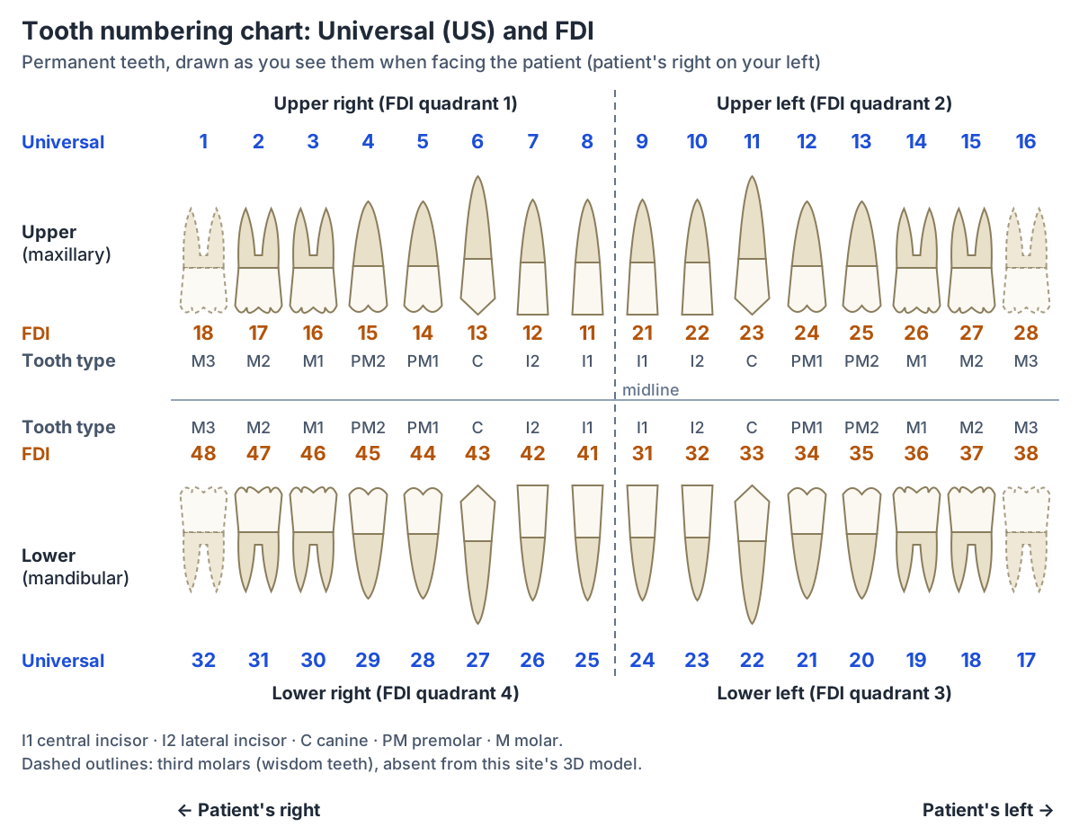
In the Universal system the permanent canines are teeth 6 (upper right), 11 (upper left), 22 (lower left) and 27 (lower right); the Radiopaedia list names each one "canine (cuspid)". In FDI notation they are 13, 23, 33 and 43. (Radiopaedia: American Dental Association Universal Numbering System.)
Permanent canines erupt on average at 11 to 12 years in the upper jaw and 9 to 10 years in the lower jaw, a few years after the incisors. (Dentalcare: Permanent Dentition.)
In teenagers with crowded teeth a canine may fail to erupt and stay buried, often in the palate or high in the buccal sulcus, and it may need surgical help to come through. (TeachMeAnatomy: Child and Adult Dentition.)
Molars, the first molar and wisdom teeth
Molars have the broadest, flattest surfaces and the largest crowns of all teeth, with 4 to 5 cusps. Upper molars usually have 3 roots and lower molars 2. In each quadrant they are named from the midline backwards as the first, second and third molar. (Dentalcare: Types of Teeth and their Functions.)
The first molar is also called the 6-year molar and the second molar the 12-year molar, after the age at which they come through. The third molar is the wisdom tooth. In the Universal system the first molars are teeth 3, 14, 19 and 30. (Radiopaedia: American Dental Association Universal Numbering System.)
The first permanent molar erupts at about 6 to 7 years, the earliest of the permanent teeth in the Dentalcare table. Third molars erupt on average between 17 and 21 years. (Dentalcare: Permanent Dentition.)
Wisdom teeth often come through late, in early adulthood, and sometimes fail to erupt and stay impacted in the bone; impacted third molars are usually removed surgically. (OpenStax Anatomy and Physiology 2e, 23.3: The Mouth, Pharynx, and Esophagus.)
Molars have deep grooves on their biting surfaces and wide contact points with their neighbours, which makes them prone to dental caries (decay). (TeachMeAnatomy: Child and Adult Dentition.)
Tooth numbering chart: Universal and FDI systems
In the Universal (American Dental Association) system, used mainly in the United States, the permanent teeth are numbered 1 to 32 as seen by a dentist looking into the open mouth, going clockwise from the back of the upper right side. Tooth 1 is the upper right third molar, 8 and 9 are the upper central incisors, 16 is the upper left third molar, 17 is the lower left third molar and 32 is the lower right third molar. (Radiopaedia: American Dental Association Universal Numbering System.)
Deciduous teeth use letters in the Universal system: A to J in the upper jaw, from the upper right second molar to the upper left second molar, and K to T in the lower jaw, from the lower left second molar to the lower right second molar. Extra (supernumerary) teeth are numbered by adding 50 to the number of the neighbouring permanent tooth. (Radiopaedia: American Dental Association Universal Numbering System.)
The FDI World Dental Federation system uses two digits. The first digit is the quadrant: 1 upper right, 2 upper left, 3 lower left and 4 lower right. The second digit is the tooth's position from the midline, from 1 (central incisor) to 8 (third molar). So 21 is the permanent upper left central incisor. (Radiopaedia: FDI World Dental Federation notation.)
For deciduous teeth the FDI quadrants are numbered 5 to 8 (5 upper right, 6 upper left, 7 lower left, 8 lower right) and the second digit runs only from 1 to 5, because there are no premolars: 82, for example, is the deciduous lower right lateral incisor. (Radiopaedia: FDI World Dental Federation notation.)
Kenhub describes FDI notation as the international standard for naming teeth. A third system, Palmer notation, is the main system used for numbering teeth in the United Kingdom. (Kenhub: Teeth.)
Tooth surfaces: mesial, distal, buccal, lingual and occlusal
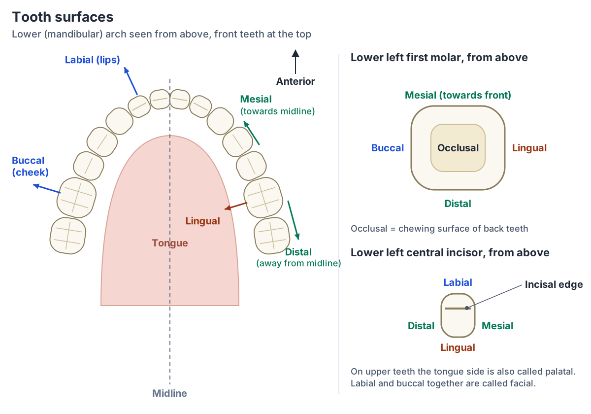
Dentists name each surface of a tooth by the direction it faces. The mesial surface is the one closest to the midline of the face and the distal surface is the one facing away from it. Neighbouring surfaces that touch, such as the distal of a lateral incisor and the mesial of a canine, are called proximal surfaces. (Dentalcare: Surfaces of the Teeth.)
The facial surface faces the lips or cheeks: it is called labial on the front teeth (towards the lips) and buccal on the back teeth (towards the cheek). The lingual surface faces the tongue. The chewing surface of a posterior tooth is the occlusal surface, and the biting edge of an anterior tooth is the incisal edge. (Dentalcare: Surfaces of the Teeth.)
On upper teeth the inner surface faces the palate, so it is often called palatal rather than lingual. Other common terms are vestibular (towards the vestibule of the mouth, the same side as labial and buccal) and cervical (towards the neck of the tooth). (Medeco Dental Atlas: Dental surface description.)
Surface names combine with tooth numbers to pinpoint a spot. Dentalcare's example is the mesio-lingual surface of tooth 3, the upper right first molar: the part of that tooth nearest the front of the mouth on the tongue side. (Dentalcare: Surfaces of the Teeth.)
Tooth anatomy: crown, root, enamel, dentin, pulp and cementum
Each tooth has a crown, which projects above the gum line, and one or more roots embedded in the bone. Running through the middle is the pulp cavity, which contains loose connective tissue, blood vessels and nerves; the part of the pulp cavity in the root is the root canal. (OpenStax Anatomy and Physiology 2e, 23.3: The Mouth, Pharynx, and Esophagus.)
Enamel covers the crown and is the hardest substance in the body. It is slightly translucent and can withstand biting forces, but once formed it cannot regrow. Dentin lies under the enamel and the cementum. It is softer than enamel and makes up most of the tooth, so decay that gets through the enamel can spread quickly in the dentin. (Dentalcare: Divisions and Components of the Teeth.)
Cementum is a thin layer covering the root. It is not as hard as enamel but is similar in hardness to bone, and fibres projecting from it anchor the tooth to the alveolar bone. Enamel and cementum meet at the cementoenamel junction (CEJ). The pulp is divided into the pulp chamber in the crown and the pulp canals in the roots. (Dentalcare: Divisions and Components of the Teeth.)
Vessels and nerves enter the pulp through the apical foramen at the tip of the root. Dentin is living and innervated, while enamel has no blood supply. (TeachMeAnatomy: Child and Adult Dentition.)
The upper teeth are supplied by the superior alveolar nerves, from the maxillary division of the trigeminal nerve (CN V2), and the lower teeth by the inferior alveolar nerve, from the mandibular division (CN V3). Their blood supply comes from the maxillary artery through the anterior superior, posterior superior and inferior alveolar arteries. (Kenhub: Teeth.)
Deciduous vs permanent teeth: counts and eruption
The deciduous (primary, baby) dentition has 20 teeth: in each arch there are two central incisors, two lateral incisors, two canines and four molars. There are no premolars and no third molars. By about age 3 all the primary teeth have erupted, and all the permanent teeth except the third molars are already forming in the jaws. (Dentalcare: Primary Dentition.)
Deciduous teeth start to appear at about 6 months of age. The 32 permanent teeth replace them between about 6 and 12 years. (OpenStax Anatomy and Physiology 2e, 23.3: The Mouth, Pharynx, and Esophagus.)
TeachMeAnatomy gives the replacement as starting at about 6 years and ending at about 13, with third molars usually erupting by about 21. (TeachMeAnatomy: Child and Adult Dentition.)
When do teeth erupt? Average eruption ages
| Tooth | Deciduous, upper | Deciduous, lower | Permanent, upper | Permanent, lower |
|---|---|---|---|---|
| Central incisor | 8 to 12 months | 6 to 10 months | 7 to 8 years | 7 to 8 years |
| Lateral incisor | 9 to 13 months | 10 to 16 months | 8 to 9 years | 8 to 9 years |
| Canine | 16 to 22 months | 17 to 23 months | 11 to 12 years | 9 to 10 years |
| First premolar | None | None | 10 to 11 years | 10 to 12 years |
| Second premolar | None | None | 10 to 12 years | 11 to 12 years |
| First molar | 13 to 19 months | 14 to 18 months | 6 to 7 years | 6 to 7 years |
| Second molar | 25 to 33 months | 22 to 31 months | 11 to 13 years | 11 to 13 years |
| Third molar | None | None | 17 to 21 years | 17 to 21 years |
Average ranges from the Dentalcare primary and permanent dentition tables. Individual children vary, and third molars may not erupt at all.
What is the curve of Spee?
The curve of Spee is the upward curve of the lower teeth seen from the side. In orthodontics it means the curve of the mandibular teeth in the sagittal plane, running from the incisal edges of the front teeth along the buccal cusp tips of the premolars and molars. (Dhiman S. Curve of Spee: from orthodontic perspective (Indian J Dent, 2015).)
It is named after Spee, who described it in 1890 from skulls with worn teeth. He drew it as an arc that touched the front of the mandibular condyle, the occlusal surface of the second molar and the incisal edges of the lower incisors. (Dhiman S. Curve of Spee: from orthodontic perspective (Indian J Dent, 2015).)
The curve is thought to support efficient chewing and to help the dentition resist the forces of occlusion. An exaggerated (deep) curve may disturb muscle balance and lead to poor functional occlusion, and a deep curve can make a normal (Class I) canine relationship nearly impossible. (Dhiman S. Curve of Spee: from orthodontic perspective (Indian J Dent, 2015).)
Occlusion and Angle's classes (class 1, class 2, class 3)
Occlusion is the way the upper and lower teeth meet when the jaws close. When teeth do not meet in the normal way, the result is called malocclusion, which can affect chewing, speech, appearance and the risk of dental disease. (Dentalcare: Occlusion.)
Angle's classification, introduced by Angle in 1899, groups occlusion by how the permanent first molars meet. It uses the Roman numerals I, II and III. (Modified Angle's Classification for Primary Dentition (PMC).)
Class I is considered normal (orthognathic) occlusion. Class II (retrognathic) and Class III (prognathic) are malocclusions. Class II has two divisions: in division 1 the upper front teeth stick out (protrude), and in division 2 one or more upper front teeth are tipped back (retruded). (Dentalcare: Occlusion.)
Teeth in the 3D model
- Lower central secondary incisor tooth
- The permanent lower central incisor, the smallest tooth in the mouth, one on each side of the midline of the lower jaw.
- Lower first secondary molar tooth
- The permanent lower first molar, the largest lower tooth and the first permanent tooth to erupt.
- Lower first secondary premolar tooth
- The permanent lower first premolar, a canine-like tooth with one large and one small cusp.
- Lower lateral secondary incisor tooth
- The permanent lower lateral incisor, a chisel-shaped cutting tooth between the central incisor and the canine.
- Lower second secondary molar tooth
- The permanent lower second molar, a four-cusped grinding tooth at the back of the lower arch in this model.
- Lower second secondary premolar tooth
- The permanent lower second premolar, a crushing tooth in front of the first molar, often with three cusps.
- Lower secondary canine tooth
- The permanent lower canine, a pointed single-cusped tooth at the corner of the lower dental arch.
- Upper central secondary incisor tooth
- The permanent upper central incisor, the largest and most prominent incisor, beside the midline of the upper jaw.
- Upper first secondary molar tooth
- The permanent upper first molar, a large three-rooted grinding tooth whose roots lie close to the maxillary sinus.
- Upper first secondary premolar tooth
- The permanent upper first premolar, a two-cusped tooth that usually has two roots.
- Upper lateral secondary incisor tooth
- The permanent upper lateral incisor, a smaller cutting tooth between the central incisor and the canine.
- Upper second secondary molar tooth
- The permanent upper second molar, a three-rooted grinding tooth at the back of the upper arch in this model.
- Upper second secondary premolar tooth
- The permanent upper second premolar, a two-cusped tooth that usually has one root.
- Upper secondary canine tooth
- The permanent upper canine (eye tooth), which has the longest root of any tooth.
Frequently asked questions
How many teeth do adults and children have?
Adults have 32 permanent teeth: 8 incisors, 4 canines, 8 premolars and 12 molars, including the 4 wisdom teeth. Children have 20 deciduous teeth: 8 incisors, 4 canines and 8 molars, with no premolars. Baby teeth start to appear at about 6 months, and the permanent teeth replace them between about 6 and 12 years of age.
What are K9 teeth?
"K9 teeth" is a common spelling for canine teeth, also called cuspids. Humans have four, one at each corner of the mouth between the lateral incisor and the first premolar. Each has a single pointed cusp for piercing and holding food and a single root, which is the longest root of any tooth. In the Universal system they are teeth 6, 11, 22 and 27.
What is the difference between central and lateral incisors?
Central incisors are the two front teeth on either side of the midline in each jaw. Lateral incisors sit next to them, between the central incisors and the canines. Both have straight cutting edges. The upper lateral incisor varies most in shape: it can be small and peg-shaped, and it is one of the teeth that may be congenitally missing.
How are teeth numbered?
In the US Universal system, adult teeth are numbered 1 to 32 clockwise as the dentist looks into the mouth, from the upper right third molar (1) to the lower right third molar (32); baby teeth are lettered A to T. In the international FDI system, each tooth has two digits: the quadrant (1 to 4, or 5 to 8 for baby teeth) and then its position from the midline.
What are the five surfaces of a tooth?
A back tooth has five surfaces: mesial (towards the midline), distal (away from the midline), buccal (towards the cheek), lingual (towards the tongue, called palatal on upper teeth) and occlusal (the chewing surface). Front teeth have a labial surface towards the lips instead of buccal and an incisal edge instead of an occlusal surface.
When does the first molar come in?
The first permanent molar erupts at about 6 to 7 years, which is why it is called the 6-year molar. The second molar follows at about 11 to 13 years and the third molar, or wisdom tooth, on average between 17 and 21 years, if it erupts at all.
What is a class 2 malocclusion?
In Angle's classification, which is based on how the upper and lower first molars meet, Class I is normal occlusion and Class II and Class III are malocclusions. Class II is described as retrognathic. In Class II division 1 the upper front teeth protrude, and in Class II division 2 one or more upper front teeth are tipped backwards.
References
- OpenStax. Anatomy and Physiology 2e, §23.3 The Mouth, Pharynx, and Esophagus. Rice University, 2022.
- TeachMeAnatomy. Child and Adult Dentition.
- Kenhub. Teeth.
- Radiopaedia. American Dental Association Universal Numbering System.
- Radiopaedia. FDI World Dental Federation notation.
- Foster VP. An Overview of Dental Anatomy: Types of Teeth, Structures, Location and Functions. Dentalcare (Procter & Gamble) continuing education course CE500.
- Foster VP. An Overview of Dental Anatomy: Types of Teeth and their Functions. Dentalcare CE500.
- Foster VP. An Overview of Dental Anatomy: Surfaces of the Teeth. Dentalcare CE500.
- Foster VP. An Overview of Dental Anatomy: Divisions and Components of the Teeth. Dentalcare CE500.
- Foster VP. An Overview of Dental Anatomy: Primary Dentition. Dentalcare CE500.
- Foster VP. An Overview of Dental Anatomy: Permanent Dentition. Dentalcare CE500.
- Foster VP. An Overview of Dental Anatomy: Occlusion. Dentalcare CE500.
- Medeco Dental Atlas. Dental surface description.
- Dhiman S. Curve of Spee: from orthodontic perspective. Indian Journal of Dentistry, 2015.
- Modified Angle's Classification for Primary Dentition. PubMed Central.
- Mitsuhashi N. et al. (2009). BodyParts3D: 3D structure database for anatomical concepts. Nucleic Acids Research 37, D782–D785. Data © The Database Center for Life Science, CC BY 4.0.
Related anatomy
- MandibleMandible anatomy with a labelled diagram: body, ramus, angle, condylar and coronoid processes, foramina, muscle…
- MaxillaMaxilla bone anatomy: the body and four processes, palatine and alveolar processes, maxillary sinus, articulations, Le…
- Muscles of masticationThe muscles of mastication explained: masseter, temporalis, medial and lateral pterygoid, with labelled diagrams, the…
- TongueTongue anatomy explained: parts and papillae of the dorsum, the underside with the frenulum and sublingual folds…
- Skull bonesLearn the 22 bones of the skull, the 8 cranial bones of the neurocranium and the coronal, sagittal, lambdoid and…
Explore the whole body in the interactive 3D human anatomy model, or browse the A–Z list of human structures.