

Overview: one artery with several names
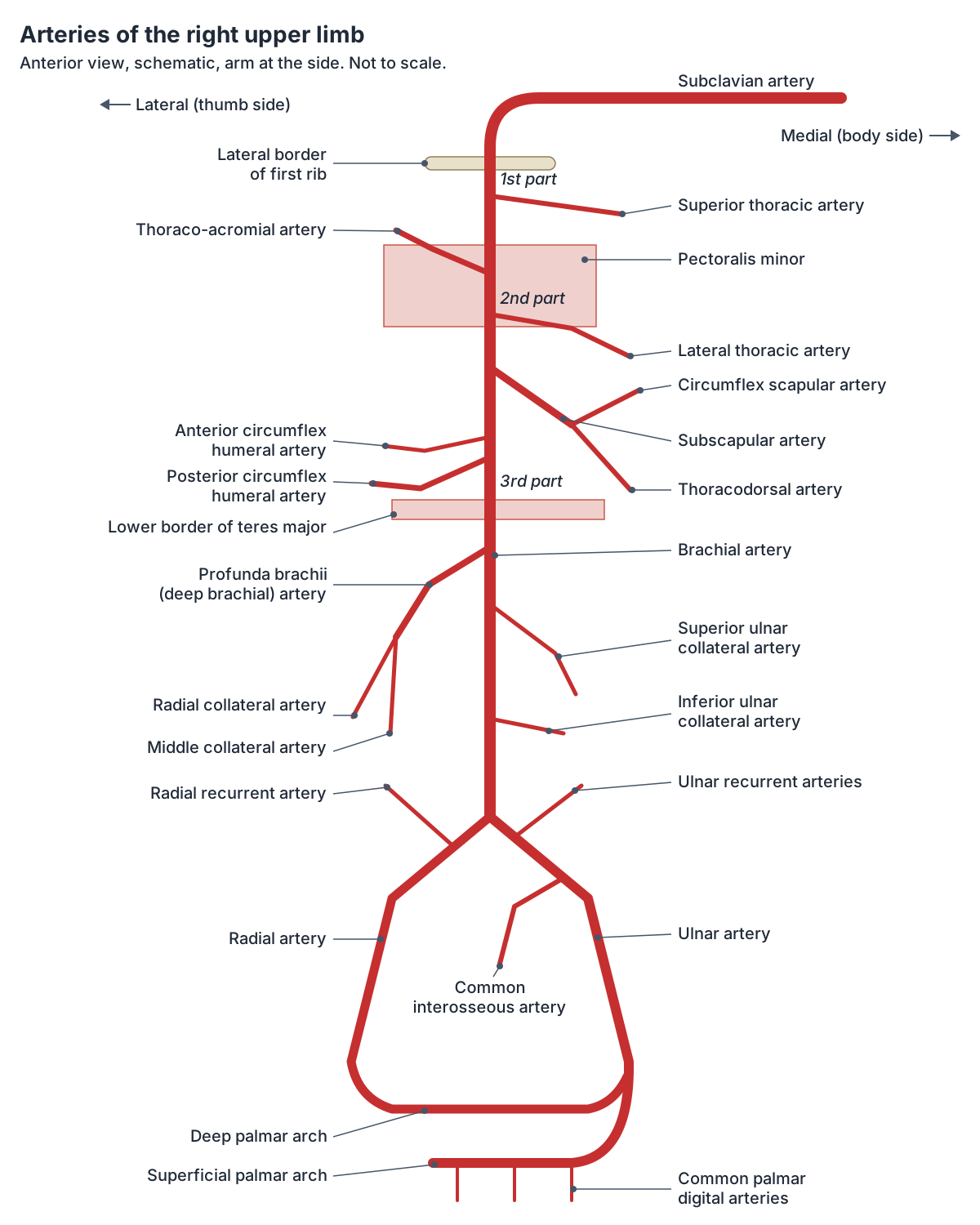
TeachMeAnatomy lists five main arteries for the upper limb, from proximal to distal: the subclavian, axillary, brachial, radial and ulnar arteries. They are not separate vessels so much as one trunk renamed at fixed landmarks, so learning the landmarks is the quickest way to learn the names. (TeachMeAnatomy: Arterial Supply to the Upper Limb.)
OpenStax describes the same chain: the subclavian artery is renamed the axillary artery as it leaves the thorax for the axillary region, the vessel continues into the upper arm (brachium) as the brachial artery, and near the elbow the brachial artery divides into the radial and ulnar arteries. These run beside their namesake bones to the wrist, where they form the superficial and deep palmar arches and then the digital arteries of the fingers. (OpenStax Anatomy and Physiology 2e, 20.5: Circulatory Pathways.)
The veins come in two layers. Deep veins lie beneath the deep fascia, usually as a pair of venae comitantes on either side of an artery, and take that artery's name (radial veins, ulnar veins, brachial veins). Superficial veins lie in the subcutaneous tissue; the two large ones are the cephalic vein on the lateral (thumb) side and the basilic vein on the medial (little finger) side. Perforating veins connect the two layers. (TeachMeAnatomy: Venous Drainage of the Upper Limb.)
The 3D view highlights the arteries and veins of the upper limb from the BodyParts3D adult male data set. Small or variable vessels, such as the superior thoracic artery, are not modelled separately, and the venous pattern at the elbow is only one of the many normal patterns; the H-shaped and M-shaped variants are described below.
Subclavian artery: the root of the arm's blood supply
The right subclavian artery is a terminal branch of the brachiocephalic artery (brachiocephalic trunk). The left subclavian artery is the third branch of the arch of the aorta, after the left common carotid artery. (Radiopaedia: Subclavian artery.)
Each subclavian artery leaves the thorax through the superior thoracic aperture, passes between the anterior and middle scalene muscles and then between the first rib and the clavicle. It is described in three parts by its relation to scalenus anterior: medial to the muscle, behind it, and lateral to it as far as the lateral border of the first rib. (Radiopaedia: Subclavian artery.)
Its branches mostly supply the neck, brain and chest wall rather than the arm. The first part gives the vertebral artery, the internal thoracic artery and the thyrocervical trunk; the second part gives the costocervical trunk; the third part often gives the dorsal scapular artery, which in some people arises from the second part or the thyrocervical trunk instead. (Radiopaedia: Subclavian artery.)
The most common variant is an aberrant right subclavian artery, which arises from the aortic arch beyond the left subclavian artery and crosses to the right behind the trachea, near the oesophagus (esophagus). It may press on these structures and cause difficulty breathing or swallowing. (Radiopaedia: Subclavian artery.)
The subclavian artery in the 3D model and its neighbouring structures. Subclavian artery.
Axillary artery: the artery in the armpit and its six branches
The axillary artery is the artery in the armpit. It begins at the lateral border of the first rib as the continuation of the subclavian artery and ends at the lower border of teres major, where it becomes the brachial artery. It runs inside the axillary sheath, deep to pectoralis minor, which divides it into three parts. (TeachMeAnatomy: Arterial Supply to the Upper Limb.)
The first part runs from the first rib to the upper border of pectoralis minor, the second part lies behind pectoralis minor, and the third part runs from the lower border of pectoralis minor to the lower border of teres major. The cords of the brachial plexus are named for where they lie around the second part: lateral, medial and posterior to the artery. Along its whole course the artery lies behind the axillary vein. (Radiopaedia: Axillary artery.)
The usual count is one branch from the first part, two from the second and three from the third. The first part gives the superior thoracic artery to the pectoral muscles. The second part gives the thoraco-acromial artery, which pierces the clavipectoral fascia and splits into four terminal branches for the clavicle, acromion, deltoid and pectoral muscles, and the lateral thoracic artery (thoracica lateralis), which follows the lower border of pectoralis minor to serratus anterior and the pectorals and is an important supply to the breast. (Radiopaedia: Axillary artery.)
The third part gives the subscapular artery, its largest branch, which divides into the thoracodorsal artery to latissimus dorsi and the circumflex scapular artery to the scapular anastomosis. It also gives the anterior and posterior circumflex humeral arteries, which ring the surgical neck of the humerus. The posterior circumflex humeral artery passes through the posterior wall of the axilla with the axillary nerve and is the main supply to the head of the humerus. (Radiopaedia: Axillary artery.)
The two circumflex humeral arteries anastomose around the surgical neck of the humerus, so a fracture there can damage them. (TeachMeAnatomy: Arterial Supply to the Upper Limb.)
Textbook diagrams show six separate branches, but real axillary arteries vary a lot. Radiopaedia lists many recorded patterns, such as a common trunk for the subscapular and posterior circumflex humeral arteries, a common trunk for the thoraco-acromial and lateral thoracic arteries, or a lateral thoracic artery that is missing altogether. (Radiopaedia: Axillary artery.)
A traditional memory aid for the branches in order is "Screw The Lawyer, Save A Patient": superior thoracic, thoraco-acromial, lateral thoracic, subscapular, anterior and posterior circumflex humeral.
Parts, branches, relations to the brachial plexus and clinical anatomy of the axillary artery. Axillary artery.
Brachial artery and the profunda brachii (deep brachial) artery
The brachial artery is the main artery of the arm. It begins at the lower border of teres major, runs down the medial side of the arm, medial to biceps brachii and in front of the medial head of triceps, then moves in front of the humerus near the elbow and passes under the bicipital aponeurosis (lacertus fibrosus) into the cubital fossa. (Radiopaedia: Brachial artery.)
Its first and largest branch is the profunda brachii artery, also called the deep brachial artery. It arises from the posteromedial side of the brachial artery just below teres major, enters the posterior compartment with the radial nerve through the triangular interval and runs in the radial groove of the humerus between the medial and lateral heads of triceps. It supplies triceps and ends as the middle collateral and radial collateral arteries. (Radiopaedia: Profunda brachii artery.)
Lower down the brachial artery gives nutrient arteries to the humerus and the superior and inferior ulnar collateral arteries, which join the elbow anastomosis. Because the collateral arteries of the arm link up with recurrent branches of the radial and ulnar arteries around the elbow, a block below the origin of the profunda brachii is usually less damaging than one above it. (Radiopaedia: Brachial artery.)
The brachial artery ends at the apex (lower point) of the cubital fossa by dividing into the radial and ulnar arteries. TeachMeAnatomy describes the cubital fossa contents from lateral to medial as the radial nerve, the biceps tendon, the brachial artery and the median nerve; the brachial pulse is felt just medial to the biceps tendon. (TeachMeAnatomy: The Cubital Fossa.)
The division sometimes happens much higher, in the arm or even the axilla. A Cureus review puts the overall incidence of this high bifurcation of the brachial artery at about 8%, and notes that it matters for arteriovenous fistula surgery, angiography and flap planning. (Tsoucalas G et al. High Bifurcation of the Brachial Artery: An Embryological Overview. Cureus. 2020.)
Where the brachial artery runs, its branches, the brachial pulse and blood pressure measurement. Brachial artery.
Radial artery and ulnar artery (arteria ulnaris) in the forearm
The radial artery runs down the lateral (thumb) side of the anterior forearm, under brachioradialis above and lateral to the tendon of flexor carpi radialis below, where its distal part lies on the radius. At the wrist it turns laterally around the joint, crosses the floor of the anatomical snuffbox and passes through the back of the first web space to reach the palm between the two heads of adductor pollicis. (Radiopaedia: Radial artery.)
The radial artery location at the wrist is why the radial pulse is so easy to find: it can be felt just lateral to the flexor carpi radialis tendon, pressed against the radius. The radial artery supplies the posterolateral forearm and joins the anastomoses around the elbow and the wrist. (TeachMeAnatomy: Arterial Supply to the Upper Limb.)
The ulnar artery (arteria ulnaris) is the other terminal branch of the brachial artery. It runs down the medial side of the forearm deep to pronator teres, flexor carpi radialis and flexor digitorum superficialis, and for most of its length lies between flexor digitorum superficialis and flexor carpi ulnaris. (Radiopaedia: Ulnar artery.)
Near its origin the ulnar artery gives the anterior and posterior ulnar recurrent arteries to the elbow anastomosis and the common interosseous artery, the source of the arteries that run on the interosseous membrane between the radius and ulna. It enters the hand with the ulnar nerve and vein through Guyon's canal, between the pisiform and the hook of the hamate. (Radiopaedia: Ulnar artery.)
Radial pulse, anatomical snuffbox, Allen test and radial artery access. Radial artery.
Arteries of the upper limb
| Artery | Begins | Ends | Main branches | Supplies |
|---|---|---|---|---|
| Subclavian artery | Brachiocephalic trunk (right); arch of the aorta (left) | Lateral border of the first rib | Vertebral, internal thoracic, thyrocervical trunk, costocervical trunk, dorsal scapular | Brain (via vertebral artery), neck, chest wall, shoulder |
| Axillary artery | Lateral border of the first rib | Lower border of teres major | Superior thoracic; thoraco-acromial, lateral thoracic; subscapular, anterior and posterior circumflex humeral | Chest wall, shoulder, breast, scapular muscles |
| Brachial artery | Lower border of teres major | Cubital fossa, about the neck of the radius | Profunda brachii, nutrient arteries, superior and inferior ulnar collateral, muscular branches | Arm muscles and humerus; forearm and hand via its terminal branches |
| Profunda brachii (deep brachial) artery | Brachial artery, just below teres major | Middle collateral and radial collateral arteries | Middle collateral, radial collateral | Triceps brachii; humerus (nutrient artery in some people) |
| Radial artery | Brachial artery in the cubital fossa | Deep palmar arch | Radial recurrent, palmar carpal, dorsal carpal, superficial palmar branch, princeps pollicis, radialis indicis | Lateral forearm, thumb, lateral index finger, wrist |
| Ulnar artery | Brachial artery in the cubital fossa | Superficial palmar arch | Anterior and posterior ulnar recurrent, common interosseous, palmar and dorsal carpal branches | Medial forearm, deep forearm (via interosseous arteries), hand |
Arteries of the hand: the superficial and deep palmar arches
The superficial palmar arch is the end of the ulnar artery. It lies deep to the palmar aponeurosis and superficial to the long flexor tendons, and it gives the common palmar digital arteries to the fingers. In about two-thirds of hands it is incomplete; in the rest it is completed by the superficial palmar branch of the radial artery or the radialis indicis artery. (Radiopaedia: Superficial palmar arch.)
The deep palmar arch is the end of the radial artery. It usually joins a branch of the ulnar artery to form a complete arch, crosses the palm about 1 cm proximal to the superficial arch, and gives three palmar metacarpal arteries that join the common palmar digital arteries near the metacarpal heads. (Radiopaedia: Deep palmar arch.)
Because the radial and ulnar arteries are linked through these arches, the hand has a double blood supply. TeachMeAnatomy notes that these rich anastomoses keep the hand perfused even when gripping squeezes the vessels. (TeachMeAnatomy: Arterial Supply to the Upper Limb.)
Veins of the arm: cephalic, basilic and median cubital veins
Both large superficial veins start in the dorsal venous network on the back of the hand. The cephalic vein begins from its radial (thumb) side and crosses the anatomical snuffbox before winding round to the front of the forearm. (TeachMeAnatomy: The Anatomical Snuffbox.)
The cephalic vein runs up the anterolateral forearm and arm, lateral to biceps, then in the deltopectoral groove between deltoid and pectoralis major. It pierces the clavipectoral fascia at the clavipectoral triangle and drains into the first part of the axillary vein. (Radiopaedia: Cephalic vein.)
The basilic vein begins from the ulnar side of the dorsal venous network, runs up the medial forearm, passes in front of the medial epicondyle and continues up the medial arm, medial to the brachial artery. About halfway up the arm it passes through the deep fascia at the basilic hiatus, and at the lower border of teres major it joins the brachial veins to form the axillary vein. (Radiopaedia: Basilic vein.)
The median cubital vein is a short connecting vein in the roof of the cubital fossa. It leaves the cephalic vein just below the lateral epicondyle, runs obliquely upwards and medially and ends in the basilic vein. The bicipital aponeurosis lies between it and the brachial artery and median nerve. (TeachMeAnatomy: Median Cubital Vein.)
The superficial veins at the elbow vary a great deal. The median antebrachial vein runs up the front of the forearm and ends in the basilic or median cubital vein; sometimes it divides into a median cephalic and a median basilic vein, giving an M-shaped pattern instead of the usual H-shaped one. (Radiopaedia: Basilic vein.)
Course of the cephalic vein, the deltopectoral groove, and cephalic vs basilic vein. Cephalic vein.
Veins of the hand
In the fingers, digital veins join in the hand to form the palmar venous arches. From these arches come the radial and ulnar veins, which follow their arteries, and the median antebrachial vein, which runs up the front of the forearm. (OpenStax Anatomy and Physiology 2e, 20.5: Circulatory Pathways.)
Blood from the palm also drains round to the back of the hand into the dorsal venous network. Its lateral part feeds the cephalic vein and its medial part feeds the basilic vein. (Radiopaedia: Cephalic vein.)
Deep veins, the axillary vein and the subclavian vein
The radial and ulnar veins unite near the cubital fossa to form the brachial veins, which run up the arm close to the brachial artery. TeachMeAnatomy calls the brachial veins the largest of the deep veins, and notes that pulsation of the neighbouring artery helps push venous blood back towards the heart. (TeachMeAnatomy: Venous Drainage of the Upper Limb.)
The axillary vein forms at the lower border of teres major where the brachial veins join the basilic vein. It runs up through the axilla on the medial side of the axillary artery, receives the cephalic vein above pectoralis minor and veins matching the axillary artery's branches, and becomes the subclavian vein at the lateral border of the first rib. Double axillary veins are found in about 14% of people. (Radiopaedia: Axillary vein.)
The axillary vein is not a mirror image of the artery. In a dissection study of 40 limbs, the brachial veins usually joined the axillary vein separately, sometimes joined the basilic vein instead, and the thoraco-acromial vein drained into the cephalic vein in 70% of limbs rather than straight into the axillary vein. (Lee H et al. The axillary vein and its tributaries are not in the mirror image of the axillary artery and its branches. PLoS One. 2019.)
The subclavian vein arches behind the medial clavicle and runs in front of scalenus anterior, while the subclavian artery runs behind it. Its only named tributary is the external jugular vein. Behind the sternoclavicular joint it joins the internal jugular vein to form the brachiocephalic vein; the thoracic duct on the left and the right lymphatic duct on the right drain into this junction. (Radiopaedia: Subclavian vein.)
The last valve on the way back from the arm usually lies in the lateral part of the subclavian vein. The veins medial to it have no valves and are counted as central veins. (Radiopaedia: Subclavian vein.)
Veins of the upper limb
| Vein | Type | Begins | Course | Ends in |
|---|---|---|---|---|
| Cephalic vein | Superficial | Lateral (radial) side of the dorsal venous network of the hand | Anatomical snuffbox, lateral forearm and arm, deltopectoral groove | Axillary vein, after piercing the clavipectoral fascia |
| Basilic vein | Superficial | Medial (ulnar) side of the dorsal venous network of the hand | Medial forearm, in front of the medial epicondyle, medial arm; goes deep at mid-arm | Joins the brachial veins at teres major to form the axillary vein |
| Median cubital vein | Superficial | Cephalic vein just below the lateral epicondyle | Obliquely across the roof of the cubital fossa, over the bicipital aponeurosis | Basilic vein |
| Median antebrachial vein | Superficial | Front of the wrist and forearm | Up the middle of the anterior forearm | Basilic or median cubital vein (variable) |
| Radial and ulnar veins | Deep (paired venae comitantes) | Palmar venous arches | Beside the radial and ulnar arteries | Unite near the cubital fossa to form the brachial veins |
| Brachial veins | Deep (paired) | Union of radial and ulnar veins | Beside the brachial artery | Axillary vein (with the basilic vein) |
| Axillary vein | Deep | Lower border of teres major | Medial to the axillary artery through the axilla | Becomes the subclavian vein at the lateral border of the first rib |
| Subclavian vein | Deep | Lateral border of the first rib | Behind the clavicle, in front of scalenus anterior | Joins the internal jugular vein to form the brachiocephalic vein |
Pulses, blood tests and clinical points
The pulse is most often checked at the radial artery at the wrist; the brachial artery in the upper arm is another site. Blood pressure is measured with a cuff around the arm at about heart level and a stethoscope over the brachial artery at the front of the elbow: the first Korotkoff sound marks the systolic pressure and the point where the sounds disappear marks the diastolic pressure. (OpenStax Anatomy and Physiology 2e, 20.2: Blood Flow, Blood Pressure, and Resistance.)
OpenStax calls the median cubital vein the most common site for drawing venous blood. TeachMeAnatomy adds that it is superficial and held in place by perforating veins that pierce the bicipital aponeurosis, which makes it easy to enter with a needle. (TeachMeAnatomy: Venous Drainage of the Upper Limb.)
A supracondylar fracture of the humerus, usually in children who fall on an outstretched hand, can injure the brachial artery in the cubital fossa. Untreated damage to the artery can lead to Volkmann's ischaemic contracture, a fixed flexion deformity caused by dead forearm muscle being replaced by scar tissue. (TeachMeAnatomy: The Cubital Fossa.)
At the apex of the axilla the vessels and nerves pass between the clavicle, the first rib and the scapula and can be compressed there, for example after a clavicle fracture or with a cervical rib. This is called thoracic outlet syndrome and causes pain, tingling, weakness and colour change in the arm. (TeachMeAnatomy: The Axilla.)
Clamping the brachial artery below the origin of the profunda brachii is generally tolerated because collateral vessels around the elbow keep blood flowing to the forearm; bleeding from the arm is best controlled by pressing the artery against the medial side of the humerus above the wound. (Kenhub: Brachial artery.)
Upper limb blood vessels in the 3D model
- Acromial branch of thoraco-acromial artery
- The acromial branch is one of four branches of the thoraco-acromial artery and runs to the acromion.
- Anterior circumflex humeral artery
- The anterior circumflex humeral artery is a small branch of the axillary artery that wraps around the front of the surgical neck of the humerus.
- Anterior interosseous artery
- The anterior interosseous artery is a branch of the common interosseous artery that runs down the front of the forearm interosseous membrane.
- Anterior ulnar recurrent artery
- The anterior ulnar recurrent artery is a small branch of the ulnar artery that supplies the elbow.
- Axillary artery
- The axillary artery is the main artery of the armpit, continuing the subclavian artery.
- Axillary vein
- The axillary vein is the main vein of the armpit and carries blood from the upper limb.
- Basilic vein
- The basilic vein is a large superficial vein on the medial side of the arm.
- Brachial artery
- The brachial artery is the main artery of the arm, continuing the axillary artery.
- Cephalic vein
- The cephalic vein is a large superficial vein on the lateral side of the upper limb.
- Circumflex scapular artery
- The circumflex scapular artery is the large branch of the subscapular artery that passes round the lateral border of the scapula.
- Common interosseous artery
- The common interosseous artery is a short branch of the ulnar artery in the forearm.
- Deep brachial artery
- The deep brachial (profunda brachii) artery is the main branch of the brachial artery in the upper arm.
- Deep palmar arch
- The deep palmar arch is an arterial arch in the palm, mainly formed by the radial artery.
- Deep palmar venous arch
- The deep palmar venous arch is a venous arch in the palm accompanying the deep palmar arterial arch.
- Deltoid branch of thoraco-acromial artery
- This is a branch of the thoraco-acromial artery that supplies the deltoid muscle.
- Dorsal venous network of hand
- The dorsal venous network is a superficial venous plexus on the back of the hand.
- Inferior ulnar collateral artery
- The inferior ulnar collateral artery is a branch of the brachial artery that supplies the elbow.
- Lateral thoracic artery
- A branch of the axillary artery that runs along the lateral chest wall.
- Medial brachial vein
- A deep vein accompanying the brachial artery on its medial side.
- Median antebrachial vein
- A superficial vein running up the front of the forearm.
- Median cubital vein
- A superficial vein across the front of the elbow linking the cephalic and basilic veins.
- Middle collateral branch of deep brachial artery
- A terminal branch of the deep brachial artery (profunda brachii) in the posterior arm.
- Pectoral branch of thoraco-acromial artery
- A branch of the thoraco-acromial artery supplying the pectoral muscles.
- Posterior circumflex humeral artery
- A branch of the axillary artery that wraps around the surgical neck of the humerus.
- Posterior ulnar recurrent artery
- A branch of the ulnar artery that ascends behind the medial epicondyle.
- Radial artery
- One of the two terminal branches of the brachial artery, running on the lateral forearm.
- Radial collateral branch of deep brachial artery
- A terminal branch of the deep brachial artery that runs with the radial nerve.
- Radial recurrent artery
- A branch of the radial artery that ascends toward the elbow.
- Radial vein
- Paired deep veins accompanying the radial artery.
- Subclavian artery
- The major artery to the upper limb, neck and part of the brain.
- Subclavian vein
- The large vein draining the upper limb into the brachiocephalic vein.
- Subscapular artery
- The largest branch of the axillary artery.
- Superficial palmar arterial arch
- An arterial arch in the palm formed mainly by the ulnar artery.
- Superficial palmar venous arch
- A venous network in the palm.
- Superior ulnar collateral artery
- A branch of the brachial artery in the arm.
- Thoracodorsal artery
- A branch of the subscapular artery to latissimus dorsi.
- Trunk of thoraco-acromial artery
- The main stem of the thoraco-acromial artery.
- Ulnar artery
- The larger terminal branch of the brachial artery.
- Ulnar vein
- Paired deep veins accompanying the ulnar artery.
Frequently asked questions
What is the main artery in the armpit?
The artery in the armpit is the axillary artery. It continues the subclavian artery from the lateral border of the first rib to the lower border of teres major, where it becomes the brachial artery. Pectoralis minor crosses in front of it and divides it into three parts, which give one, two and three branches respectively: six branches in all.
What are the main veins of the arm?
The main superficial veins are the cephalic vein on the thumb side and the basilic vein on the little-finger side, joined at the elbow by the median cubital vein. The deep veins are paired radial, ulnar and brachial veins that run beside the arteries. All of them end in the axillary vein, which becomes the subclavian vein at the first rib.
Where is the radial artery located?
The radial artery runs down the thumb side of the front of the forearm, under brachioradialis and then just lateral to the flexor carpi radialis tendon, where it lies on the radius at the wrist. This is where the radial pulse is felt. It then winds round the wrist through the anatomical snuffbox into the palm to form the deep palmar arch.
What is the profunda brachii artery?
The profunda brachii, or deep brachial artery, is the first and largest branch of the brachial artery. It arises just below teres major, travels with the radial nerve in the radial groove on the back of the humerus, supplies triceps and ends as the middle and radial collateral arteries, which join the network of vessels around the elbow.
Which vein is used to take blood from the arm?
The median cubital vein at the front of the elbow is the most common site for drawing venous blood. It is superficial, fairly fixed in place and separated from the brachial artery and median nerve by the bicipital aponeurosis. The cephalic and basilic veins are also used for blood tests and cannulas.
Where does the subclavian vein go?
The subclavian vein continues the axillary vein from the lateral border of the first rib. It passes behind the clavicle and in front of scalenus anterior, receives the external jugular vein, and behind the sternoclavicular joint joins the internal jugular vein to form the brachiocephalic vein, which drains into the superior vena cava.
References
- TeachMeAnatomy. Arterial Supply to the Upper Limb.
- TeachMeAnatomy. Venous Drainage of the Upper Limb.
- TeachMeAnatomy. The Axilla.
- TeachMeAnatomy. The Cubital Fossa.
- TeachMeAnatomy. The Anatomical Snuffbox.
- TeachMeAnatomy. Median Cubital Vein.
- OpenStax. Anatomy and Physiology 2e, §20.5 Circulatory Pathways. Rice University, 2022.
- OpenStax. Anatomy and Physiology 2e, §20.2 Blood Flow, Blood Pressure, and Resistance. Rice University, 2022.
- Radiopaedia. Subclavian artery.
- Radiopaedia. Axillary artery.
- Radiopaedia. Brachial artery.
- Radiopaedia. Profunda brachii artery.
- Radiopaedia. Radial artery.
- Radiopaedia. Ulnar artery.
- Radiopaedia. Superficial palmar arch.
- Radiopaedia. Deep palmar arch.
- Radiopaedia. Axillary vein.
- Radiopaedia. Subclavian vein.
- Radiopaedia. Cephalic vein.
- Radiopaedia. Basilic vein.
- Radiopaedia. Median cubital vein.
- Radiopaedia. Brachial veins.
- Kenhub. Brachial artery.
- Lee H, Bang J, Kim S, Yang H. The axillary vein and its tributaries are not in the mirror image of the axillary artery and its branches. PLoS One. 2019;14(1):e0210464.
- Tsoucalas G, Eleftheriou A, Panagouli E. High Bifurcation of the Brachial Artery: An Embryological Overview. Cureus. 2020;12(2):e7097.
- Mitsuhashi N. et al. (2009). BodyParts3D: 3D structure database for anatomical concepts. Nucleic Acids Research 37, D782–D785. Data © The Database Center for Life Science, CC BY 4.0.
Related anatomy
- Brachial arteryWhere is the brachial artery? Its course down the arm, relations to the median nerve, branches, the brachial pulse…
- Radial arteryRadial artery anatomy: its course down the forearm, where to feel the radial pulse, the anatomical snuffbox, branches…
- Axillary arteryAxillary artery anatomy: where it starts and ends, its three parts around pectoralis minor, all six branches…
- Cephalic veinCephalic vein anatomy: its course from the hand through the deltopectoral groove to the axillary vein, cephalic vs…
- Basilic veinBasilic vein anatomy: its course up the medial forearm and arm, where it goes deep, how it forms the axillary vein…
- Upper limb nervesBrachial plexus diagram with roots, trunks, cords and branches; axillary, musculocutaneous, median, ulnar and radial…
Explore the whole body in the interactive 3D human anatomy model, or browse the A–Z list of human structures.