

Femoral artery at a glance
- What it is
- The femoral artery is the main artery of the thigh, continuing the external iliac artery.
- Location
- It begins at the inguinal ligament at the mid-inguinal point, runs through the femoral triangle and the adductor canal and ends at the adductor hiatus, where it becomes the popliteal artery. Its branches include the superficial epigastric, superficial circumflex iliac, external pudendal, deep femoral and descending genicular arteries.
- Function
- It supplies the anterior abdominal wall below the umbilicus, the external genitalia and the thigh, and continues to the leg.
- Body system
- Cardiovascular · paired (left and right)
- FMA ID
- FMA70250 (Foundational Model of Anatomy)
Key facts
- Its pulse is felt at the mid-inguinal point.
- The deep femoral artery supplies most of the thigh muscles.
- In the femoral triangle it lies between the femoral nerve laterally and the femoral vein medially (NAVY, from lateral to medial).
Clinical relevance
It is a common access site for cardiac catheterization.
Where is the femoral artery?
The femoral artery begins beneath the inguinal ligament, medial to the midpoint of the ligament and to the deep inguinal ring. This point, the mid-inguinal point, lies roughly halfway between the anterior superior iliac spine (the bony point at the front of the hip) and the pubic symphysis. At this level the femoral vein lies on its medial side. (Radiopaedia: Femoral artery.)
From the groin it descends through the femoral triangle into the adductor (subsartorial) canal on the medial side of the thigh. It leaves the canal through the adductor hiatus, a gap in adductor magnus just above the knee, and passes to the back of the thigh, where it is renamed the popliteal artery. (Kenhub: Femoral artery.)
OpenStax summarises the name change simply: once the external iliac artery passes through the body wall into the thigh, it is called the femoral artery. The femoral artery supplies most of the thigh and, through the popliteal artery, the leg and foot. (OpenStax Anatomy and Physiology 2e, 20.5: Circulatory Pathways.)
In the 3D model the femoral artery is highlighted with the external iliac artery above it, the popliteal artery below it and the femoral vein beside it. The deep femoral artery is not part of the BodyParts3D data set, so it appears only in the diagram and text.
Common femoral vs superficial femoral artery
Doctors often divide the vessel into two parts. The common femoral artery runs from the inguinal ligament to the origin of the deep femoral artery. The superficial femoral artery runs from there to the adductor hiatus. The word superficial only separates it from the deep femoral artery: in the middle of the thigh it actually lies deep under sartorius. (Radiopaedia: Femoral artery.)
Terminologia Anatomica and many anatomists do not use this split, and simply call the whole vessel the femoral artery. You will meet both naming systems: anatomy textbooks tend to use femoral artery, while ultrasound and vascular surgery reports use common and superficial femoral artery. (Radiopaedia: Femoral artery.)
The femoral triangle
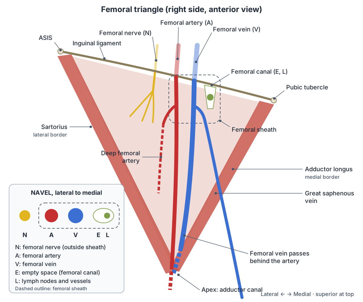
The femoral triangle is a hollow in the upper front of the thigh. Its upper border (base) is the inguinal ligament, running from the anterior superior iliac spine to the pubic tubercle. Its lateral border is the medial edge of sartorius and its medial border is the medial edge of adductor longus. The roof is the fascia lata and the floor is formed by iliopsoas, pectineus and adductor longus. (TeachMeAnatomy: The Femoral Triangle.)
From lateral to medial the triangle contains the femoral nerve, the femoral artery, the femoral vein and the femoral canal, which holds deep lymph nodes and lymph vessels. The great saphenous vein drains into the femoral vein inside the triangle. (TeachMeAnatomy: The Femoral Triangle.)
In the triangle the artery lies under the skin, superficial fascia, the superficial inguinal lymph nodes and the fascia lata, which is why it is easy to feel and to reach with a needle. At the apex of the triangle the femoral vein moves from the medial side of the artery to lie behind it, and the medial femoral cutaneous nerve crosses in front of the artery. (Kenhub: Femoral artery.)
Sartorius, adductor longus and the other muscles that frame the femoral triangle are covered on the Thigh muscles.
The femoral artery in the adductor canal
Below the apex of the femoral triangle the artery enters the adductor canal, also called the subsartorial canal. Sartorius lies in front and medially, vastus medialis laterally, and adductor longus and adductor magnus behind. The canal ends at the adductor hiatus. (TeachMeAnatomy: The Adductor Canal.)
The canal carries the femoral artery, the femoral vein behind the artery, the nerve to vastus medialis and the saphenous nerve. The saphenous nerve, the largest skin branch of the femoral nerve, runs with the artery and is the target of the adductor canal block used for knee and lower leg surgery. (TeachMeAnatomy: The Adductor Canal.)
Radiopaedia describes the vein lower in the canal as lying behind and then lateral to the artery: the vein is medial to the artery at the inguinal ligament, behind it near the apex of the triangle, and posterolateral to it at the lower end of the canal. (Radiopaedia: Femoral vein.)
Branches of the femoral artery
Kenhub lists six named branches from top to bottom: the superficial epigastric, superficial circumflex iliac, superficial external pudendal, deep external pudendal, deep femoral (profunda femoris) and descending genicular arteries, plus muscular branches. (Kenhub: Femoral artery.)
The superficial epigastric artery arises about 1 cm below the inguinal ligament. It and the superficial circumflex iliac artery, the smallest branch, supply the skin, superficial fascia and superficial inguinal lymph nodes. The superficial and deep external pudendal arteries supply the skin of the lower abdomen, perineum and external genitalia. (Kenhub: Femoral artery.)
The deep femoral artery is the largest branch. Radiopaedia places its origin 2 to 5 cm below the inguinal ligament, from the back and outer side of the femoral artery, and Kenhub gives about 3.5 cm. It runs behind the femoral vessels to the inner side of the femur, gives the medial and lateral circumflex femoral arteries and the perforating arteries, and supplies most of the thigh muscles. (Radiopaedia: Profunda femoris artery.)
The medial circumflex femoral artery does not always come from the deep femoral artery. In about 39% of people it arises straight from the common femoral artery. This matters because it is a main supply of the femoral neck, and its injury can lead to avascular necrosis of the femoral head. (Radiopaedia: Medial circumflex femoral artery.)
The descending genicular artery is the lowest branch, arising in the adductor canal. It supplies vastus medialis, adductor magnus and the skin on the upper inner side of the leg. (Kenhub: Femoral artery.)
Branches of the femoral artery
| Branch (top to bottom) | Notes | Main supply |
|---|---|---|
| 1. Superficial epigastric | Arises about 1 cm below the inguinal ligament | Skin and superficial fascia of the lower abdomen, superficial inguinal lymph nodes |
| 2. Superficial circumflex iliac | Smallest branch | Skin, superficial fascia and superficial inguinal lymph nodes |
| 3. Superficial external pudendal | Third branch | Skin of the lower abdomen and external genitalia |
| 4. Deep external pudendal | Fourth branch | Skin of the perineum and external genitalia |
| 5. Deep femoral (profunda femoris) | Largest branch, 2 to 5 cm below the inguinal ligament | Most thigh muscles via the circumflex femoral and perforating arteries |
| 6. Descending genicular | Lowest branch, in the adductor canal | Vastus medialis, adductor magnus, skin of the upper medial leg |
Based on Kenhub and Radiopaedia (see references).
How to feel the femoral pulse
The femoral pulse is felt in the groin just below the inguinal ligament at the mid-inguinal point, midway between the anterior superior iliac spine and the pubic symphysis. (TeachMeAnatomy: Arterial Supply to the Lower Limb.)
A weak femoral pulse can mean narrowing higher up, in the common or external iliac artery, or coarctation (narrowing) of the aorta. (Kenhub: Femoral artery.)
Femoral artery catheter access
Because the femoral artery is superficial and large, it is used for coronary angiography and angioplasty. A catheter is passed up through the external iliac and common iliac arteries and the aorta to the coronary arteries, and contrast dye is injected so that narrowing shows on X-ray. (TeachMeAnatomy: The Femoral Triangle.)
The ideal puncture site is the common femoral artery, between the loop of the inferior epigastric artery above and the femoral bifurcation below. A puncture that is too high raises the risk of bleeding into the retroperitoneum (behind the abdominal cavity), where it cannot be compressed. A puncture that is too low raises the risk of false aneurysm (pseudoaneurysm), haematoma, arteriovenous fistula or limb ischaemia. (Ahn HY et al. Assessment of the Optimal Site of Femoral Artery Puncture and Angiographic Anatomical Study of the Common Femoral Artery. J Korean Neurosurg Soc. 2014.)
In a study of 109 angiograms, every inferior epigastric loop lay above the centre of the femoral head, and the bifurcation lay above that centre in only 4.6% of cases. The authors concluded that a fluoroscopy-guided puncture just below the centre of the femoral head usually hits the right segment. They also cited earlier work showing that the skin crease of the groin lies below the bifurcation in about 72% of limbs, so the crease is a poor guide. (Ahn HY et al. Assessment of the Optimal Site of Femoral Artery Puncture and Angiographic Anatomical Study of the Common Femoral Artery. J Korean Neurosurg Soc. 2014.)
Clinical relevance: PAD, injury and compression
Peripheral arterial disease, usually caused by atherosclerosis, can block the femoral artery. The typical symptom is intermittent claudication: cramp-like calf pain brought on by walking and eased by rest. (Kenhub: Femoral artery.)
The femoral artery's superficial position makes it vulnerable to cuts and stab wounds of the front of the thigh, and the femoral vein next to it is often injured at the same time. (Kenhub: Femoral artery.)
Adductor canal compression syndrome is a rare entrapment of the femoral vessels and saphenous nerve in the canal, usually by enlarged neighbouring muscle such as vastus medialis. It is most common in young men and more often presents with claudication from femoral artery blockage than with nerve symptoms. (TeachMeAnatomy: The Adductor Canal.)
Structures next to the femoral artery
The nearest named structures in the 3D model, measured from the centre of each part:
- Adductor longus · muscular
- Femoral vein · cardiovascular
- Deep femoral vein · cardiovascular
- Adductor brevis · muscular
- Sartorius · muscular
- Femur · skeletal
- Testis · reproductive
- Descending branch of lateral circumflex femoral artery · cardiovascular
- Epididymis · reproductive
- Adductor magnus · muscular
Frequently asked questions
Where is the femoral artery located?
The femoral artery starts in the groin, just below the inguinal ligament at the mid-inguinal point, halfway between the front of the hip bone (anterior superior iliac spine) and the pubic symphysis. It runs down the front and inner thigh through the femoral triangle and adductor canal, then passes to the back of the thigh just above the knee to become the popliteal artery.
What does NAVEL stand for in the femoral triangle?
NAVEL lists the contents of the femoral triangle from lateral to medial: femoral Nerve, femoral Artery, femoral Vein, Empty space (the femoral canal) and Lymphatics (deep inguinal lymph nodes and vessels in the canal). The artery, vein and canal are inside the femoral sheath; the nerve lies outside it, on the lateral side.
What is the difference between the femoral artery and the deep femoral artery?
The femoral artery is the main trunk that runs down the thigh and continues to the knee as the popliteal artery. The deep femoral (profunda femoris) artery is its largest branch, arising a few centimetres below the inguinal ligament. It stays in the thigh, running close to the femur, and supplies most of the thigh muscles through its circumflex and perforating branches.
Why is the femoral artery used for heart catheterisation?
In the groin the femoral artery is large and close to the surface, and its common femoral segment lies over the femoral head, a landmark that shows on X-ray. A catheter can be passed from it up the iliac arteries and aorta to the heart. The puncture should be in the common femoral artery, neither too high nor too low.
Where does the femoral artery become the popliteal artery?
The femoral artery becomes the popliteal artery at the adductor hiatus, a gap in adductor magnus at the lower end of the adductor canal, at about the junction of the middle and lower thirds of the thigh. From there the vessel passes behind the knee into the popliteal fossa.
Is the superficial femoral artery actually superficial?
No. Superficial femoral artery is a clinical name for the femoral artery below the origin of the deep femoral artery. The word only distinguishes it from the deeper profunda femoris. In the middle of the thigh it lies deep under sartorius in the adductor canal, and anatomical terminology does not use the term.
References
- Femoral artery. Radiopaedia reference article.
- Femoral sheath. Radiopaedia reference article.
- Femoral vein. Radiopaedia reference article.
- Profunda femoris artery. Radiopaedia reference article.
- Medial circumflex femoral artery. Radiopaedia reference article.
- Jones O. The Femoral Triangle. TeachMeAnatomy.
- Jones O. The Adductor Canal. TeachMeAnatomy.
- Jones O. Arterial Supply to the Lower Limb. TeachMeAnatomy.
- Femoral artery. Kenhub anatomy library.
- OpenStax. Anatomy and Physiology 2e, §20.5 Circulatory Pathways. Rice University, 2022.
- Ahn HY, Lee HJ, Lee HJ, Yang JH, Yi JS, Lee IW. Assessment of the optimal site of femoral artery puncture and angiographic anatomical study of the common femoral artery. J Korean Neurosurg Soc. 2014;56(2):91-97.
- Foundational Model of Anatomy: Femoral artery (FMA70250). EMBL-EBI Ontology Lookup Service.
- Mitsuhashi N. et al. (2009). BodyParts3D: 3D structure database for anatomical concepts. Nucleic Acids Research 37, D782–D785. Data © The Database Center for Life Science, CC BY 4.0.
Related anatomy
- Lower limb arteriesArteries of the leg explained: common, internal and external iliac, femoral, deep femoral, popliteal, tibial, fibular…
- Popliteal arteryPopliteal artery anatomy: course through the popliteal fossa, genicular branches, how to feel the popliteal pulse…
- Great saphenous veinSaphenous vein anatomy: course of the great and small saphenous veins, saphenofemoral junction, valves, saphenous…
- Thigh musclesThigh muscles explained: quadriceps and sartorius at the front, adductor muscles of the inner thigh, hamstrings at the…
- Femoral veinFemoral vein: The femoral vein is the main deep vein of the thigh and continues the popliteal vein. See where it is in…
- Lower limb nervesLower limb nerves explained: lumbar and sacral plexus diagram, femoral, saphenous, sciatic, tibial, common peroneal…
Explore the whole body in the interactive 3D human anatomy model, or browse the A–Z list of human structures.