

Arteries of the leg: the main pathway
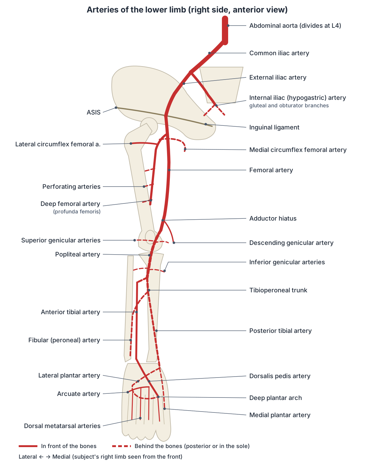
All the blood for one lower limb passes through a single chain of named vessels. The abdominal aorta divides at about the level of vertebra L4 into a left and a right common iliac artery. The common iliac arteries supply the pelvis and, through their branches, the lower limbs. (OpenStax Anatomy and Physiology 2e, 20.5: Circulatory Pathways.)
Each common iliac artery ends by splitting into an internal iliac artery, which mainly supplies the pelvis, and the larger external iliac artery, which supplies the lower limb. Once the external iliac artery leaves the abdomen under the inguinal ligament, it is renamed the femoral artery. (OpenStax Anatomy and Physiology 2e, 20.5: Circulatory Pathways.)
The femoral artery runs down the front and inner thigh, passes through the adductor canal and turns backwards through the adductor hiatus, a gap in adductor magnus, to become the popliteal artery behind the knee. At the lower border of the popliteus muscle the popliteal artery divides into the anterior tibial artery and the tibioperoneal trunk, and the trunk divides into the posterior tibial and fibular (peroneal) arteries. (TeachMeAnatomy: Arterial Supply to the Lower Limb.)
In the foot, the anterior tibial artery continues as the dorsalis pedis artery on the top of the foot, and the posterior tibial artery divides into the medial and lateral plantar arteries in the sole. Branches of the two systems meet in the deep plantar arch, so the foot has two routes of supply. (TeachMeAnatomy: Arterial Supply to the Lower Limb.)
Common iliac artery
The right and left common iliac arteries begin at the aortic bifurcation in front of L4. Each enters the pelvis on the medial side of the psoas major muscle and ends at the pelvic brim, in front of the sacroiliac joint, by dividing into the internal and external iliac arteries. The left common iliac artery is shorter than the right. (Radiopaedia: Common iliac artery.)
The ureter crosses in front of the artery at its bifurcation. The common iliac veins and psoas major lie behind the artery. The right common iliac artery passes in front of the left common iliac vein, a relation that matters for the venous problem described in the veins section below. (Radiopaedia: Common iliac artery.)
Apart from its two terminal branches, the common iliac artery gives only small branches to the peritoneum, psoas, ureter and nearby nerves. Occasionally the iliolumbar artery or an extra renal artery arises from it. (Radiopaedia: Common iliac artery.)
Internal iliac (hypogastric) artery
The internal iliac artery is also called the hypogastric artery; internal iliac is the Terminologia Anatomica name. It is about 4 cm long. It runs backwards and medially towards the greater sciatic foramen, where it divides into an anterior and a posterior division. (Radiopaedia: Internal iliac artery.)
The posterior division gives only branches to the body wall: the iliolumbar artery, the lateral sacral arteries and the superior gluteal artery. The anterior division supplies the pelvic organs (umbilical, superior and inferior vesical, uterine, vaginal and middle rectal arteries) and also gives the obturator, internal pudendal and inferior gluteal arteries. In about a quarter of people the obturator artery arises instead from the inferior epigastric artery. (Radiopaedia: Internal iliac artery.)
Three internal iliac branches reach the lower limb itself. The superior and inferior gluteal arteries leave the pelvis through the greater sciatic foramen, above and below piriformis, to supply the buttock, and the inferior gluteal also helps supply the back of the thigh. The obturator artery passes through the obturator canal into the medial thigh, where its anterior branch supplies pectineus, obturator externus, the adductors and gracilis. (TeachMeAnatomy: Arterial Supply to the Lower Limb.)
External iliac artery
The external iliac artery is the larger of the two terminal branches of the common iliac artery. It runs along the medial side of the iliopsoas muscle, with the iliac fascia and psoas behind it, and passes under the inguinal ligament into the thigh, where it continues as the (common) femoral artery. (Radiopaedia: External iliac artery.)
Just before it leaves the abdomen it gives two named branches: the inferior epigastric artery and the deep circumflex iliac artery. (Radiopaedia: External iliac artery.)
Femoral artery, deep femoral artery and circumflex femoral arteries
The femoral artery begins below the inguinal ligament roughly halfway between the anterior superior iliac spine and the pubic symphysis, with the femoral vein on its medial side. Clinicians often call the part above the origin of the deep femoral artery the common femoral artery and the part below it, down to the adductor hiatus, the superficial femoral artery. Besides the leg, it gives small branches to the anterior abdominal wall and the superficial pelvis. (Radiopaedia: Femoral artery.)
The deep femoral artery (profunda femoris artery, arteria profunda femoris, deep artery of the thigh) is the largest branch. It arises from the back and outer side of the femoral artery in the upper femoral triangle, about 2 to 5 cm below the inguinal ligament, passes behind the femoral artery and vein, and descends behind adductor longus close to the femur. It ends in the lower third of the thigh as the last perforating artery. (Radiopaedia: Profunda femoris artery.)
The deep femoral artery gives the medial and lateral circumflex femoral arteries and usually three or four perforating arteries. The perforating arteries pierce adductor magnus to supply the muscles of the medial and back of the thigh, so most of the thigh muscles depend on the deep femoral artery rather than on the femoral artery itself. (TeachMeAnatomy: Arterial Supply to the Lower Limb.)
The medial circumflex femoral artery usually arises from the deep femoral artery, but Radiopaedia reports a direct origin from the common femoral artery in about 39% of people. It winds backwards round the femoral neck between pectineus and the psoas tendon. Because it is a main supply to the femoral neck and head, damage to it, for example in a femoral neck fracture, can cause avascular necrosis of the femoral head. (Radiopaedia: Medial circumflex femoral artery.)
The lateral circumflex femoral artery runs laterally in front of the femoral neck and usually passes between the branches of the femoral nerve. It has ascending, transverse and descending branches. The ascending and transverse branches of both circumflex arteries join the trochanteric and cruciate anastomoses around the hip. (Radiopaedia: Lateral circumflex femoral artery.)
Popliteal artery and genicular arteries
The popliteal artery begins at the adductor hiatus, at the junction of the middle and lower thirds of the thigh, and runs through the popliteal fossa as its deepest structure, lying on the femur, the knee joint capsule and popliteus. It ends at the lower border of popliteus by dividing into the anterior tibial artery and the tibioperoneal trunk. About 90% of people have this usual pattern. (Radiopaedia: Popliteal artery.)
Five genicular arteries arise from the popliteal artery: the superior lateral, superior medial, middle, inferior lateral and inferior medial genicular arteries. They form a network around the knee, the genicular anastomosis, which supplies the joint capsule and ligaments. The middle genicular artery pierces the back of the capsule to supply the cruciate ligaments. Two sural arteries supply gastrocnemius, soleus and plantaris. (Kenhub: Popliteal artery.)
Branches from below join the network too: the anterior tibial recurrent artery leaves the anterior tibial artery straight after its origin, climbs up through tibialis anterior and joins the lateral genicular branches of the popliteal artery. (Radiopaedia: Anterior tibial artery.)
Popliteal fossa, popliteal pulse, aneurysm and entrapment in detail on the Popliteal artery.
Anterior tibial, posterior tibial and fibular (peroneal) arteries
The anterior tibial artery arises in the popliteal fossa and passes forwards through a gap above the interosseous membrane into the anterior compartment of the leg. It descends on the front of the membrane, beside tibialis anterior, and crosses in front of the ankle joint to continue as the dorsalis pedis artery. Its branches include the anterior tibial recurrent artery and the anterior medial and lateral malleolar arteries. (Radiopaedia: Anterior tibial artery.)
The posterior tibial artery (arteria tibialis posterior) arises from the tibioperoneal trunk and runs down through the posterior compartment of the leg, with the posterior tibial veins beside it. It passes behind the medial malleolus and divides in the tarsal tunnel, about halfway between the medial malleolus and the heel, into the medial and lateral plantar arteries. It also gives the nutrient artery of the tibia and calcaneal branches to the heel. (Radiopaedia: Posterior tibial artery.)
The fibular (peroneal) artery arises from the tibioperoneal trunk about 2.5 cm below popliteus. It runs down along the medial crest of the fibula, between tibialis posterior and flexor hallucis longus or within flexor hallucis longus, and ends near the ankle as calcaneal branches. It supplies the fibula through a nutrient artery and sends perforating branches through the intermuscular septum to the lateral compartment. (Radiopaedia: Peroneal (fibular) artery.)
Some textbooks, such as OpenStax, describe the fibular artery simply as a branch of the posterior tibial artery. Most anatomy references describe it as one of the two branches of the tibioperoneal trunk, the short common stem it shares with the posterior tibial artery. The two descriptions refer to the same vessel. (OpenStax Anatomy and Physiology 2e, 20.5: Circulatory Pathways.)
The branching pattern below the knee varies. Radiopaedia lists a true trifurcation with no tibioperoneal trunk in about 2% of people, a high origin of the anterior tibial artery in about 2.5% and hypoplastic leg arteries in about 4%. (Radiopaedia: Popliteal artery.)
Arteries of the foot: dorsalis pedis, arcuate artery and plantar arch
The dorsalis pedis artery starts in front of the ankle, midway between the malleoli, as the continuation of the anterior tibial artery. It runs across the talus, navicular and intermediate cuneiform towards the first space between the metatarsals, lying between the tendon of extensor hallucis longus and the most medial tendon of extensor digitorum longus, with the deep fibular nerve beside it. (Kenhub: Dorsalis pedis artery.)
Its branches are the lateral and medial tarsal arteries, the arcuate artery and the first dorsal metatarsal artery. It ends by diving into the sole as the deep plantar artery between the two heads of the first dorsal interosseous muscle, where it joins the lateral plantar artery to complete the deep plantar arch. (Kenhub: Dorsalis pedis artery.)
The arcuate artery arises from the dorsalis pedis near the medial cuneiform and arches laterally across the bases of the metatarsals, under the extensor digitorum brevis tendons. It gives the second, third and fourth dorsal metatarsal arteries, each of which divides into dorsal digital arteries for the adjoining sides of the toes. (Kenhub: Arcuate artery of the foot.)
In the sole, the medial plantar artery supplies the medial side of the foot, including abductor hallucis and flexor digitorum brevis. The lateral plantar artery crosses the sole obliquely, supplies the lateral side of the foot and forms the deep plantar arch, which gives the plantar metatarsal arteries to the toes. (Radiopaedia: Posterior tibial artery.)
The dorsalis pedis artery is congenitally absent in about 2% of healthy people, so an absent dorsalis pedis pulse on its own does not always mean disease. In rare cases a perforating branch of the fibular artery takes its place. (Radiopaedia: Dorsalis pedis artery.)
Main arteries of the lower limb
| Artery | Origin | Course and end | Main branches or supply |
|---|---|---|---|
| Common iliac | Aortic bifurcation at L4 | To the pelvic brim, in front of the sacroiliac joint | Internal and external iliac arteries |
| Internal iliac (hypogastric) | Common iliac | About 4 cm; divides at the greater sciatic foramen | Pelvic organs; superior and inferior gluteal and obturator arteries |
| External iliac | Common iliac | Along iliopsoas to the inguinal ligament | Inferior epigastric, deep circumflex iliac; continues as femoral |
| Femoral | External iliac at the inguinal ligament | Femoral triangle, adductor canal, ends at the adductor hiatus | Superficial branches, deep femoral, descending genicular |
| Deep femoral (profunda femoris) | Femoral, 2 to 5 cm below the inguinal ligament | Behind adductor longus; ends as last perforating artery | Medial and lateral circumflex femoral, perforating arteries |
| Popliteal | Femoral at the adductor hiatus | Popliteal fossa to the lower border of popliteus | Five genicular arteries, sural arteries; anterior tibial and tibioperoneal trunk |
| Anterior tibial | Popliteal | Through the interosseous membrane, down the anterior compartment | Recurrent and malleolar branches; continues as dorsalis pedis |
| Posterior tibial | Tibioperoneal trunk | Posterior compartment, behind the medial malleolus | Nutrient artery of tibia, calcaneal branches; medial and lateral plantar |
| Fibular (peroneal) | Tibioperoneal trunk, about 2.5 cm below popliteus | Along the fibula | Fibula, lateral compartment (perforating branches), heel |
| Dorsalis pedis | Anterior tibial in front of the ankle | Across the dorsum to the first intermetatarsal space | Tarsal, arcuate, first dorsal metatarsal, deep plantar arteries |
Compiled from Radiopaedia, TeachMeAnatomy, Kenhub and OpenStax (see references).
Pulse points of the lower limb
OpenStax lists four pulse points in the lower limb: the femoral artery in the groin, the popliteal artery behind the knee, the posterior tibial artery near the medial ankle and the dorsalis pedis artery on the top of the foot. (OpenStax Anatomy and Physiology 2e, 20.2: Blood Flow, Blood Pressure, and Resistance.)
The femoral pulse is felt at the mid-inguinal point, midway between the anterior superior iliac spine and the pubic symphysis. The popliteal pulse is the hardest to feel and needs deep pressure; bending the knee slightly relaxes the popliteal fascia. The posterior tibial pulse is felt below and behind the medial malleolus, and the dorsalis pedis pulse just lateral to the extensor hallucis longus tendon. (TeachMeAnatomy: Arterial Supply to the Lower Limb.)
Clinicians check these pulses in a peripheral vascular examination. A weak or absent dorsalis pedis pulse usually suggests some degree of arterial insufficiency, although, as noted above, the artery is missing in a small number of healthy people. (Kenhub: Dorsalis pedis artery.)
Pulse points of the lower limb
| Pulse | Artery | Where to feel it |
|---|---|---|
| Femoral | Femoral artery | Just below the inguinal ligament at the mid-inguinal point, midway between the ASIS and pubic symphysis |
| Popliteal | Popliteal artery | Deep in the popliteal fossa behind the knee, with the knee slightly bent |
| Posterior tibial | Posterior tibial artery | Below and behind the medial malleolus |
| Dorsalis pedis | Dorsalis pedis artery | Top of the foot, just lateral to the extensor hallucis longus tendon |
Veins of the lower limb: deep, superficial and iliac veins
The lower limb has two sets of veins. Deep veins lie under the deep fascia, run with the main arteries and share their names: anterior tibial, posterior tibial and fibular veins in the leg, the popliteal vein behind the knee and the femoral vein in the thigh. Superficial veins run in the fat under the skin and drain into the deep veins. (TeachMeAnatomy: Venous Drainage of the Lower Limb.)
The anterior tibial, posterior tibial and fibular veins join behind the knee to form the popliteal vein. It continues through the adductor canal as the femoral vein, which receives the deep femoral vein, becomes the common femoral vein and passes under the inguinal ligament to become the external iliac vein. Radiopaedia advises against the old term superficial femoral vein: this is a deep vein, and the misleading name has caused confusion when deep vein thrombosis is reported. (Radiopaedia: Femoral vein.)
The two main superficial veins are the great saphenous vein, which begins at the medial end of the dorsal venous arch, passes in front of the medial malleolus and ends in the femoral vein just below the inguinal ligament, and the small saphenous vein, which begins at the lateral side of the arch, passes behind the lateral malleolus and up the back of the calf, and usually ends in the popliteal vein. (TeachMeAnatomy: Venous Drainage of the Lower Limb.)
The small saphenous vein runs with the sural nerve. Its ending varies: it does not always drain into the popliteal vein, and an extension up the back of the thigh, the vein of Giacomini, is common. (Radiopaedia: Small saphenous vein.)
The external and internal iliac veins join in front of the sacroiliac joint to form the common iliac vein. The two common iliac veins unite at about the level of L5 to form the inferior vena cava. The left common iliac vein also receives the median sacral vein, and the common iliac veins have no valves in most people. (Radiopaedia: Common iliac vein.)
Because the right common iliac artery crosses in front of the left common iliac vein, the vein can be compressed. When this causes symptoms it is called May-Thurner syndrome, a cause of left leg swelling and deep vein thrombosis, seen mainly in women aged 20 to 40. Some compression is found in about a quarter of people without symptoms. (Radiopaedia: May-Thurner syndrome.)
Leg veins contain one-way valves. When the calf and thigh muscles contract, they squeeze the deep veins and the valves below close, so blood is pushed towards the heart. This skeletal muscle pump helps the low-pressure veins work against gravity. (OpenStax Anatomy and Physiology 2e, 20.2: Blood Flow, Blood Pressure, and Resistance.)
Course, saphenofemoral junction, varicose veins and graft use on the Great saphenous vein.
What the 3D model shows and leaves out
The 3D view uses BodyParts3D, an adult male data set. It includes the aorta, the common, internal and external iliac arteries, the femoral and popliteal arteries, the lateral circumflex femoral artery and its descending branch, the five genicular arteries, the tibial arteries and the main arteries of the foot. It does not include the deep femoral (profunda femoris) artery, the medial circumflex femoral artery, the fibular artery or the gluteal and obturator arteries, so these are shown in the diagram instead.
When you study the model, start at the aortic bifurcation in front of L4. Follow each common iliac artery along the medial side of psoas major to the pelvic brim, where it divides in front of the sacroiliac joint; the ghosted skeleton makes these bony landmarks easy to find. (Radiopaedia: Common iliac artery.)
Lower limb arteries in the 3D model
- Abdominal aorta
- The abdominal aorta is the part of the aorta that runs through the abdomen and supplies the abdominal organs, pelvis and lower limbs.
- Anterior tibial artery
- The anterior tibial artery is the artery of the front compartment of the leg and the main source of the dorsalis pedis artery.
- Anterior tibial recurrent artery
- The anterior tibial recurrent artery is a small branch of the anterior tibial artery that runs up toward the knee.
- Arcuate artery
- The arcuate artery here is the artery of the dorsum of the foot, a branch of the dorsalis pedis artery.
- Common iliac artery
- The common iliac arteries are the two terminal branches of the abdominal aorta.
- Deep plantar artery
- The deep plantar artery is a branch of the dorsalis pedis that passes to the sole of the foot.
- Descending branch of lateral circumflex femoral artery
- This is the long branch of the lateral circumflex femoral artery that runs down the front of the thigh.
- Descending genicular artery
- The descending genicular artery is a branch of the femoral artery that supplies the knee region.
- Dorsalis pedis artery
- The dorsalis pedis artery is the continuation of the anterior tibial artery onto the top of the foot.
- External iliac artery
- The external iliac artery is the main artery of the lower limb in the pelvis and becomes the femoral artery.
- Femoral artery
- The femoral artery is the main artery of the thigh, continuing the external iliac artery.
- Inferior lateral genicular artery
- The inferior lateral genicular artery is a branch of the popliteal artery around the knee.
- Inferior medial genicular artery
- The inferior medial genicular artery is a branch of the popliteal artery around the knee.
- Internal iliac artery
- The internal iliac artery supplies the pelvis.
- Lateral circumflex femoral artery
- The lateral circumflex femoral artery supplies the lateral thigh.
- Lateral plantar artery
- The lateral plantar artery is the larger terminal branch of the posterior tibial artery in the sole.
- Lateral superior genicular artery
- The lateral superior genicular artery is a branch of the popliteal artery around the knee.
- Medial plantar artery
- The smaller terminal branch of the posterior tibial artery in the sole of the foot.
- Medial superior genicular artery
- A branch of the popliteal artery that wraps around the medial side of the knee.
- Middle genicular artery
- A branch of the popliteal artery supplying the inside of the knee joint.
- Plantar arch
- The arterial arch of the sole formed mainly by the lateral plantar artery.
- Popliteal artery
- The continuation of the femoral artery behind the knee.
- Posterior tibial artery
- The larger terminal branch of the popliteal artery in the posterior leg.
Frequently asked questions
What are the main arteries of the leg?
Each leg is supplied by one chain: the external iliac artery becomes the femoral artery at the groin, which becomes the popliteal artery behind the knee. Below the knee it splits into the anterior tibial, posterior tibial and fibular arteries. The anterior tibial becomes the dorsalis pedis on the foot, and the posterior tibial gives the plantar arteries of the sole.
What is the difference between the internal and external iliac arteries?
Both are branches of the common iliac artery at the pelvic brim. The internal iliac (hypogastric) artery is short and mainly supplies the pelvic organs, the buttock through the gluteal arteries and the medial thigh through the obturator artery. The larger external iliac artery runs to the inguinal ligament and continues as the femoral artery, the main supply of the leg.
What does the deep femoral artery supply?
The deep femoral (profunda femoris) artery supplies most of the thigh. Its medial and lateral circumflex femoral branches wrap around the top of the femur and supply the hip and femoral neck, and its three or four perforating branches pierce adductor magnus to supply the adductor and hamstring muscles at the back and inner side of the thigh.
Where can you feel pulses in the leg?
There are four standard pulse points: the femoral pulse at the mid-inguinal point in the groin, the popliteal pulse deep behind the slightly bent knee, the posterior tibial pulse below and behind the medial ankle bone, and the dorsalis pedis pulse on the top of the foot just lateral to the big toe extensor tendon.
Is the fibular artery the same as the peroneal artery?
Yes. Fibular artery is the current Terminologia Anatomica name and peroneal artery is the older name, still common in clinical use. It arises from the tibioperoneal trunk just below the knee, runs down along the back of the fibula and supplies the fibula, the lateral compartment muscles through perforating branches and the outer heel.
Where does the common iliac vein join the inferior vena cava?
The external and internal iliac veins join in front of the sacroiliac joint to form each common iliac vein. The right and left common iliac veins then unite at about the level of the fifth lumbar vertebra to form the inferior vena cava. For comparison, the aorta divides into the common iliac arteries at about L4.
What is the arcuate artery of the foot?
The arcuate artery is a branch of the dorsalis pedis artery that arches laterally across the top of the foot over the bases of the metatarsal bones. It gives the second, third and fourth dorsal metatarsal arteries, which split into dorsal digital arteries for the toes, and it links with the lateral tarsal and plantar arteries.
References
- OpenStax. Anatomy and Physiology 2e, §20.5 Circulatory Pathways. Rice University, 2022.
- OpenStax. Anatomy and Physiology 2e, §20.2 Blood Flow, Blood Pressure, and Resistance. Rice University, 2022.
- Jones O. Arterial Supply to the Lower Limb. TeachMeAnatomy.
- Jones O. Venous Drainage of the Lower Limb. TeachMeAnatomy.
- Common iliac artery. Radiopaedia reference article.
- Internal iliac artery. Radiopaedia reference article.
- External iliac artery. Radiopaedia reference article.
- Femoral artery. Radiopaedia reference article.
- Profunda femoris artery. Radiopaedia reference article.
- Medial circumflex femoral artery. Radiopaedia reference article.
- Lateral circumflex femoral artery. Radiopaedia reference article.
- Popliteal artery. Radiopaedia reference article.
- Anterior tibial artery. Radiopaedia reference article.
- Posterior tibial artery. Radiopaedia reference article.
- Peroneal artery. Radiopaedia reference article.
- Dorsalis pedis artery. Radiopaedia reference article.
- Common iliac vein. Radiopaedia reference article.
- Femoral vein. Radiopaedia reference article.
- May-Thurner syndrome. Radiopaedia reference article.
- Small saphenous vein. Radiopaedia reference article.
- Dorsalis pedis artery. Kenhub anatomy library.
- Arcuate artery of the foot. Kenhub anatomy library.
- Popliteal artery. Kenhub anatomy library.
- Mitsuhashi N. et al. (2009). BodyParts3D: 3D structure database for anatomical concepts. Nucleic Acids Research 37, D782–D785. Data © The Database Center for Life Science, CC BY 4.0.
Related anatomy
- Femoral arteryFemoral artery anatomy: where it is, the femoral triangle and sheath (NAVEL), adductor canal, branches, deep femoral…
- Popliteal arteryPopliteal artery anatomy: course through the popliteal fossa, genicular branches, how to feel the popliteal pulse…
- Great saphenous veinSaphenous vein anatomy: course of the great and small saphenous veins, saphenofemoral junction, valves, saphenous…
- AortaAorta anatomy explained: ascending aorta, aortic arch and its three branches, thoracic aorta, and every abdominal…
- Inferior vena cavaInferior vena cava anatomy: course, relations, tributaries by vertebral level, embryology, variants and IVC filters…
- Lower limb nervesLower limb nerves explained: lumbar and sacral plexus diagram, femoral, saphenous, sciatic, tibial, common peroneal…
Explore the whole body in the interactive 3D human anatomy model, or browse the A–Z list of human structures.