

Cat anatomy at a glance
A cat's body has the same organ systems as ours: skin, skeleton, muscles, nerves and senses, heart and vessels, lungs, gut, kidneys, hormone glands and reproductive organs. Each has its own color in the 3D viewer. The reference animal is the domestic cat, Felis catus, modelled as a short-haired adult; breeds differ mainly in coat, from long-haired Persians to short-haired Abyssinians.[3] Vets use direction words for four-legged animals: cranial (towards the head), caudal (towards the tail), dorsal (the back) and ventral (the belly).
Cat body by the numbers
| Feature | Domestic cat | Source |
|---|---|---|
| Vertebrae | 7 neck, 13 chest, 7 lumbar, 3 fused sacral, 22–23 tail | [1] |
| Teeth | 26 milk teeth, 30 adult teeth | [14, 21, 1] |
| Resting heart rate | 120–140 a minute; about 132 at home, 150 or more at the vet | [15, 41] |
| Resting breathing rate | 16–40 breaths a minute | [16] |
| Body temperature | 38.1–39.2 °C (100.5–102.5 °F), rectal | [17] |
| Hearing range | 48 Hz to 85 kHz | [35] |
External anatomy: the parts of a cat
The external parts of a cat are the ears (pinnae), eyes, nose, whiskers, furred skin, four legs with padded, clawed paws, and a long tail. The nose, lips and paw pads are hairless, and a cat has long hairs above the eyes but no true eyelashes.[2, 1] Oil glands are plentiful on the face, chin, paws and tail, and a cat marks objects by rubbing its face on them, leaving scent from facial pheromones.[6]
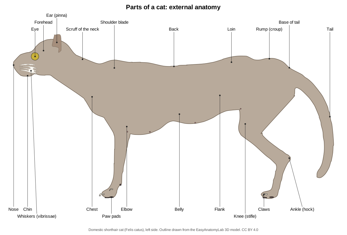
Whiskers (vibrissae) are long, stiff touch hairs: about a dozen on each upper lip, with more on the cheeks and above the eyes.[2, 1] Sensory cells around their roots make them very sensitive, and cats use them to judge the size of openings.[6, 3]
Cat ears and eyes
The upright pinna funnels sound down an ear canal deeper than ours to the eardrum; the middle ear holds three tiny bones, and the inner ear holds the cochlea and the organs of balance.[5] Cats hear from 48 Hz to 85 kHz, one of the broadest ranges of any mammal.[35] Their highly developed balance organs help a falling cat land feet first (the righting reflex).[3]
Cats see about six times better than people in dim light.[3] The retina has many light-collecting rod cells,[4] and behind it the tapetum lucidum, a reflective layer of the choroid, bounces light back for a second pass and makes the eyes shine at night.[36, 2] The pupil is round in the dark but closes to a vertical slit in bright light.[2] A whitish-pink third eyelid sits in the inner corner of each eye. Whether cats see colors is uncertain.[4]
Cat paw anatomy: pads and retractable claws
Each paw has one large pad under the sole and a small pad under each toe, and each front leg has an extra carpal pad behind the wrist. The pads are cushions of skin, elastic tissue and fat.[1] Cats sweat through their paw pads when excited.[6] The front paw has five toes, including the dewclaw, and the hind paw four.[2]
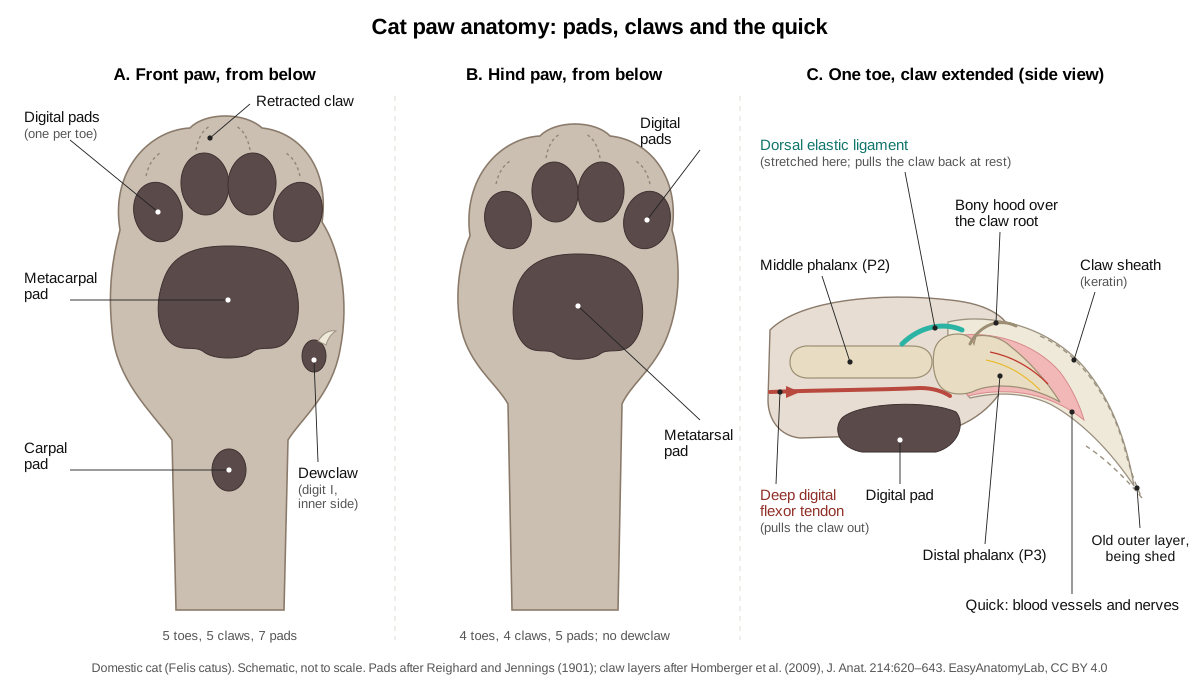
Each claw grows over the last toe bone, the distal phalanx. A very elastic ligament on top of the toe keeps that bone rolled back, so at rest the claw is held up off the ground. To unsheathe it, the cat pulls on the flexor tendon under the toe, which stretches the ligament and swings the claw down.[2] Dogs cannot retract their claws.[3]
The claw is a keratin sheath made by living tissue around the bone; its outer layers crack and peel off with use, which keeps the tip sharp.[39] Scratching strips these old sheaths and leaves scent from glands in the paws. The pink quick inside holds blood vessels and nerves and bleeds if cut.[24, 3] Declawing is an amputation of the bones at the tips of the toes,[24] and in one study 63% of 137 declawed cats still had fragments of that bone on X-rays.[40]
Skeleton and muscles
The spine has 7 cervical, 13 thoracic, 7 lumbar and 3 fused sacral vertebrae, plus 22 or 23 in the tail. The collarbone is a slender rod buried in the shoulder muscles,[1] and the shoulder girdle is attached to the spine by soft tissues alone.[2] For every bone, see the cat skeleton guide.
A large, thin skin muscle, the cutaneus maximus, covers most of the side of the trunk, and several muscles are split into named parts: the trapezius into the clavotrapezius, acromiotrapezius and spinotrapezius.[1] The cat dissection guide covers every muscle a lab asks for; in the viewer, switch on Muscular to see them.
Cat internal anatomy: where the organs are
The chest holds the heart and lungs, and the diaphragm separates them from the abdomen, where the liver, stomach and spleen lie in front, the small intestine fills most of the space, and the kidneys sit under the lumbar spine.[1]
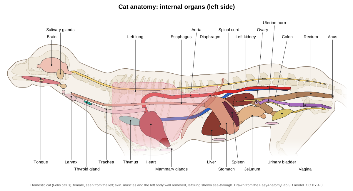
| Organ | Where it is | Notes |
|---|---|---|
| Heart | Lower chest, about the 4th or 5th to 8th rib | Apex points down, back and left, touching the diaphragm |
| Lungs | Either side of the heart | Right lung 4 lobes; left lung 2[2] |
| Liver | Against the diaphragm, mostly right | Five lobes; gallbladder in the right median lobe |
| Stomach | Left front of the abdomen | Narrow pyloric end crosses to the right |
| Spleen | Along the stomach's outer curve | Deep red lymphatic organ |
| Kidneys | Under the 3rd to 5th lumbar vertebrae | Right one 1–2 cm further forward |
Table source: Reighard and Jennings.[1]
Digestive system: built for meat
Cats are obligate carnivores: they rely on nutrients found only in animal products.[23] An adult has 30 teeth, 2 × (I 3/3, C 1/1, P 3/2, M 1/1).[1, 20] The tongue's sharp, backward-facing papillae have hollow tips that wick saliva into the fur during grooming,[38] and they push loose hair down the throat, which is how hairballs start.[22] Cats ignore sugar because one of their two sweet-receptor genes is broken.[37]
The stomach is a simple, curved sac with its broad end on the left.[1, 2] Sources disagree on gut length: Reighard and Jennings give the small intestine as about three times the body length and the large intestine about half, while Mivart put the whole intestine at about five times.[1, 2] The large intestine starts with a small cecum, a rudiment of an appendix, and two anal sacs open into the anus.[1]
Breathing, heart and circulation
Air passes the larynx into the trachea, which splits into the right and left bronchi.[9] A small vomeronasal (Jacobson's) organ on the floor of the nasal cavity opens into the roof of the mouth.[1] Old anatomy texts say purring is made by vibration of the false vocal cords in the larynx.[1, 2]
The heart has two atria and two ventricles. Blood from the body enters the right side, is pumped to the lungs, returns to the left side and leaves through the aorta.[8] The aortic arch gives off only two trunks: the brachiocephalic artery, which supplies both carotid arteries and the right forelimb, and the left subclavian artery.[2] A resting heart beats 120–140 times a minute;[15] monitored cats averaged 132 at home, 150 at the clinic and 187 while held for a scan.[41] Hypertrophic cardiomyopathy, a thickening of the left ventricle, is the most common heart disease of cats.[25]
Kidneys, nerves and glands
The kidneys filter wastes, balance water and salts, and make the hormones erythropoietin and renin.[10] Chronic kidney disease affects up to 40% of cats over 10 and 80% over 15.[27] The male's urethra is longer and narrower than the female's, so males are more likely to suffer a blockage.[26]
The brain has a cerebrum, cerebellum and brainstem.[7] The spinal cord thickens where the leg nerves leave it and tapers behind the seventh lumbar vertebra into a thread reaching the tail.[1]
The two-lobed thyroid lies beside the trachea just behind the larynx;[1] an enlarged, overactive thyroid is common in middle-aged and older cats.[28] The pancreas makes insulin,[11] and 1 in 100 to 1 in 500 cats develop diabetes.[29] The thymus, in front of the heart, is largest in kittens and shrinks in adults.[1]
Male vs female cat anatomy
A male (tom) has two testes in a scrotum just below the anus and a penis below that, pointing backwards; a female (queen) has two ovaries just behind the kidneys, a Y-shaped uterus and a vulva just below the anus.[1, 2] Use the Female/Male switch in the viewer to compare them.
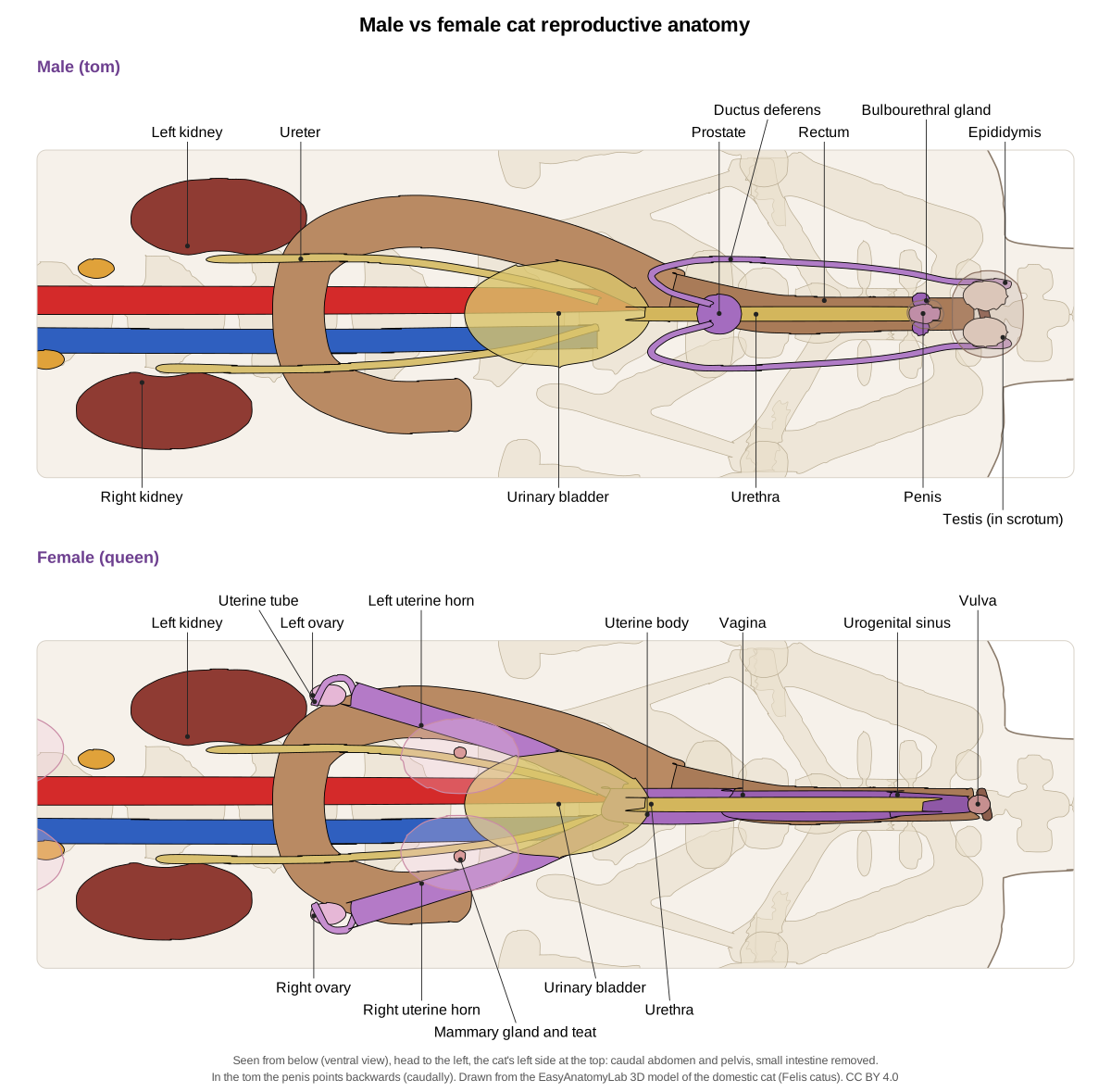
| Male cat (tom) | Female cat (queen) | |
|---|---|---|
| Gonads | Two testes in the scrotum | Two ovaries behind the kidneys |
| Ducts | Epididymis and ductus deferens | Uterine tubes, two uterine horns, short uterine body |
| Glands | Prostate, two bulbourethral glands; no seminal vesicles | Four or five pairs of mammary glands |
| Special features | Penis bone; spines on the glans | Ovulation triggered by mating |
| Urethra | Long and narrow | Short, opening into the urogenital sinus |
Table sources: Reighard and Jennings; Mivart; MSD Veterinary Manual.[1, 2, 12]
Male cat anatomy
The testes descend into the scrotum by two months of age.[33] Sperm pass from each testis through the epididymis and ductus deferens to the urethra,[12] past the two-lobed prostate and the bulbourethral glands.[1] Cats have no seminal vesicles.[2] The penis points backwards when relaxed, so toms urinate backwards; it contains a small bone, the os penis, and its tip is covered with small, hard spines.[2, 1] The spines depend on testosterone and disappear within weeks of neutering.[34] Toms reach puberty at 4 to 12 months, and intact males often spray urine to mark territory.[32] Male cats also have nipples over rudimentary mammary glands.[1, 2]
Female cat anatomy
The ovaries make eggs, estrogen and progesterone.[12] The uterus has a body about 4 cm long and two horns running forwards to the uterine tubes. The vagina joins the bladder neck in a short urogenital sinus that opens at the vulva.[1] Queens are induced ovulators: mating triggers ovulation.[12, 31] Puberty comes at about six months (range 4–12), and from spring to early autumn a queen comes into heat every 2–3 weeks.[31, 18] Pregnancy lasts about 60–65 days according to the MSD manual, or 64–71 (average 65) according to VCA.[13, 31]
Mivart counted eight nipples, four on each side; Reighard and Jennings found five on each side, so the number varies.[2, 1] Spaying removes the uterus and both ovaries.[30]
Telling males from females: lift the tail. In a male the scrotum lies between the anus and the prepuce, so the two openings are well apart; in a female the vulva is directly below the anus.[1]
Cat vs dog anatomy
Cats and dogs share the same organ systems, but they differ in claws, teeth, heart rate and reproduction. Both have a tapetum lucidum, as carnivores do.[36]
| Feature | Cat | Dog |
|---|---|---|
| Claws | Retractable | Not retractable[3] |
| Adult teeth | 30 | 42[19] |
| Resting heart rate | 120–140 a minute | 70–120 a minute[15] |
| Resting breathing rate | 16–40 a minute | 18–34 a minute[16] |
| Heat cycle | Every 14–21 days in season; mating triggers ovulation | Every 6–7 months, all year[18] |
| Male accessory glands | Prostate and bulbourethral glands | Prostate only[43] |
| Humerus | Supracondylar foramen for the median nerve and brachial artery | No such foramen[42] |
For a dog in 3D, see dog anatomy.
Cat vs human anatomy
Cat anatomy labeled: every structure in the 3D model
This is the text version of the 3D cat: all 193 labeled structures, grouped by body system, with what each does. Structures marked "males only" or "females only" appear when you switch sex in the viewer.
Skin & external
- Skin and fur (Integument)
- The furred skin that covers the whole body; it is loosely attached to the muscles underneath. Protects the body, keeps it warm, senses touch and carries scent glands.
- Pinna (ear flap) (Auricle, external ear)
- The upright, triangular flap of the external ear, supported by elastic cartilage. Collects sound and funnels it into the ear canal. Many small ear muscles turn each pinna independently towards a sound.
- Eye (Eyeball, bulbus oculi)
- A very large eyeball for the size of the head, set facing forwards. Vision, especially in dim light: a reflective layer behind the retina (tapetum lucidum) bounces light back through it and makes the eyes shine at night.
- Vibrissae (whiskers) (Sinus hairs, mystacial whiskers)
- Long, stiff touch hairs in rows on the upper lip, with smaller tufts above the eyes and on the chin. Each whisker sits in a follicle surrounded by a blood sinus and many nerve endings, so the slightest bend is felt. Cats use them to judge gaps and to sense prey at close range.
- Paw pads (Digital, metacarpal/metatarsal and carpal pads)
- Thick, hairless cushions of skin and fat under the toes and the sole. Absorb shock and give grip. The cat walks on its toes (digitigrade stance), so its weight rests on these pads.
- Claws (Ungues)
- Curved, sharp, keratin sheaths that cover the bony ungual process of each distal phalanx. Climbing, holding prey and fighting. At rest they are pulled back (retracted) into a skin sheath so they stay sharp and do not touch the ground.
- Nipples (mammary papillae) (Teats)
- Small projections of skin in two rows along the chest and belly. In females each nipple carries the openings of the milk ducts of one mammary gland. Males have small, rudimentary nipples.
- Scrotum (Scrotal sac) · males only
- A furred sac of skin holding the two testes, just below the anus. Keeps the testes slightly cooler than the body, which sperm production needs.
- Vulva (External genitalia, urogenital opening) · females only
- The vertical, slit-like external opening of the urogenital sinus. External opening of the reproductive and urinary tracts.
Skeletal
- Incisive bone (Premaxilla)
- The paired bone at the very front of the upper jaw that holds the upper incisor teeth. Carries the six upper incisors and forms the front of the hard palate and nasal aperture.
- Nasal bones
- Two narrow bones that form the roof (bridge) of the nose. Roof over the nasal cavity, which holds the scroll-like turbinate bones that warm air and carry the smell receptors.
- Maxilla (Upper jaw bone)
- The large paired bone of the upper jaw and side of the face. Holds the upper canine and cheek teeth, forms most of the hard palate, and gives origin to whisker-pad and lip muscles.
- Zygomatic arch (Cheek bone (zygomatic bone + zygomatic process of the temporal bone))
- A strong, wide bony arch on each side of the skull, below the orbit. Anchors the masseter muscle and guards the eye from below. Its wide flare makes room for large jaw muscles, which give cats a strong bite at the canines.
- Frontal bones
- The paired bones of the forehead, between and above the orbits. Forms the inner wall and roof of each orbit and the front of the braincase; contains an air space (frontal sinus).
- Parietal bones
- The paired bones forming most of the rounded roof and sides of the braincase. Protect the brain and give origin to the temporalis muscle.
- Interparietal bone
- A small single bone in the midline between the two parietal bones and the occipital bone. Part of the braincase roof. In adult cats it fuses with its neighbours and is hard to see.
- Occipital bone
- The bone at the back and base of the skull that surrounds the foramen magnum. Its two occipital condyles articulate with the atlas (first vertebra), and the spinal cord passes through the foramen magnum.
- Temporal bone and tympanic bulla (Squamous, petrous and tympanic parts)
- The bone on the side and underside of the braincase that houses the ear, including the large rounded tympanic bulla. Holds the middle and inner ear, forms the socket for the jaw joint and carries the back root of the zygomatic arch.
- Mandible (Lower jaw (two halves joined at the chin))
- The lower jaw: two halves joined at the chin by a fibrous joint (mandibular symphysis). Holds the lower teeth and gives attachment to the masseter, temporalis and digastric muscles.
- Incisors
- Twelve tiny front teeth: three on each side, top and bottom. Nibble, groom fur and pull meat from bones.
- Canine teeth (Fangs)
- Four long, conical, slightly curved fangs. Grip and kill prey. Domestic cats usually kill small prey with a bite to the neck, driving the canines between the vertebrae.
- Premolars and molars (carnassials) (Cheek teeth)
- Blade-like cheek teeth: three premolars and one molar in each upper jaw, two premolars and one molar in each lower jaw. Slice meat like scissors. The largest pair, the carnassials (upper fourth premolar and lower first molar), shear past each other.
- Hyoid apparatus (Hyoid bone)
- A chain of small bones that slings the tongue and larynx from the skull. Supports the tongue and larynx and gives attachment to swallowing muscles.
- Atlas (C1) (First cervical vertebra)
- The ring-shaped first neck vertebra with broad, flat wings. The atlanto-occipital joint lets the cat nod its head up and down.
- Axis (C2) (Second cervical vertebra, epistropheus)
- The longest neck vertebra, with a tall blade-like spinous process and a peg (dens) that projects into the atlas. The atlas rotates around the dens, letting the cat turn its head from side to side.
- Cervical vertebrae C3–C7 (Neck vertebrae)
- The five remaining neck vertebrae; with the atlas and axis there are seven, as in almost all mammals. Support and move the neck. Their transverse processes are flattened and pierced by the transverse foramina for the vertebral artery.
- Thoracic vertebrae (13)
- Thirteen vertebrae, each bearing a pair of ribs; humans have twelve. Form the roof of the rib cage. The tall spinous processes of the first few form the withers and anchor shoulder and back muscles.
- Lumbar vertebrae (7)
- Seven large vertebrae of the lower back, with long transverse processes; humans have five. Carry the weight of the abdomen and anchor the powerful back and abdominal muscles. Flexing and extending this long lumbar region lengthens the cat's stride when it gallops or springs.
- Sacrum
- Three sacral vertebrae fused into a single bone. Joins the spine to the pelvis at the sacroiliac joints, so thrust from the hindlimbs reaches the body.
- Caudal vertebrae (tail) (Coccygeal vertebrae)
- The tail vertebrae: about 22 or 23 in a typical long-tailed cat, getting smaller and simpler towards the tip. Support the tail, which the cat uses for balance on narrow perches and for signalling (an upright tail is a friendly greeting).
- Ribs (13 pairs) (Costae)
- Thirteen pairs of curved bones forming the sides of the chest. Protect the heart and lungs and move during breathing.
- Costal cartilages
- Bars of flexible cartilage that continue the ribs ventrally. Let the chest wall flex during breathing and when the cat squeezes through gaps.
- Sternum (Breastbone: manubrium, body (sternebrae), xiphoid process)
- A rod of bony segments (sternebrae) along the floor of the chest. Anchors the costal cartilages and the pectoral muscles.
- Scapula (Shoulder blade)
- A broad, flat, triangular bone lying on the side of the chest, with a tall spine on its outer surface. The shoulder is attached to the body only by muscles, so the scapula slides freely, lengthening the stride and absorbing landing shock.
- Clavicle (floating collarbone) (Collarbone)
- A small, thin, curved rod of bone that forms no joint with any other bone. Gives attachment to the clavotrapezius and clavodeltoid muscles. Because it does not link the shoulder to the sternum, the forelimbs can move closer together and the shoulders can squeeze through narrow gaps.
- Humerus (Upper arm bone)
- The long bone of the upper arm, from the shoulder to the elbow. Lever for the shoulder and elbow muscles.
- Radius
- The forearm bone that carries most of the weight to the wrist. Its round head turns against the ulna, so cats can rotate the forearm and turn the paw over (supination), unlike dogs, which can do this only a little.
- Ulna
- The forearm bone that forms the point of the elbow. Its olecranon process is the lever of the triceps, which straightens the elbow.
- Carpal bones (7) (Wrist bones)
- Seven small bones in two rows: three in the proximal row and four in the distal row. Allow the wrist to flex and give spring to each step.
- Metacarpals (5) (Palm bones)
- Five bones of the palm; the first (for the dewclaw) is short. Lengthen the limb and act as springs in the digitigrade stance.
- Phalanges of the forepaw (Toe bones (proximal, middle and distal phalanges))
- Each of the four main front toes has three phalanges: proximal, middle and distal. The distal phalanx carries the claw. At rest it is pulled up beside the middle phalanx, keeping the claw off the ground.
- Dewclaw (digit I) (Pollex, first digit)
- The thumb-like first toe on the inside of each forepaw, with only two phalanges. Grips prey and helps when climbing.
- Ilium
- The long, narrow, blade-like front part of the hip bone (os coxae). Joins the hip bone to the sacrum (sacroiliac joint) and anchors the gluteal, sartorius and tensor fasciae latae muscles.
- Ischium
- The caudal part of the hip bone, ending in the ischial tuberosity. The ischial tuberosity anchors the hamstring muscles (biceps femoris, semitendinosus, semimembranosus).
- Pubis
- The ventral part of the hip bone that meets its partner at the pelvic symphysis. Closes the pelvic ring ventrally and anchors the gracilis and adductor muscles.
- Femur (Thigh bone)
- The long, straight thigh bone, from the hip to the knee (stifle). Lever for the powerful thigh muscles that drive jumping.
- Patella (Kneecap)
- A small sesamoid bone in the tendon of the quadriceps. Protects the tendon and increases the leverage of the quadriceps that straighten the knee.
- Tibia (Shin bone)
- The main bone of the lower leg (crus), slightly longer than the femur. Carries weight to the foot; its crest anchors the sartorius, gracilis and hamstrings.
- Fibula
- A thin bone lying alongside the tibia. Anchors lower-leg muscles; its lower end (lateral malleolus) steadies the ankle.
- Calcaneus (Heel bone, os calcis)
- The longest tarsal bone, projecting backwards to form the point of the hock. Lever for the calf muscles: the calcaneal (Achilles) tendon pulls on it to extend the hock and push the cat off the ground.
- Other tarsal bones (Talus, central (navicular), cuboid and three cuneiforms)
- Six small ankle bones besides the calcaneus: seven tarsals in all. The talus forms the pulley-like hinge of the ankle with the tibia; the others transfer weight to the metatarsals.
- Metatarsals
- Four long bones of the sole (II–V), plus a tiny remnant of the first. Form the long lever of the hind foot used in jumping.
- Phalanges of the hindpaw
- Each of the four hind toes has three phalanges. As in the forepaw, the distal phalanx carries a retractile claw.
- Os penis (Baculum) · males only
- A tiny bone in the tip of the penis. Stiffens the tip of the penis.
Muscular
- Masseter
- A thick, powerful jaw-closing muscle on the cheek. Closes the jaw (elevates the mandible).
- Temporalis
- The largest jaw-closing muscle, fan-shaped on the side of the skull. Closes the jaw and holds it shut on struggling prey.
- Digastric
- A strap muscle under the lower jaw. Opens the jaw (depresses the mandible).
- Mylohyoid
- A thin sheet of transverse muscle fibres that forms the floor of the mouth. Raises the floor of the mouth and the tongue during swallowing.
- Sternomastoid (Sternocleidomastoid (human))
- A strap muscle running diagonally up the side of the neck. Turns and flexes the head and neck.
- Cleidomastoid (Clavicular part of the human sternocleidomastoid)
- A narrow, flat neck muscle running from the skull to the clavicle. Pulls the clavicle forwards when the head is fixed; turns the head and lowers the snout when the clavicle is fixed.
- Sternohyoid
- A narrow strap muscle along the ventral midline of the neck. Pulls the hyoid and tongue back during swallowing.
- Clavotrapezius (Cranial part of the human trapezius (with clavodeltoid = brachiocephalicus))
- A broad, flat muscle covering the side and back of the neck. Draws the clavicle and the front of the shoulder forwards and upwards.
- Acromiotrapezius (Middle part of the human trapezius)
- A thin, square muscle over the shoulders. Holds the two scapulae together and pulls them towards the midline.
- Spinotrapezius (Caudal part of the human trapezius)
- A flat triangular muscle behind the acromiotrapezius. Pulls the scapula backwards and upwards.
- Clavodeltoid (Clavobrachialis, cleidobrachialis)
- A band of muscle down the front of the shoulder and arm. Flexes the elbow and pulls the forelimb forwards.
- Acromiodeltoid
- A short, thick muscle over the point of the shoulder. Flexes and rotates the humerus outwards (lateral rotation).
- Spinodeltoid
- A flat band behind the acromiodeltoid. Flexes the shoulder and rotates the humerus, together with the acromiodeltoid.
- Latissimus dorsi
- A large, flat, triangular muscle covering the side of the chest behind the shoulder. Pulls the forelimb backwards and upwards: the power stroke of each stride.
- Levator scapulae ventralis
- A narrow strap from the head to the shoulder blade; humans have no equivalent. Pulls the scapula forwards.
- Rhomboideus (Rhomboideus major, minor and capitis)
- Deep muscles that tie the dorsal border of the scapula to the spine. Pulls the scapula towards the spine and holds it against the body.
- Splenius
- A broad, thin, deep muscle of the back of the neck. Raises the head and turns it to the side.
- Supraspinatus
- A thick muscle filling the fossa in front of the scapular spine. Extends the shoulder and steadies the joint.
- Infraspinatus
- A muscle filling the fossa behind the scapular spine. Rotates the humerus outwards and stabilises the shoulder.
- Teres major
- A thick muscle along the caudal (axillary) border of the scapula. Pulls the humerus backwards and rotates it inwards.
- Serratus ventralis (Serratus anterior (human))
- A large fan of muscle slips between the scapula and the rib cage. Slings the trunk between the forelimbs, carrying the weight of the chest.
- Scalenus (Scalene muscles (scalenus medius, posterior and anterior))
- A long, complex muscle running from the ribs forwards along the side of the chest to the neck. Bends the neck to the side and down; with the neck fixed it draws the ribs forwards, helping breathing.
- Intercostal muscles (External and internal intercostals)
- Two thin layers of muscle filling the spaces between the ribs. Move the ribs during breathing and stiffen the chest wall.
- Pectoantebrachialis
- The most superficial chest muscle: a narrow band from the sternum to the forearm. Humans have no equivalent. Pulls the forelimb towards the chest (adduction).
- Pectoralis major
- A broad chest muscle from the sternum to the humerus. Adducts the forelimb and pulls it backwards.
- Pectoralis minor
- A large fan-shaped chest muscle caudal to the pectoralis major. Adducts the forelimb and pulls it backwards.
- Xiphihumeralis
- The most caudal chest muscle, a thin band from the xiphoid process. Adducts the forelimb.
- Biceps brachii
- A spindle-shaped muscle on the front of the upper arm. Flexes (bends) the elbow.
- Triceps brachii
- The large muscle mass on the back of the upper arm, with long, lateral and medial heads. Extends (straightens) the elbow; the long head also flexes the shoulder.
- Epitrochlearis (Extensor antibrachii longus)
- A thin, flat muscle on the inner side of the upper arm. Helps extend the elbow.
- Brachioradialis (Supinator longus)
- A very thin ribbon of muscle on the front of the forearm. Turns the paw palm-up (supination).
- Forearm extensors (Extensor carpi radialis, extensor digitorum communis and lateralis, extensor carpi ulnaris)
- The group of muscles on the dorsal (front) side of the forearm. Extend the wrist and toes.
- Forearm flexors (Flexor carpi radialis and ulnaris, flexor digitorum superficialis and profundus, palmaris longus)
- The group of muscles on the palmar (back) side of the forearm. Flex the wrist and toes. The deep digital flexor (flexor digitorum profundus) pulls the distal phalanges down to unsheathe the claws.
- External oblique (Obliquus externus abdominis)
- The outermost, thin sheet of the abdominal wall. Compresses the abdomen and bends the trunk.
- Internal oblique
- The middle sheet of the abdominal wall. Compresses the abdomen.
- Transversus abdominis
- The innermost sheet of the abdominal wall. Compresses the abdomen.
- Rectus abdominis
- A long, straight band either side of the ventral midline. Flexes the trunk and supports the abdominal organs.
- Sartorius
- A wide, flat strap across the front (cranial) and inner side of the thigh. Adducts and rotates the thigh and extends the knee.
- Gracilis
- A broad, thin muscle covering the caudal half of the medial thigh. Adducts the thigh and pulls it backwards.
- Tensor fasciae latae
- A triangular muscle on the front of the lateral thigh. Tightens the fascia lata, extends the knee and flexes the thigh.
- Gluteus medius
- The largest gluteal muscle in the cat, over the ilium. Abducts and extends the thigh.
- Gluteus maximus
- A small, thin muscle over the hip. Abducts the thigh.
- Caudofemoralis
- A slender muscle from the tail to the knee. Humans have no equivalent. Abducts the thigh and helps extend the knee.
- Tenuissimus (Abductor cruris caudalis)
- A very slender ribbon of muscle, only 3–4 mm wide, under the biceps femoris. Helps the biceps femoris abduct the thigh and flex the knee.
- Biceps femoris
- A very large, wide muscle covering most of the lateral thigh. Abducts the thigh, extends the hip and flexes the knee (hamstring muscle).
- Semitendinosus
- A narrow hamstring muscle on the back (caudal edge) of the thigh. Flexes the knee and extends the hip.
- Semimembranosus
- A thick hamstring muscle on the medial side of the back of the thigh. Extends the hip and flexes the knee.
- Adductor femoris (Adductor magnus et brevis)
- A large adductor muscle of the medial thigh. Adducts the thigh (pulls it towards the midline).
- Adductor longus
- A thin, narrow adductor muscle of the medial thigh. Adducts the thigh (pulls it towards the midline).
- Vastus lateralis
- The largest part of the quadriceps, on the lateral thigh. Extends the knee.
- Rectus femoris
- The spindle-shaped part of the quadriceps on the front of the thigh. Extends the knee and flexes the hip.
- Gastrocnemius
- The large calf muscle on the back of the lower leg. Extends the hock (ankle) and flexes the knee: the push-off for running and jumping.
- Soleus
- A flat calf muscle under the lateral head of the gastrocnemius. Extends the hock.
- Tibialis cranialis (Tibialis anterior (human))
- The muscle on the front of the lower leg, beside the tibial crest. Flexes the hock (lifts the foot).
- Extensor digitorum longus
- A spindle-shaped muscle on the craniolateral side of the lower leg. Extends (straightens) the four hind toes and flexes the hock.
- Fibularis (peroneus) muscles (Peroneus longus, brevis and tertius)
- A group of slender muscles on the lateral side of the lower leg. Flex the hock and turn the foot outwards (eversion); the peroneus tertius also extends the fifth toe.
- Deep digital flexors of the hindlimb (Flexor digitorum longus and flexor hallucis longus)
- Deep muscles on the caudal (plantar) side of the lower leg that flex the toes. Flex the toes and pull the claws out (protract them), like the deep digital flexor of the forelimb.
- Calcaneal tendon (Achilles tendon, common calcanean tendon)
- The strong cord of tendon at the back of the hock. Transmits the pull of the calf muscles to the heel bone.
Nervous
- Brain
- The brain, with large olfactory bulbs, folded cerebral hemispheres and a cerebellum behind them. Controls behaviour, the senses, balance and movement.
- Spinal cord
- The cord of nerve tissue inside the vertebral canal. Carries signals between the brain and body and runs reflexes.
- Brachial plexus
- A network of large nerves in the armpit that supplies the forelimb. Sorts the nerve fibres from the spinal cord into the named nerves of the forelimb.
- Radial nerve
- The largest nerve of the brachial plexus. Supplies the triceps and the extensors of the wrist and toes.
- Median nerve
- A nerve running down the medial arm with the brachial artery. Supplies most of the forearm flexors and the paw.
- Ulnar nerve
- A nerve passing behind the medial epicondyle of the humerus. Supplies the flexors on the ulnar side and the paw.
- Musculocutaneous nerve
- A smaller nerve to the front of the arm. Supplies the biceps and brachialis (elbow flexors).
- Sciatic nerve
- The largest nerve of the body, a thick white cord in the back of the thigh. Supplies the hamstrings and everything below the knee.
- Vagus nerve (Cranial nerve X (vagosympathetic trunk in the neck))
- A long cranial nerve running down the neck beside the common carotid artery and on into the thorax and abdomen. Parasympathetic control of the heart, lungs and digestive tract.
- Phrenic nerves
- Thin nerves running over the sides of the heart to the diaphragm. Control the diaphragm, and so breathing.
Respiratory
- Larynx (Voice box (with epiglottis))
- A box of cartilages at the top of the trachea; the epiglottis is the flap at its front. Closes the airway during swallowing (epiglottis) and produces the voice. Purring is made by rapid twitching of the laryngeal muscles.
- Trachea (Windpipe)
- The air tube from the larynx to the chest, stiffened by C-shaped rings of cartilage. Conducts air to the lungs; the rings keep it open.
- Bronchi (Primary bronchi)
- The two branches of the trachea, one to each lung. Carry air into each lung, where they branch into smaller and smaller airways.
- Right lung
- The larger lung, with four lobes: cranial, middle, caudal and accessory (mediastinal). Gas exchange: oxygen into the blood, carbon dioxide out.
- Left lung
- The smaller lung, with a cranial lobe (two parts) and a caudal lobe. Gas exchange.
- Diaphragm
- The dome-shaped sheet of muscle and tendon that separates the thoracic and abdominal cavities. The main breathing muscle: contracting flattens the dome and draws air into the lungs.
Cardiovascular
- Heart (Cor: right and left atria and ventricles)
- A four-chambered muscular pump in a sac (pericardium) in the middle of the chest. The right side pumps blood to the lungs; the left side pumps it to the body.
- Pulmonary trunk and arteries
- The large vessel leaving the right ventricle that divides into right and left pulmonary arteries. Carries oxygen-poor blood to the lungs.
- Pulmonary veins
- Short veins from the lungs to the left atrium. Return oxygen-rich blood from the lungs.
- Aorta (ascending and aortic arch)
- The main artery leaving the left ventricle, curving to the left and back over the heart. Distributes oxygen-rich blood to the whole body.
- Brachiocephalic trunk (Innominate artery)
- The first, large branch of the aortic arch. Gives off the left and right common carotid arteries and continues as the right subclavian artery.
- Left subclavian artery
- The second branch of the aortic arch. Supplies the left forelimb, the neck (vertebral artery) and the chest wall (internal thoracic artery).
- Common carotid arteries
- Paired arteries running up the neck beside the trachea. Supply the head and brain.
- Subclavian, axillary and brachial arteries
- The continuous artery of each forelimb, named by region. Supply the shoulder, arm and forearm.
- Thoracic aorta (Descending aorta (thoracic part))
- The aorta running back along the vertebral column in the chest. Gives intercostal arteries to the chest wall and small branches to the bronchi and oesophagus.
- Abdominal aorta
- The aorta in the abdomen, lying on the lumbar vertebrae. Supplies the abdominal organs, body wall and hindlimbs.
- Celiac trunk (Coeliac artery)
- The first unpaired branch of the abdominal aorta. Divides into the hepatic, left gastric and splenic arteries for the liver, stomach, spleen and pancreas.
- Cranial mesenteric artery (Superior mesenteric artery (human))
- A large unpaired artery that fans out in the mesentery. Supplies the small intestine, pancreas, cecum and first part of the colon.
- Renal arteries
- Paired arteries from the aorta to the kidneys. Bring blood to the kidneys for filtering.
- Caudal mesenteric artery (Inferior mesenteric artery (human))
- A small unpaired artery from the caudal abdominal aorta. Supplies the descending colon and rectum.
- External iliac and femoral arteries
- The main artery of each hindlimb. Supplies the hindlimb.
- Internal iliac arteries (Hypogastric arteries)
- Paired arteries to the pelvis, branching from the aorta just behind the external iliacs. Supply the pelvic organs, rump and tail muscles.
- Cranial vena cava (Precava, superior vena cava)
- The large vein returning blood from the head, neck and forelimbs. Drains the front half of the body into the heart.
- Caudal vena cava (Postcava, inferior vena cava)
- The large vein returning blood from the abdomen and hindlimbs. Drains the back half of the body into the heart.
- Brachiocephalic veins (Innominate veins)
- Short, wide veins that join to form the cranial vena cava. Each collects the external jugular, internal jugular and subclavian veins of its side.
- External jugular veins
- Large superficial veins on each side of the neck. Drain the face and head.
- Internal jugular veins
- Small, deep veins of the neck that drain the brain. Drain blood from the brain and deep structures of the head and neck.
- Azygos vein
- A single vein running along the right side of the thoracic vertebrae. Drains the intercostal spaces and the dorsal wall of the trunk.
- Hepatic portal vein
- The vein carrying blood from the gut and spleen to the liver. Brings absorbed nutrients to the liver to be processed before they reach the rest of the body.
- Renal veins
- Paired veins from the kidneys to the caudal vena cava. Return filtered blood from the kidneys.
- Common iliac and femoral veins
- The veins of the hindlimbs and pelvis. Return blood from the hindlimbs and pelvis to the caudal vena cava.
Digestive
- Tongue
- A muscular tongue covered with backward-pointing, hook-like papillae. Grooms the fur, laps water and rasps meat from bones.
- Salivary glands (Parotid, mandibular, sublingual and molar glands)
- Glands that make saliva: the cat has parotid, mandibular, sublingual and molar glands (plus a small zygomatic gland). Moisten food for swallowing.
- Oesophagus (Esophagus)
- The muscular tube from the pharynx to the stomach. Moves swallowed food to the stomach by waves of muscle contraction.
- Stomach (Cardiac, fundic, body and pyloric regions)
- A J-shaped muscular sac on the left side of the cranial abdomen. Stores food and begins protein digestion with acid and enzymes.
- Pyloric sphincter (Pylorus)
- A thick ring of smooth muscle at the outlet of the stomach. Controls how fast stomach contents enter the small intestine.
- Duodenum
- The first part of the small intestine, forming a U-shaped loop. Receives bile and pancreatic juice and continues digestion.
- Jejunum
- The long, coiled middle part of the small intestine. Digestion and absorption of nutrients through its velvety villi.
- Ileum
- The last part of the small intestine. Absorbs nutrients and bile salts.
- Cecum (Caecum)
- A small, blind pouch at the start of the large intestine. Minor in carnivores; some fermentation by bacteria.
- Colon (Ascending, transverse and descending colon)
- The main part of the large intestine: short ascending and transverse parts and a longer descending colon. Absorbs water and forms faeces.
- Rectum
- The last straight part of the large intestine. Stores faeces before they are expelled.
- Anus
- The opening of the digestive tract under the base of the tail. Expels faeces; controlled by the anal sphincter muscles.
- Liver (Hepar: right lateral, right medial, left lateral, left medial and caudate lobes)
- The largest organ in the abdomen: dark red-brown, with five main lobes. Makes bile, stores glycogen, processes nutrients from the portal vein and removes toxins.
- Gallbladder
- A small greenish sac that stores bile. Stores and concentrates bile and releases it into the duodenum after a meal.
- Common bile duct (Ductus choledochus (cystic + hepatic ducts))
- The duct that carries bile from the liver and gallbladder to the duodenum. Delivers bile, which breaks fat into small droplets for digestion.
- Pancreas
- A pale, lobulated gland with two limbs: one in the loop of the duodenum and one along the stomach towards the spleen. Releases digestive enzymes into the duodenum and the hormones insulin and glucagon into the blood.
- Spleen
- A long, flat, dark red organ along the stomach. Filters the blood, removes old red blood cells and stores blood; part of the immune (lymphoid) system.
- Greater omentum
- A lacy, fat-streaked double sheet of peritoneum hanging like an apron over the intestines. Stores fat, protects the gut and walls off infections.
- Lesser omentum
- A thin sheet of peritoneum between the liver and the stomach. Holds the stomach close to the liver and carries the bile duct and portal vein in its free edge.
- Mesentery
- The fan-shaped double sheet of peritoneum that suspends the small intestine from the dorsal body wall. Holds the intestines in place and carries their blood vessels, nerves and lymph vessels.
Urinary
- Kidneys
- Two bean-shaped organs high in the back of the abdomen. Filter the blood to make urine, and control water and salt balance and blood pressure.
- Ureters
- Thin tubes from each kidney to the urinary bladder. Carry urine to the bladder.
- Urinary bladder
- A pear-shaped muscular sac. Stores urine.
- Urethra
- The tube that carries urine out of the bladder. Drains urine; in males it also carries semen.
Reproductive
- Testes · males only
- Two oval organs in the scrotum. Produce sperm and the hormone testosterone.
- Epididymis · males only
- A coiled duct on the surface of each testis. Stores sperm while they mature.
- Ductus deferens and spermatic cord (Vas deferens) · males only
- The sperm duct, running from the epididymis inside the spermatic cord into the abdomen. Carries sperm to the urethra.
- Prostate gland · males only
- A small gland around the urethra just behind the bladder. Adds fluid to the semen.
- Bulbourethral glands (Cowper's glands) · males only
- Two pea-sized glands at the root of the penis. Secrete fluid into the urethra before mating.
- Penis · males only
- A short penis that points backwards (caudally) at rest. Copulation and urination.
- Ovaries · females only
- Two small, oval organs just behind the kidneys. Produce eggs and the hormones oestrogen and progesterone.
- Uterine tubes (Oviducts, fallopian tubes) · females only
- Small, coiled tubes curving around each ovary. Catch the eggs and carry them to the uterus; fertilisation happens here.
- Uterine horns (Cornua uteri) · females only
- The two long branches of the Y-shaped (bicornuate) uterus. The kittens develop in the horns, spaced along both of them.
- Uterine body and cervix · females only
- The short, single part of the uterus where the horns join, ending at the cervix. Connects the horns to the vagina; the cervix closes it during pregnancy.
- Vagina · females only
- The muscular tube from the cervix to the urogenital sinus. Receives the penis and forms the birth canal.
- Urogenital sinus (Vestibule) · females only
- The shared chamber that receives both the vagina and the urethra. Common passage for urine and for mating and birth.
- Mammary glands · females only
- Two long rows of milk glands under the skin of the chest and belly. Produce milk for the kittens.
Endocrine & lymphoid
- Thyroid gland
- Two small, elongated lobes on the sides of the trachea. Makes thyroid hormones that set the metabolic rate.
- Thymus
- A soft, pale, lobed organ in the front of the chest, large in kittens and small in adults. Trains T lymphocytes of the immune system.
- Adrenal glands (Suprarenal bodies)
- Two small, pale, oval glands. The cortex makes cortisol and aldosterone; the medulla makes adrenaline.
Frequently asked questions
Where is a cat's heart located?
A cat's heart lies in the lower chest between the two lungs, roughly from the fourth or fifth to the eighth rib. Its base faces forwards and upwards, and its tip (apex) points down, back and slightly to the left, touching the diaphragm.
How many nipples does a cat have?
A cat usually has eight nipples (teats), four on each side, in two rows from the chest to the groin; that is the count in Mivart's classic description of the cat. The number varies: Reighard and Jennings counted five on each side in their lab cats. Male cats have nipples too, with small rudimentary mammary glands beneath them.
Do male cats have a penis bone?
Yes. A small bone, the os penis, lies in the tip of the tom's penis. The penis points backwards (caudally) when relaxed, below the scrotum, and the glans is covered with tiny backward-pointing spines that depend on testosterone and fade after neutering.
How can you tell a male from a female cat?
Look under the tail. In a male the scrotum lies just below the anus and the small opening of the prepuce sits below that, so the two openings are well apart. In a female the vulva lies directly below the anus, so the two openings are close together.
How many teeth does a cat have?
An adult cat has 30 teeth: 12 incisors, 4 canines, 10 premolars and 4 molars, giving the dental formula 2 × (I 3/3, C 1/1, P 3/2, M 1/1). Kittens have 26 milk teeth, which are replaced by the adult set by about six months of age.
Why are cats' claws retractable?
An elastic ligament on top of each toe holds the last toe bone, and the claw on it, pulled up and back at rest, so the claw stays sheathed and sharp. To extend the claw the cat contracts the deep digital flexor muscle, whose tendon pulls that bone down and forwards. Dogs cannot retract their claws.
Why do cats' eyes glow in the dark?
Behind the retina, a cat's eye has a reflective layer called the tapetum lucidum. It bounces light back through the retina, giving the light-sensitive cells a second chance to catch it, and the reflected light makes the eyes shine. It is one reason cats see about six times better than people in dim light.
Can cats taste sweet food?
Probably not. The sweet-taste receptor is built from two proteins, and in cats the gene for one of them (Tas1r2) is broken, with a deletion and stop codons, so the receptor cannot form. Tigers and cheetahs share the same defect, which fits the cat family's all-meat diet.
References
Every source below was opened and checked on 24 October 2026.
- Reighard, J. & Jennings, H. S. (1901). Anatomy of the Cat. New York: Henry Holt (Cornell University Library copy, Internet Archive)
- Mivart, St G. J. (1881). The Cat: An Introduction to the Study of Backboned Animals, Especially Mammals. London: John Murray (Internet Archive)
- MSD Veterinary Manual (pet owner version): Physical description of cats
- MSD Veterinary Manual (pet owner version): Eye structure and function in cats
- MSD Veterinary Manual (pet owner version): Ear structure and function in cats
- MSD Veterinary Manual (pet owner version): Structure of the skin in cats
- MSD Veterinary Manual (pet owner version): Parts of the nervous system in cats
- MSD Veterinary Manual (pet owner version): Introduction to heart and blood vessel disorders in cats
- MSD Veterinary Manual (pet owner version): Introduction to lung and airway disorders of cats
- MSD Veterinary Manual (pet owner version): The urinary system of cats
- MSD Veterinary Manual (pet owner version): Introduction to hormonal disorders of cats
- MSD Veterinary Manual (pet owner version): The gonads and genital tract of cats
- MSD Veterinary Manual (pet owner version): Management of reproduction of cats
- MSD Veterinary Manual (pet owner version): Dental development of cats
- MSD Veterinary Manual: Table, resting heart rates
- MSD Veterinary Manual: Table, resting respiratory rates
- MSD Veterinary Manual: Table, normal rectal temperature ranges
- Merck Veterinary Manual: Table, features of the reproductive cycle
- Merck Veterinary Manual: Dentition and dental nomenclature of animals
- Catanzaro, T. E. Appendix U: Dental scoring (feline and canine dental formulas), VIN
- Cornell Feline Health Center: When kitty needs a dentist
- Cornell Feline Health Center: The danger of hairballs
- Cornell Feline Health Center: Feeding your cat
- Cornell Feline Health Center: Feline behavior problems, destructive behavior
- Cornell Feline Health Center: Hypertrophic cardiomyopathy
- Cornell Feline Health Center: Feline lower urinary tract disease
- Cornell Feline Health Center: Chronic kidney disease
- Cornell Feline Health Center: Hyperthyroidism in cats
- Cornell Feline Health Center: Feline diabetes
- Cornell Feline Health Center: Spaying and neutering
- VCA Animal Hospitals: Estrus cycles in cats
- VCA Animal Hospitals: Neutering in cats
- VCA Animal Hospitals: Cryptorchidism in cats
- University of Illinois, ANSC 207 lecture notes: Glans penis of the tom cat
- Heffner, R. S. & Heffner, H. E. (1985). Hearing range of the domestic cat. Hearing Research 19, 85–88 (Europe PMC)
- Ollivier, F. J. et al. (2004). Comparative morphology of the tapetum lucidum (among selected species). Veterinary Ophthalmology 7, 11–22 (Europe PMC)
- Li, X. et al. (2005). Pseudogenization of a sweet-receptor gene accounts for cats' indifference toward sugar. PLoS Genetics 1, e3 (PMC1183522)
- Noel, A. C. & Hu, D. L. (2018). Cats use hollow papillae to wick saliva into fur. PNAS 115, 12377–12382 (PMC6298077)
- Homberger, D. G. et al. (2009). The structure of the cornified claw sheath in the domesticated cat (Felis catus). Journal of Anatomy 214, 620–643 (PMC2736126)
- Martell-Moran, N. K., Solano, M. & Townsend, H. G. G. (2018). Pain and adverse behavior in declawed cats. Journal of Feline Medicine and Surgery 20, 280–288 (PMC11129215)
- Abbott, J. A. (2005). Heart rate and heart rate variability of healthy cats in home and hospital environments. Journal of Feline Medicine and Surgery 7, 195–202 (PMC10832729)
- AO Foundation VET surgery reference: Anatomical differences between dogs and cats (humerus)
- University of Illinois ANSC 207: Reproductive anatomy of the male dog
- OpenStax Anatomy and Physiology 2e, 8.1: The pectoral girdle
- OpenStax Anatomy and Physiology 2e, 22.2: The lungs
- OpenStax Anatomy and Physiology 2e, 23.3: The mouth, pharynx, and esophagus
More interactive anatomy models
- Cat skeletonThe feline skeleton bone by bone: floating clavicle, flexible spine and tail.
- Cat dissectionGuided cat dissection: muscles, digestive organs, vessels and reproductive system.
- Human anatomy1,000+ bones, muscles, organs, vessels and nerves from real anatomical data.
- Octopus anatomyThree hearts, a ring-shaped brain, eight arms and a beak, in 3D.
- Squid anatomyPen, eight arms, two tentacles, giant axons and three hearts of the longfin squid.
- Squid dissectionStep-by-step guided squid dissection with every structure labeled.