

Cow anatomy at a glance
A cow has the same organ systems as other mammals, arranged around one very large stomach. The rumen and the rest of the stomach fill almost three-quarters of the abdomen, nearly all of the left side, so most other abdominal organs are pushed to the right.[23, 24] This page describes an adult Holstein dairy cow, a breed whose mature cows weigh about 1,500 lb (680 kg) and are black and white or red and white.[12, 13] Beef breeds and bulls have the same organs; the bull differs in its reproductive organs and has no udder. Vets use four direction words for a standing animal: cranial (towards the head), caudal (towards the tail), dorsal (the back) and ventral (the belly).
Cattle body by the numbers
| Feature | Adult cattle | Source |
|---|---|---|
| Vertebrae | 7 neck, 13 chest, 6 lumbar, 5 fused sacral, 18–20 tail | [1, 17] |
| Ribs | 13 pairs | [1] |
| Teeth | 32 permanent (no upper incisors); 20 milk teeth | [18] |
| Toes | 2 weight-bearing claws and 2 dewclaws per foot | [6] |
| Stomach | 1 stomach with 4 compartments | [23, 24] |
| Resting heart rate | 48–84 a minute (dairy cow); 45–85 (adult cattle) | [27, 26] |
| Resting breathing rate | 26–50 a minute (dairy cow); 15–50 (adult cattle) | [28, 26] |
| Rectal temperature | 38.0–39.3 °C (dairy cow); 37.8–39.4 °C (adult cattle) | [29, 26] |
| Estrous cycle | 21 days on average (17–24 or 18–24) | [42, 44] |
| Gestation | About 283 days (about 9 months) | [45, 46] |
External anatomy: the parts of a cow
The external parts of a cow, often called the points of a cow, are the named regions farmers, judges and vets use to describe the body: the poll and muzzle on the head, the dewlap and brisket in front, the withers, back and loin along the top, the hooks, thurl, pins and tailhead on the rump, and the knee, hock, pastern, dewclaws and hooves on the legs.[10, 11]
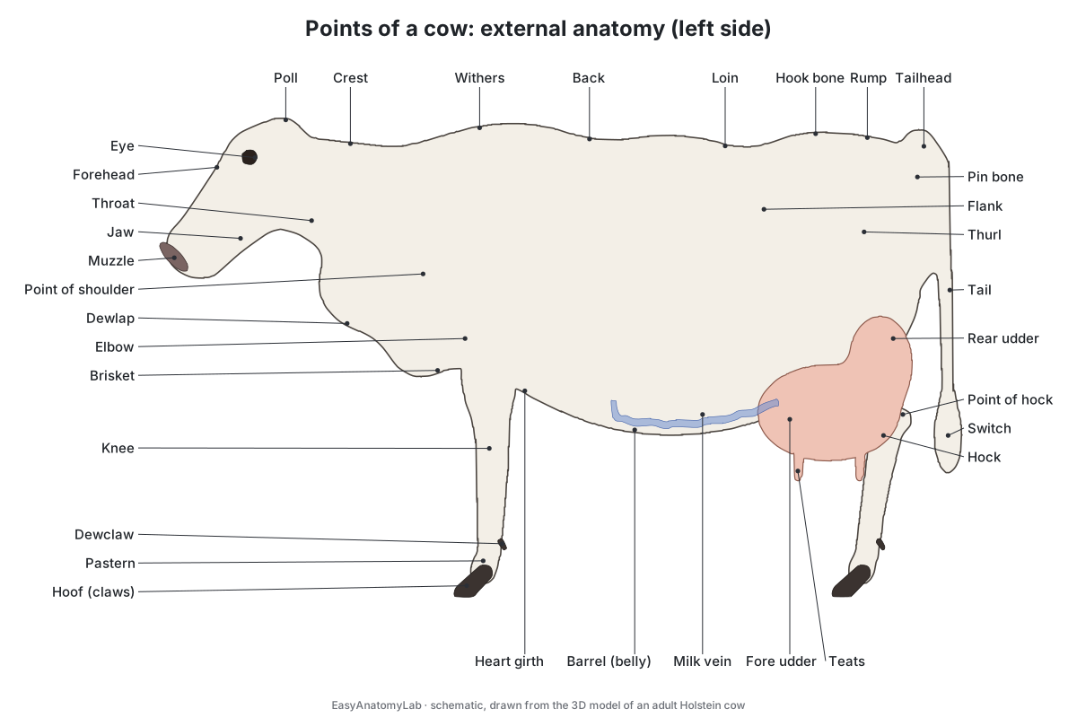
| Point | Where it is | What lies beneath |
|---|---|---|
| Poll | Top of the head, between and behind the horns | Frontal bone; horn cores in horned cattle[10, 5] |
| Muzzle (muffle) | Hairless pad around the nostrils and upper lip | Rigid nasolabial plate[10, 5] |
| Dewlap | Pendulous fold of skin under the neck | Skin only[10] |
| Brisket | Lower chest between the forelegs | Front of the sternum[10, 1] |
| Withers | Rise where the neck meets the back | First 3–4 thoracic vertebrae[14, 10] |
| Loin | Each side of the spine between the last rib and the hip | Lumbar vertebrae[10] |
| Flank | Hollow between the last rib and the hook bone | Paralumbar fossa: last rib, lumbar transverse processes, tuber coxae[10, 16] |
| Hook bones (hooks) | Front corners of the rump | Tubera coxae of the ilium[3, 10] |
| Thurl | Midway between hooks and pins | Hip joint[10] |
| Pin bones (pins) | Either side of the tail root | Tubera ischii, the widest point of the pelvic outlet[3] |
| Tailhead and switch | Base of the tail; tuft of hair at its tip | Tail vertebrae[10, 15] |
| Knee | Front leg, above the cannon | Carpus (wrist)[10] |
| Hock and point of hock | Hind leg joint; its point at the back | Tarsus; the tuber calcis of the heel bone[10] |
| Pastern and dewclaws | Below the fetlock; two small claws behind it | Phalanges; digits 2 and 5[10, 6] |
The hooks, pins, thurl, tailhead and short ribs are also the points used to score body condition, from 1 (emaciated) to 5 (obese) in quarter points.[15] A cow has an udder with four teats and visible milk veins running forward under the belly to the milk wells; a bull has a scrotum and a sheath (prepuce) instead.[10]
Cow internal organs: where everything is
Inside a cow, the chest holds the heart, the lungs and the start of the great vessels, and the steeply slanted diaphragm lets the abdomen push forwards under the last ribs.[1] In the abdomen the rumen fills the left side; the omasum, abomasum, liver and intestines lie mainly on the right, and the reproductive tract and bladder sit at the pelvic inlet under the rectum.[16, 43]
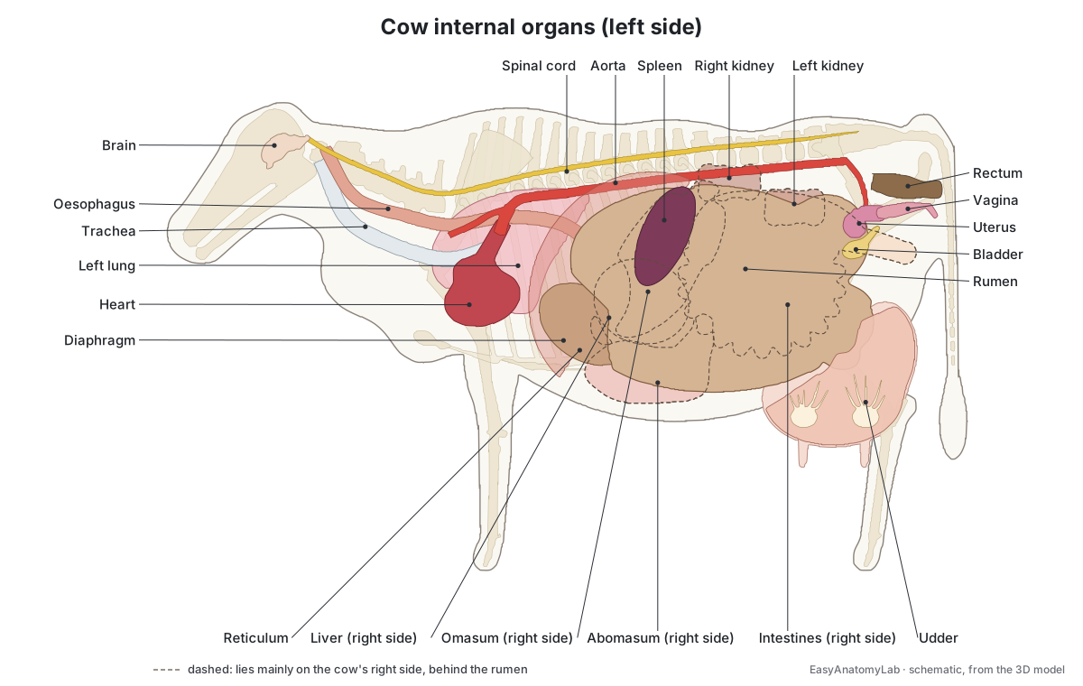
| Organ | Where it is |
|---|---|
| Heart | Low in the chest at the elbows; heard best on the left, 3rd–5th intercostal spaces[26] |
| Lungs | Chest; the lung field reaches back only to the 11th intercostal space dorsally[26] |
| Rumen and reticulum | Left abdomen; the reticulum against the diaphragm, across from the heart[23] |
| Spleen | On the left of the rumen, base at ribs 12–13, apex at rib 9[16] |
| Omasum | Right of the midline, opposite ribs 7–11[16] |
| Liver and gallbladder | Right side inside the rib cage; gallbladder near the 12th intercostal space[16] |
| Abomasum | Floor of the right lower abdomen, from the 11th rib to the xiphoid[16] |
| Intestines | Right of the rumen, filling the right flank[16] |
| Kidneys | Right kidney under L1–L4; left kidney pushed right of the midline under L3–L5[16, 2] |
| Uterus and ovaries | Over the pelvic brim, below the rectum; ovaries just cranial to the pelvic inlet[16, 4] |
| Bladder | Pelvic floor at the pelvic inlet, reaching forwards when full[16, 2] |
Cow skeleton
A cow's spine has 7 cervical, 13 thoracic, 6 lumbar and 5 fused sacral vertebrae, followed by 18 to 20 tail vertebrae, and its 13 pairs of ribs are wide and flat.[1, 17] The chest is so deep that the sternum lies below the elbow, and its front end forms the brisket.[1] Like other ungulates, cattle have no collarbone: the shoulder blade is held to the trunk by muscles alone.[8]
The cannon bone of the foreleg is the fused third and fourth metacarpals, and each foot ends in two digits of three phalanges each; digits 2 and 5 survive only as the dewclaws.[6, 7] The pelvis carries the visible hooks and pins, and its outlet is widest between the pins, which dairy cows are selected for.[3] In horned cattle the horns grow on bony cornual processes of the frontal bone, hollowed by the frontal sinus; polled cattle lack them.[5]
Cow muscles and cuts of beef
The main muscles of a cow are the same ones named on beef cuts. The table gives each muscle's anatomy first and the cut it becomes second; switch on the Muscles chip in the viewer to see them.
| Muscle | Where it is and what it does | Cut |
|---|---|---|
| Longissimus | Along the back either side of the spine, dorsal to the transverse processes[1] | Ribeye, loin eye[21, 22] |
| Psoas major | Under the lumbar vertebrae to the femur; flexes the hip[7] | Tenderloin[21, 22] |
| Gluteus medius | Rump; extends the hip[7] | Top sirloin[21, 22] |
| Gluteobiceps | Side of the thigh; biceps femoris fused with the superficial gluteal in cattle[7] | Bottom (outside) round[21] |
| Semitendinosus | Back of the thigh; extends hip, stifle and hock[7] | Eye of round[21] |
| Infraspinatus | Outer face of the shoulder blade; braces the shoulder joint[8] | Top blade, flat iron[21, 22] |
| Trapezius | Over the neck and withers; raises the scapula[8] | Part of the outside chuck[21] |
| Deep pectoral | Chest floor; one of the muscles joining the foreleg to the trunk[1] | Brisket[21] |
Cow teeth and the dental pad
An adult cow has 32 teeth and no upper front teeth: the front of the upper jaw is a tough dental pad, and the eight lower incisor-like teeth bite grass against it.[18, 19, 5] The outer pair of these eight are really canines shaped like incisors.[5, 18] Further back, each side of each jaw has three premolars and three molars, high-crowned (hypsodont) teeth that keep erupting as they wear.[18, 20]
| Permanent lower front teeth | USDA FSIS | University of Minnesota |
|---|---|---|
| Central pair (I1) | 18–24 months | 1–1½ years |
| Second pair (I2) | 24–30 months | 2–2½ years |
| Third pair (I3) | 36 months | 3 years |
| Corner pair (I4, canine) | 42–48 months | 3½–4 years |
Eruption ages vary between animals, breeds and diets, which is why the sources differ; FutureBeef's averages for British-breed cattle run from 24 months for the first pair to 46 months for a full mouth.[18, 5, 19]
Digestive system in brief
A cow has one stomach with four compartments. Swallowed feed goes into the rumen and reticulum, where microbes ferment it; the cow brings it back up as cud to chew again, then the omasum absorbs water and the abomasum, the true stomach, digests it with acid and pepsin.[24, 23] Swallowed nails and wire collect in the reticulum, the "hardware stomach", and can pierce through towards the heart.[24, 1] For every compartment, the rumen layers and how cud chewing works, see the cow digestive system guide.
Where is a cow's heart? Heart and blood vessels
A cow's heart lies low in the chest at the level of the elbows and is heard best from the left side: the pulmonic valve at the 3rd intercostal space, the aortic valve at the 4th and the mitral valve at the 5th.[26] Only the diaphragm separates it from the reticulum, which is how a swallowed wire can cause pericarditis.[1, 23]
The aortic arch gives off a single brachiocephalic trunk, which supplies both forelegs and, through the bicarotid trunk, both common carotid arteries; the descending aorta runs under the spine to the iliac arteries.[1] The cranial and caudal venae cavae return blood to the right atrium, and the external jugular veins in the jugular grooves are the usual sites for blood sampling.[1, 10] Cattle also have two small bones in the heart's fibrous skeleton, the ossa cordis. DocCheck gives the larger right bone as 50–60 mm long, while a study of bulls measured about 37 mm, so size varies.[30, 31]
Cow lungs and breathing
A cow's left lung has two lobes (a cranial lobe in two parts and a caudal lobe) and its right lung four (cranial, middle, caudal and accessory), and the right cranial lobe gets its own tracheal bronchus straight from the trachea.[1] Connective-tissue septa divide bovine lungs into obvious lobules with little or no air flow between them, and in pneumonia exudate settles in the cranioventral lung.[1, 32, 33] On a live cow the lung field is small: it ends at the 11th intercostal space dorsally, the 9th at mid-chest and the 7th at the elbow.[26]
Udder anatomy: quarters, cisterns and teats
A cow's udder is four separate mammary glands, or quarters, each draining through its own teat; milk made in one quarter cannot pass to another.[3, 37] A thick, elastic median suspensory ligament divides the right and left halves and attaches to the abdominal wall, while less elastic lateral laminae support the sides.[3, 38] The fore and rear quarters are only diffusely separated, and the larger rear quarters give about 60% of the milk (55–60% in a University of Wisconsin fact sheet).[40, 39, 37]
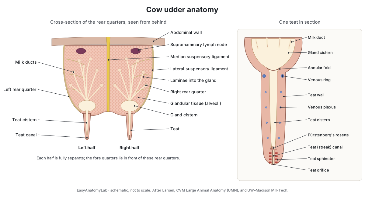
Milk is made in microscopic alveoli and collects through ducts into the gland cistern, which holds about half a litre, then the teat cistern; only 20–40% of the milk sits in the cisterns before let-down.[38, 40, 37] The teat canal at the tip is about 1 cm long, or about 9–11 mm by ultrasound in Holsteins, and its sphincter and keratin lining keep bacteria out between milkings.[40, 41, 38] About 500 litres of blood pass through the udder for each litre of milk; it drains through the external pudendal veins and, once a cow has been pregnant, also through the winding milk veins.[40, 37, 3] Empty, the udder weighs about 10–30 kg; with milk and blood it can reach 50 kg or more.[40, 37]
Female reproductive anatomy of the cow
The cow's reproductive tract runs from the vulva through the vagina, the firm cervix with its interlocking folds, a short uterine body and two long uterine horns to the oviducts and almond-shaped ovaries.[42, 43, 4] It lies directly below the rectum, which is why vets can feel it by rectal palpation for pregnancy checks and artificial insemination.[43]
| Part | What it does |
|---|---|
| Vulva and vestibule | External opening; swells at heat and before calving[42, 43] |
| Vagina | Receives the penis; semen is left near the cervix[43] |
| Cervix | Thick-walled seal with interlocking rings; closed in pregnancy[42, 43] |
| Uterine body | Short; where semen is placed in artificial insemination[4, 43] |
| Uterine horns | Coiled; the calf grows here, attached at placentomes[4, 43] |
| Oviducts | Infundibulum, ampulla, isthmus; fertilisation at the ampulla–isthmus junction[43] |
| Ovaries | Follicles and the progesterone-making corpus luteum[42] |
Heifers reach puberty at about 10–12 months, cows cycle all year about every 21 days, and pregnancy lasts about 283 days.[44, 42, 45]
Bull reproductive anatomy
A bull's two testes hang in the scrotum and make sperm and testosterone; sperm mature in the epididymis and travel up the ductus deferens to the urethra.[47, 4] The accessory sex glands are the paired ampullae and vesicular glands, which make most of the seminal fluid, a small ring-shaped prostate and small bulbourethral glands.[4] The penis is fibroelastic: it is folded into an S-shaped sigmoid flexure just behind the scrotum, held there by the retractor penis muscles, and erection mainly straightens the fold.[4, 48, 47]
| Bull's age | Minimum scrotal circumference |
|---|---|
| 15 months or less | 30 cm |
| 16–18 months | 31 cm |
| 19–21 months | 32 cm |
| 22–23 months | 33 cm |
| 2 years and over | 34 cm |
Minimums to pass a breeding soundness examination, from the Merck Veterinary Manual; South Dakota State University Extension gives the same values with age bands drawn a month differently.[48, 49] Beef bulls reach puberty at about 12 months on average, but only about 35% of 12-month-old bulls produce good semen, rising to 95% at 16 months.[47, 49]
Kidneys, spleen, brain and lymph nodes
Cattle kidneys are lobulated, divided into many lobes on the surface.[2] The right kidney lies under the first four lumbar transverse processes; the left kidney is pushed across by the rumen and hangs loosely against the rumen's right side.[16, 2] The spleen is fused to the left side of the rumen.[2]
The brain has a folded cerebrum, a cerebellum that coordinates balance and movement, and a brainstem whose medulla controls breathing and circulation.[5] Listeriosis, often called circling disease, is an infection of the brainstem that usually travels there along the trigeminal nerve.[50] The prescapular, prefemoral and supramammary lymph nodes lie close enough to the skin to be felt in a physical exam.[26, 8]
Cow vs horse anatomy
Cows and horses are both large grazers, but a cow ferments its food before the true stomach and a horse ferments it after the small intestine, in a huge caecum and colon.[25, 24] Their skeletons, teeth and reproductive organs differ too.
| Feature | Cow | Horse |
|---|---|---|
| Toes that bear weight | 2 per foot, plus 2 dewclaws | 1 per foot[6] |
| Thoracic vertebrae and rib pairs | 13 | 18[1, 17] |
| Upper incisors | None (dental pad) | 6[52] |
| Adult teeth | 32 | 36–44[52, 53] |
| Stomach | 4 compartments | Simple stomach[25] |
| Gallbladder | Yes | No[9] |
| Lung lobules and tracheal bronchus | Obvious lobules; tracheal bronchus | No obvious lobules; no tracheal bronchus[1] |
| Udder | 4 quarters, 4 teats, 1 canal each | 2 glands, 2 teats, 2 openings each[3] |
| Resting heart rate | 48–84 a minute (dairy cow) | 28–40 a minute[27] |
| Resting breathing rate | 26–50 a minute (dairy cow) | 10–14 a minute[28] |
| Estrous cycle | About 21 days, all year | About 21 days, spring to summer[44] |
| Gestation | About 283 days | About 340 days[45] |
For the horse in 3D, see horse anatomy.
Cow anatomy labeled: every structure in the 3D model
This is the text version of the 3D cow: all 93 labeled structures, grouped by body system, with where each is and what it does. Structures marked "females only" or "males only" appear when you switch sex in the viewer. Studying the eye? See the cow eye dissection guide.
External
- Hide (Skin and coat)
- The skin and hair covering the body; Holsteins are black and white or red and white (University of Kentucky). Protects the body. The bony landmarks felt through it (withers, loin, hooks, pins, tailhead) are the 'points' used to describe, judge and condition-score cattle.
- Muzzle (Nasolabial plate, muffle)
- The rigid, hairless pad around the nostrils and upper lip (UMN; BASU). Forms the firm front of the nose and upper lip. A horse's upper lip, by contrast, is very mobile (UMN).
- Eyes
- The eyes sit on the sides of the head, each inside a complete bony ring, the orbit (UMN). Vision. For the inside of the eye (cornea, lens, retina), see the cow eye dissection guide.
- Hooves (claws) (Cloven hooves, digits 3 and 4)
- Each foot ends in two weight-bearing claws on the third and fourth digits (UMN). Carry the cow's weight: cattle stand on two digits per foot, horses on one (UMN).
- Dewclaws (Digits 2 and 5)
- Two small horny digits on the back of each foot, above the claws (BASU; UMN). Underdeveloped second and fifth digits; the cow walks on digits 3 and 4 (UMN).
Skeleton
- Skull
- The bones of the head. In horned cattle, cornual processes of the frontal bone form the bony core of each horn (UMN). Protects the brain and holds the eyes, upper cheek teeth and large air sinuses.
- Mandible (Lower jaw)
- The lower jaw, which carries all the front teeth, because cattle have no upper incisors (USDA FSIS). Holds the lower incisors, which bite against the toothless dental pad, and the lower cheek teeth (UMN).
- Cervical vertebrae (Neck bones (7))
- Seven neck vertebrae, the same number as in a horse (UMN; IMAIOS). Support the head and neck. In the middle of the neck the oesophagus lies on the left, dorsolateral to the trachea (UMN).
- Thoracic vertebrae (Chest vertebrae (13))
- Thirteen vertebrae, one for each pair of ribs (UMN; IMAIOS). Carry the ribs. Their tall spinous processes lean back towards the loin.
- Lumbar vertebrae (Loin vertebrae (6))
- Six vertebrae in the loin, between the last rib and the hips (UMN; IMAIOS). Support the loin. The right kidney lies under the first few lumbar transverse processes (UMN; Hamilton).
- Sacrum
- Five fused vertebrae wedged between the hip bones (UMN; IMAIOS). Joins the spine to the pelvis and forms the roof of the pelvic canal.
- Caudal vertebrae (Tail bones (18–20))
- Eighteen to twenty tail vertebrae (UMN; IMAIOS). Support the tail, which ends in the switch, a tuft of long hair (BASU).
- Ribs (13 pairs)
- Thirteen pairs of wide, flat ribs (UMN). Protect the heart and lungs, and also the liver, spleen and reticulum, because the abdominal organs push forwards under the last ribs (UMN; Hamilton).
- Sternum (Breastbone)
- The breastbone along the floor of the chest. Its cranial end forms the brisket (UMN). Anchors the lower ends of the ribs that reach it.
- Scapula (Shoulder blade)
- The flat shoulder blade on the side of the chest. Joins the foreleg to the trunk by muscles alone: ungulates have no collarbone (UMN).
- Forelimb bones (Humerus, radius and ulna, carpus, cannon bone, phalanges)
- The bones of the front legs, ending in two digits. Support the front of the body. What farmers call the knee of the foreleg is the carpus (BASU).
- Pelvis (Hip bones: ilium, ischium, pubis)
- The hip bones. The hooks and pins you can see on a cow are parts of them (UMN; BASU). Joins the hind legs to the sacrum and surrounds the birth canal.
- Hindlimb bones (Femur, patella, tibia, tarsus, cannon bone, phalanges)
- The bones of the hind legs, ending in two digits. Push the cow forward. The stifle (knee) is formed by the femur, patella and tibia (BASU).
Muscles
- Trapezius
- A thin, flat muscle over the neck and withers, with cervical and thoracic parts (UMN). Raises the scapula; the cervical part can advance the foreleg and the thoracic part helps swing it back (UMN).
- Latissimus dorsi
- A broad, flat muscle fanning from the back to the upper foreleg, one of the muscles joining the foreleg to the trunk (UMN). Pulls the trunk forward over a foreleg planted in front, working against the brachiocephalicus (UMN).
- Infraspinatus
- The muscle on the outer face of the shoulder blade, below its spine. Its tendon acts as the lateral collateral ligament of the shoulder joint, helped by the supraspinatus (UMN).
- Longissimus (Longissimus dorsi)
- The long back muscle on each side of the spinous processes (UMN). An epaxial muscle: with the iliocostalis (lateral to it) and the transversospinalis (medial) it runs along the vertebral column (UMN).
- Gluteus medius (Middle gluteal muscle)
- The thick muscle of the rump, over the wing of the ilium (UMN). Extends the hip and can move the thigh outwards (UMN).
- Gluteobiceps (Biceps femoris + superficial gluteal)
- In cattle the superficial gluteal and biceps femoris are fused into one muscle, the gluteobiceps (UMN). The biceps femoris is one of the main extensors of the hip (UMN).
- Semitendinosus
- A long, round muscle down the back of the thigh (UMN). Extends the hip; with the foot on the ground it extends the stifle, and through the common calcanean tendon it extends the hock (UMN).
- Psoas major
- The muscle under the lumbar vertebrae that, with the iliacus, forms the iliopsoas (UMN). Flexes the hip, bringing the hind leg forward (UMN).
Digestive system
- Dental pad
- A tough pad on the front of the upper jaw where other animals have upper incisors (UMN; FutureBeef). The lower incisors press grass against it.
- Lower incisors (8 front teeth (6 incisors + 2 incisor-like canines))
- Eight shovel-shaped lower front teeth; the outer pair are canines that look like incisors (UMN; USDA FSIS). Bite grass against the dental pad. Their eruption is used to estimate a cow's age (USDA FSIS).
- Cheek teeth (Premolars and molars (24))
- Three premolars and three molars on each side of each jaw: 24 grinding teeth (USDA FSIS). Grind feed when the cow eats and again when it chews cud.
- Tongue
- A long muscular tongue with a raised hump (torus linguae) behind a transverse crease (lingual fossa) (UMN). A muscular organ supplied by the lingual artery and moved by the hypoglossal nerve (cranial nerve XII) (UMN).
- Salivary glands (Parotid and mandibular glands)
- Large glands behind and below the jaw that make saliva (UMN). Saliva's most important job in a ruminant is buffering the pH of the rumen and reticulum (MSU Extension).
- Oesophagus (Esophagus, gullet)
- The muscular tube from the throat to the stomach. In ruminants it works in both directions (MSU Extension). Carries feed down, and brings cud back up to the mouth to be chewed again (MSU Extension).
- Reticulum (Honeycomb, 'hardware stomach')
- The honeycomb-lined compartment against the diaphragm, across from the heart (VIVO). Works with the rumen as one chamber. Heavy items such as nails and wire fall into it (VIVO; MSU Extension).
- Rumen (Paunch)
- By far the largest compartment: a fermentation vat filling nearly all of the left side of the abdomen (VIVO; MSU Extension). Microbes ferment plant fibre; the volatile fatty acids they make are absorbed through its papillae (VIVO).
- Omasum (Manyplies, 'the book')
- A round compartment packed with leaves like the pages of a book (VIVO). Absorbs water and passes small particles on to the abomasum (VIVO; MSU Extension).
- Abomasum (True stomach)
- The glandular 'true stomach', very like the stomach of monogastric animals such as pigs (VIVO). Secretes hydrochloric acid and enzymes such as pepsin, which breaks down protein (MSU Extension).
- Small intestine (Duodenum, jejunum, ileum)
- The long, narrow part of the gut, coiled in the right side of the abdomen. Absorbs nutrients, including protein that escapes the rumen. Bile and pancreatic juice raise the pH of the acid digesta (MSU Extension).
- Caecum and colon (Large intestine, spiral colon)
- The caecum and a colon whose ascending part is coiled into a flat spiral. The colon absorbs most of the water from the digesta (MSU Extension).
- Rectum and anus
- The end of the gut, in the pelvic canal. The reproductive tract lies directly beneath it (UT Extension). Stores and passes dung. Because the uterus and ovaries lie just below it, vets feel them through its wall (UT Extension; UMN).
- Liver
- The large gland on the right side, lying inside the rib cage (Hamilton). Makes bile and processes nutrients absorbed from the gut (OpenStax). In ruminants it turns volatile fatty acids into glucose (MSU Extension).
- Gallbladder
- The sac that stores bile, under the liver near the 12th intercostal space on the right (Hamilton). Stores, concentrates and releases bile (OpenStax).
- Pancreas
- A pale gland beside the duodenum. Makes digestive enzymes and bicarbonate for the gut and the hormones insulin and glucagon for the blood (OpenStax).
Heart & vessels
- Heart
- The four-chambered heart, low in the chest at the level of the elbows (Breuer). Pumps blood: the right ventricle to the lungs through the pulmonary trunk, the left ventricle to the body through the aorta (UMN).
- Aorta
- The main artery, from the left ventricle over the heart and back under the spine. Carries oxygenated blood to the body. Its arch gives off one large brachiocephalic trunk, which supplies both forelegs and, through the bicarotid trunk, the head (UMN).
- Caudal vena cava
- The large vein returning blood from the abdomen and hind body to the right atrium (UMN). Returns blood from the hind body, including the udder's first-formed drainage route via the external pudendal and external iliac veins (UMN).
- Jugular veins (External jugular veins and cranial vena cava)
- Large veins just under the skin of the neck, in the jugular grooves (BASU; UMN). Drain the head and neck into the cranial vena cava, which empties into the right atrium (UMN).
- Common carotid arteries
- The paired arteries to the head, beside the trachea deep in the neck. Supply the head. In cattle the outer part of the internal carotid artery shrinks to a strand of connective tissue (UMN).
- Pulmonary trunk (Pulmonary arteries)
- The artery leaving the right ventricle, splitting into the right and left pulmonary arteries (UMN). Carries deoxygenated blood through the pulmonary valve to the lungs (UMN).
Lungs & airway
- Larynx (Voice box)
- The cartilage box at the start of the trachea, opening into the pharynx (UMN). Forms the entrance to the airway, built from the epiglottis and the thyroid, cricoid and arytenoid cartilages (UMN).
- Trachea (Windpipe)
- The windpipe, running down the neck below the oesophagus (UMN). Carries air to the lungs.
- Bronchi (Including the tracheal bronchus)
- The branching airways of the lungs, including the tracheal bronchus to the right cranial lobe (UMN). Carry air to each lobe; lobes are named by the bronchi that supply them (UMN).
- Left lung
- Two lobes: a cranial lobe (in two parts) and a caudal lobe (UMN). Gas exchange.
- Right lung
- Four lobes: cranial (in two parts), middle, caudal and accessory (UMN). Gas exchange.
- Diaphragm
- The muscular partition between the chest and the abdomen (UMN). The main breathing muscle, supplied by the phrenic nerves (UMN).
Kidneys & bladder
- Left kidney
- A lobulated kidney pushed across to the right of the midline by the rumen (UMN). Filters blood and makes urine (OpenStax).
- Right kidney
- A lobulated kidney under the loin on the right (UMN). Filters blood and makes urine (OpenStax).
- Ureters
- Two thin tubes carrying urine from the kidneys to the bladder (UMN). Carry urine from each renal pelvis to the bladder (UMN).
- Urinary bladder
- The bladder, the most ventral organ at the back of the abdomen (UMN). Stores urine.
- Urethra · females only
- The short female urethra, opening into the floor of the vestibule. Carries urine out of the bladder.
Reproductive organs
- Vulva · females only
- The external opening of the reproductive tract, below the anus (NMSU; BASU). Protects the tract. It swells before calving and when the cow is in heat (UT Extension).
- Vagina (With the vestibule) · females only
- The passage from the vulva to the cervix (UT Extension). Receives the bull's penis; semen is deposited near the cervix (UT Extension).
- Cervix · females only
- A thick-walled, firm passage with interlocking internal folds, or rings (NMSU; UT Extension). Seals the uterus: tightly closed with thick mucus in pregnancy, it relaxes and its mucus thins at heat (UT Extension).
- Uterine body · females only
- The short common part of the uterus where the horns meet (NMSU; UMN). The usual place to deposit semen in artificial insemination (UT Extension).
- Uterine horns · females only
- Two long horns coiled like a ram's horns, joined by intercornual ligaments (UMN). Where the calf develops. Caruncles on their lining bond with the cotyledons of the placenta to form placentomes (UMN; UT Extension).
- Oviducts (Uterine (Fallopian) tubes) · females only
- Thin, coiled tubes from the tips of the horns to the ovaries (UT Extension). Catch the egg in the funnel-shaped infundibulum; fertilisation happens at the junction of the ampulla and isthmus (UT Extension).
- Ovaries · females only
- Two almond-shaped organs just cranial to the pelvic inlet (UMN). Release eggs and make hormones: follicles grow on them and become a progesterone-making corpus luteum after ovulation (NMSU).
- Scrotum · males only
- The pendulous skin sac holding the testes between the hind legs (BASU). Protects the testes from knocks and temperature extremes (Oregon State). Its tunica dartos and the cremaster muscle pull the testes closer to the body for warmth, because sperm production needs a set temperature (UMN).
- Testes (Testicles) · males only
- The paired glands that make sperm and testosterone (Oregon State). Produce sperm and the male hormone testosterone (Oregon State).
- Epididymis · males only
- A coiled duct with a head, body and tail on each testis (Oregon State; UMN). Sperm mature in the head and body and are stored in the tail (Oregon State).
- Ductus deferens (Vas deferens, with the ampulla) · males only
- The duct carrying sperm from the tail of the epididymis to the urethra (Oregon State; UMN). Transports sperm; its end widens into the ampulla, which ruminants have (UMN).
- Vesicular glands (Seminal vesicles) · males only
- Paired lobulated glands beside the ampullae, dorsal to the bladder neck (UMN). Produce most of the seminal fluid in the bull (UMN).
- Prostate · males only
- A small, ring-shaped gland at the start of the pelvic urethra (UMN). Adds fluid to the semen (Oregon State).
- Bulbourethral glands (Cowper's glands) · males only
- Small paired glands near the ischial arch (UMN). Add fluid to the semen (Oregon State).
- Penis (With the pelvic urethra and sigmoid flexure) · males only
- A fibroelastic penis folded into an S-shaped sigmoid flexure behind the scrotum (UMN; Merck). Erection mostly straightens the sigmoid flexure, because the fibrous penis cannot widen much (UMN).
- Retractor penis muscles · males only
- Paired muscle bands alongside the penis that hold it folded in the sigmoid flexure (Oregon State; UMN). Keep the penis inside the body; they relax during mating so it can extend (Oregon State).
- Prepuce (Sheath) · males only
- The fold of skin enclosing the free end of the penis along the belly (BASU). Protects the penis when it is retracted.
Udder
- Udder (skin) (Mammary gland) · females only
- Four mammary glands (quarters), each with its own teat, between the hind legs (UMN; UW–Madison). Makes and stores milk. About 500 litres of blood flow through it for every litre of milk (FAO; Cornell).
- Fore quarters · females only
- The two front mammary glands, left and right (UW–Madison). Each quarter is a separate gland: milk made in one cannot pass to another (Cornell; UMN).
- Rear quarters · females only
- The two hind mammary glands, larger than the fore quarters (UMN; FAO). Make about 60% of the milk: 55–60% according to UW–Madison Extension (FAO; UW–Madison Extension).
- Teats · females only
- Four teats, one per quarter, each with a single teat canal (UMN). Let milk out. Their skin has sensory nerves that respond to a calf suckling or to milking (Cornell; UW–Madison).
- Gland cisterns and milk ducts (Gland sinus, lactiferous ducts) · females only
- The milk-collecting space above each teat, fed by large milk ducts (UMN). Store milk between milkings. The gland cistern can hold about half a litre (FAO).
- Teat cistern and teat canal (Teat sinus, streak canal (papillary duct)) · females only
- The hollow inside each teat and the narrow canal at its tip (UMN). The canal's smooth-muscle sphincter and keratin lining keep bacteria out between milkings (UW–Madison; UMN).
- Median suspensory ligament · females only
- A thick, yellow, elastic sheet in the midline separating the left and right halves (UMN). The main support of the udder. If it loses elasticity the udder sags and the teats point outwards (UMN; Cornell).
- Lateral suspensory ligaments · females only
- Strong, less elastic collagen sheets around the sides of the udder (UMN; UW–Madison). Work with the median ligament as a sling holding the udder (FAO).
- Mammary lymph nodes (Supramammary (superficial inguinal) nodes) · females only
- Large paired lymph nodes at the back of the udder's base (UMN). Filter lymph from the udder (OpenStax). They can enlarge greatly in mastitis (UMN).
- Milk veins (Subcutaneous abdominal veins) · females only
- Large, winding veins under the skin of the belly from the udder forwards (UMN). A second drainage route for the udder's large blood flow, alongside the external pudendal veins (UMN).
Brain & spinal cord
- Cerebrum
- The two cerebral hemispheres, their surface folded into gyri and sulci (UMN). Frontal lobes plan movement, occipital lobes integrate vision and piriform lobes, with the olfactory bulbs, handle smell (UMN).
- Cerebellum
- The 'little brain' behind the cerebrum (UMN). Regulates balance and coordinates movement (UMN).
- Brainstem
- Diencephalon, midbrain, pons and medulla, joining the brain to the spinal cord (UMN). Relays signals and, in the medulla, regulates breathing, circulation and feeding responses (UMN).
- Spinal cord
- The cord of nerve tissue in the vertebral canal, wrapped in three meninges (UMN). Carries signals between brain and body; sensory fibres enter by the dorsal roots and motor fibres leave by the ventral roots (UMN).
Spleen & lymph nodes
- Spleen
- A flat organ fused to the left side of the rumen (UMN). Filters blood, removing worn-out red cells and microbes, and mounts immune responses (OpenStax).
- Prescapular lymph nodes (Superficial cervical lymph nodes)
- Lymph nodes just in front of the shoulder (UMN). Filter lymph from the neck and forelimb (OpenStax).
- Prefemoral lymph nodes (Subiliac lymph nodes)
- Lymph nodes at the front edge of the thigh (UMN). Filter lymph from the flank and hind limb (OpenStax).
Frequently asked questions
How many teeth does a cow have?
An adult cow has 32 teeth: eight lower front teeth (six incisors and two incisor-like canines) and 24 cheek teeth, three premolars and three molars on each side of each jaw. There are no upper front teeth; a tough dental pad takes their place. A calf has 20 milk teeth, and the permanent front teeth come in between roughly one and four years of age.
Do cows have top teeth?
Only at the back. A cow's upper jaw has no incisors or canines, just a hard dental pad that the lower front teeth bite grass against. The upper jaw does carry premolars and molars, so cows have upper and lower cheek teeth for grinding feed, including the cud they bring back up to chew again.
How many teats does a cow have?
A cow normally has four teats, one for each quarter of the udder, and each teat has a single teat canal. The four quarters are separate glands, so milk made in one cannot reach another. Extra teats (supernumerary teats) are fairly common; they are often removed from young calves to help protect against mastitis.
Where is a cow's heart located?
A cow's heart sits low in the chest at the level of the elbows. Vets listen to it from the left side, pushing the stethoscope forward under the elbow: the pulmonic valve is heard at the 3rd intercostal space, the aortic at the 4th and the mitral at the 5th. Only the diaphragm separates it from the reticulum.
How many ribs does a cow have?
A cow has 13 pairs of ribs, one pair for each of its 13 thoracic vertebrae. The ribs are wide and flat. Because the abdominal organs push forwards under the last ribs, the rib cage also shields the liver, spleen and reticulum. A horse usually has 18 pairs of narrower ribs.
What are the hooks and pins on a cow?
Hooks and pins are the bony points of the pelvis you can see on a cow's rump. The hooks (hook bones) are the tubera coxae, the outer corners of the ilium in front; the pins (pin bones) are the tubera ischii either side of the tail. They are used to judge conformation and to score body condition.
What is the difference between cow and bull anatomy?
A cow is an adult female and a bull an intact adult male. Their skeletons, muscles and four-compartment stomachs are the same; the differences are reproductive. A cow has ovaries, a uterus with two coiled horns, a cervix, vagina and vulva, plus an udder. A bull has two testes in a scrotum, accessory sex glands and a penis folded into a sigmoid flexure inside a sheath.
How long is a cow pregnant?
About 283 days, or roughly nine months. The calf grows in one of the two uterine horns, where button-like caruncles on the uterine lining bond with cotyledons on the placenta to form placentomes. Throughout pregnancy the cervix stays tightly closed and sealed with thick mucus.
References
Every source below was opened and checked on 25 October 2026. In the 3D viewer, each structure's facts name their source in brackets (UMN = the University of Minnesota's CVM Large Animal Anatomy).
- Larsen, R. CVM Large Animal Anatomy (University of Minnesota open textbook), ch. 5: Neck and Thorax
- Larsen, R. CVM Large Animal Anatomy (University of Minnesota open textbook), ch. 7: Abdomen 2, Bovine and Camelid
- Larsen, R. CVM Large Animal Anatomy (University of Minnesota open textbook), ch. 8: Pelvis and Perineum (bovine pelvis and udder)
- Larsen, R. CVM Large Animal Anatomy (University of Minnesota open textbook), ch. 9: Reproductive Tracts and Genitalia
- Larsen, R. CVM Large Animal Anatomy (University of Minnesota open textbook), ch. 10: Head
- Larsen, R. CVM Large Animal Anatomy (University of Minnesota open textbook), ch. 2: Distal Limb
- Larsen, R. CVM Large Animal Anatomy (University of Minnesota open textbook), ch. 3: Pelvic Limb
- Larsen, R. CVM Large Animal Anatomy (University of Minnesota open textbook), ch. 1: Thoracic Limb
- Larsen, R. CVM Large Animal Anatomy (University of Minnesota open textbook), ch. 6: Abdomen 1, Equine and Porcine
- Sahu, S. P. Identification of Body Parts of Farm Animals. Bihar Veterinary College, Bihar Animal Sciences University (BASU)
- University of Kentucky Animal & Food Sciences: Dairy Cattle Discovery, Parts
- University of Kentucky Animal & Food Sciences: Dairy Cattle Discovery, Breeds
- TheDairySite: Holstein breed profile
- DocCheck Flexikon: Widerrist (withers)
- Jones, C. M. & Heinrichs, J. Learn to Score Body Condition. Penn State Extension
- Hamilton, G. F. Laparotomy (bovine abdominal anatomy). The Bovine Proceedings 13
- IMAIOS vet-Anatomy: Thoracic vertebrae (vertebral formulas by species)
- USDA Food Safety and Inspection Service: Using Dentition to Age Cattle (copy hosted by NCDA&CS)
- FutureBeef: Ageing cattle by their teeth
- Wright, A. Dentition and Dental Nomenclature of Animals. Merck Veterinary Manual
- Calkins, C. R. & Sullivan, G. Ranking of Beef Muscles for Tenderness. University of Nebraska
- Texas A&M Meat Science: Retail Cut Muscle ID (2021 skillathon guide)
- Bowen, R. Digestive Anatomy in Ruminants. VIVO Pathophysiology, Colorado State University
- Parish, J. A., Rivera, J. D. & Boland, H. T. Understanding the Ruminant Animal Digestive System. Mississippi State University Extension P2503
- Bowen, R. Digestive Anatomy and Function of Horses. VIVO Pathophysiology, Colorado State University
- Breuer, R. M. (2023). 10 pro techniques: mastering the art of the bovine physical exam. AABP Recent Graduate Conference Proceedings 56(1)
- MSD Veterinary Manual: Table, resting heart rates
- MSD Veterinary Manual: Table, resting respiratory rates
- MSD Veterinary Manual: Table, normal rectal temperature ranges
- DocCheck Flexikon: Herzskelett (cardiac skeleton)
- Own, H. A. & Kadhim, A. B. Os cordis in local adult male ruminants: histomorphological and histochemical study. Iraqi Journal of Veterinary Sciences 39(4)
- Ardans, A. A. Pulmonary structure, function and defense mechanism. AABP Proceedings
- Pierson, R. E. & Kainer, R. A. (1980). Clinical classification of pneumonias in cattle. The Bovine Practitioner 15
- OpenStax Anatomy and Physiology 2e, 23.6: Accessory organs in digestion
- OpenStax Anatomy and Physiology 2e, 21.1: Anatomy of the lymphatic and immune systems
- OpenStax Anatomy and Physiology 2e, 25.1: Physical characteristics of urine
- Anatomy of the Mammary Gland (adapted from DeLaval). Cornell Cooperative Extension, NY dairy program
- MilkTech International: The Cow, Introduction to Udder Anatomy. University of Wisconsin–Madison
- Rivera, H., Schlesser, H. & Stuttgen, S. The Udder of the Cow. University of Wisconsin–Madison Extension
- FAO Small-Scale Dairy Farming Manual, Unit 7.1: Physiology of milk production
- Štrapák, P. et al. (2017). The influence of milking on the teat canal of dairy cows determined by ultrasonographic measurements. Czech Journal of Animal Science 62(2)
- Parker, R. & Mathis, C. Reproductive Tract Anatomy and Physiology of the Cow. New Mexico State University Guide B-212
- Carter, C. & Rhinehart, J. Reproductive Anatomy of the Cow/Heifer. University of Tennessee Extension W 934
- Merck Veterinary Manual: Table, features of the reproductive cycle
- Turner, J. L. & Painter, C. The Julian Date Calendar (gestation table). New Mexico State University Guide B-130
- MSD Veterinary Manual: Table, approximate gestation periods
- Cooke, R. Basics of Reproductive Function in Bulls. Oregon State University Extension BEEF 057
- Bedford-Guaus, S. J. Breeding Soundness Examination of Bulls. Merck Veterinary Manual
- Perry, G., Walker, J. & Daly, R. Herd Bull Fertility, ch. 32. South Dakota State University Extension
- Depenbrock, S. Listeriosis in Animals. Merck Veterinary Manual
- Canadian Food Inspection Agency: Appendix B, cattle vertebral column
- MSD Veterinary Manual (horse owner version): Dental development of horses
- AAEP Proceedings 68 (2022): In-depth, preventative medicine (equine dentition)
More interactive anatomy models
- Cow digestive systemThe ruminant stomach: rumen, reticulum, omasum and abomasum, in 3D.
- Human anatomy1,000+ bones, muscles, organs, vessels and nerves from real anatomical data.
- Octopus anatomyThree hearts, a ring-shaped brain, eight arms and a beak, in 3D.
- Squid anatomyPen, eight arms, two tentacles, giant axons and three hearts of the longfin squid.
- Squid dissectionStep-by-step guided squid dissection with every structure labeled.
- Fish anatomyExternal and internal anatomy of a bony fish: fins, gills, swim bladder, heart and brain.