

What is comparative anatomy?
The Gale Encyclopedia of Science describes comparative anatomy as comparing the structural similarities of organisms to work out their evolutionary relationships. The comparisons cover bones, muscles, organs and embryos, and the results feed into how organisms are classified and into the evidence for evolution. (Gale Encyclopedia of Science, Anatomy, Comparative (Encyclopedia.com).)
Similarity alone settles nothing; the question is why two structures look alike. Structures that share an embryonic origin are homologous, while structures that merely share a function are analogous. The more complex a shared feature is, the more likely the overlap comes from a common evolutionary past rather than coincidence. (OpenStax Biology 2e, 20.2 Determining Evolutionary Relationships.)
It is practical too: a 2022 Journal of Anatomy review argues that teaching comparative anatomy with One Health, the link between human, animal and environmental health, helps medical students understand disease. (Bhattacharjee et al. (2022), integrating comparative anatomy and One Health in anatomy education, J Anat 240 (PMC).)
Homologous vs analogous vs vestigial structures
Homologous structures are similar because they were inherited from a common ancestor. The tetrapod limb is the standard example: the ancestor of tetrapods evolved four limbs, and birds, bats, mice and crocodiles all inherited them, while sharks and bony fish did not. (Understanding Evolution (UC Berkeley), Homologies and analogies.)
Analogous structures do the same job but evolved separately, under similar environmental demands, so they say nothing about close kinship. Insect and bird wings both power flight, but an insect wing contains no bone and has a completely different structure and embryonic origin. Biologists also call this kind of similarity homoplasy. (OpenStax Biology 2e, 20.2 Determining Evolutionary Relationships.)
One organ can be both. Bird and bat forelimbs are homologous, because both groups inherited forelimbs from a common ancestor, but their wings are analogous: birds and bats did not inherit wings from a winged ancestor. A bat flies on skin stretched between its finger and arm bones, a bird on feathers along its arm. Analogies like this are the result of convergent evolution. (Understanding Evolution (UC Berkeley), Homologies and analogies.)
Convergence can produce strikingly similar organs. Squid and octopuses have a camera eye with an iris, a nearly round lens and a retina, so like the vertebrate eye that it is treated as a classic example of convergent evolution. (Yoshida & Ogura (2011), evolution of the cephalopod camera eye, BMC Evol Biol 11: 180 (PMC).)
Vestigial structures are still present, often reduced in size, although they no longer serve their original function. (Gale Encyclopedia of Science, Anatomy, Comparative (Encyclopedia.com).)
Textbook examples are the wings of flightless birds, the leaves of some cacti and the hind-leg bones of whales, remnants of parts that worked in an ancestor. (OpenStax Biology 2e, 18.1 Understanding Evolution.)
Snakes are thought to descend from lizards, and boas and pythons still carry vestigial hind limbs as pelvic spurs. (OpenStax Biology 2e, 29.4 Reptiles.)
Vestigial does not mean useless. Whale pelvic bones, long described as relics of a land-living past, anchor the paired muscles that control the penis. Dines and colleagues found that males of whale species with stronger competition for mates have relatively larger pelvic bones, whose shape also evolves faster, a sign that sexual selection still shapes them. (Dines et al. (2014), sexual selection targets cetacean pelvic bones, Evolution 68 (PMC).)
Darwin called the human appendix a useless vestige, but it holds dense lymphoid tissue and may shelter gut bacteria, and a 2021 analysis found that a cecal appendix arose independently at least 16 times in mammals and was lost only once. (Collard et al. (2021), the cecal appendix is correlated with greater maximal longevity in mammals, J Anat 239 (PMC).)
Compare the cephalopod camera eye with a vertebrate eye on the octopus anatomy model.
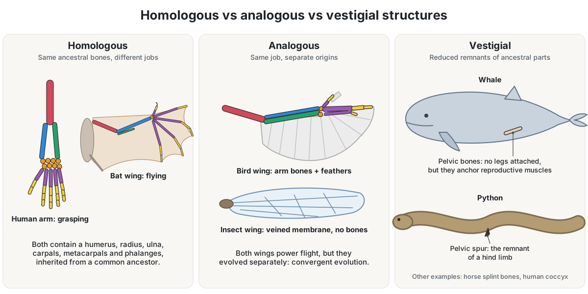
| Homologous | Analogous | Vestigial | |
|---|---|---|---|
| What is shared | Origin: the same ancestral structure and embryonic tissue | Function only | Origin, but the original function is lost or reduced |
| Why they are similar | Inherited from a common ancestor | Convergent evolution under similar demands | Inherited, then reduced |
| Tells you about ancestry? | Yes: evidence of common descent | No: can mislead classification | Yes: points to an ancestor that used the structure |
| Examples | Human arm, cat and horse forelegs, whale flipper, bat and bird wing bones; mammal middle-ear bones and reptile jaw bones | Bird vs insect wings; octopus vs vertebrate camera eyes; four-chambered hearts of birds and mammals; white winter coats of arctic fox and ptarmigan | Whale pelvic bones, python pelvic spurs, wings of flightless birds, horse splint bones, human coccyx |
The vertebrate forelimb: homologous bones in six animals
The forelimb is the textbook example of comparative anatomy. A human arm, a cat's foreleg, a horse's foreleg, a whale's flipper, a bat's wing and a bird's wing look nothing alike and do different jobs, yet each is built from a humerus, a radius and ulna, wrist bones, palm bones and finger bones, joined in the same order. In the diagram each of those bones has one colour in every animal.
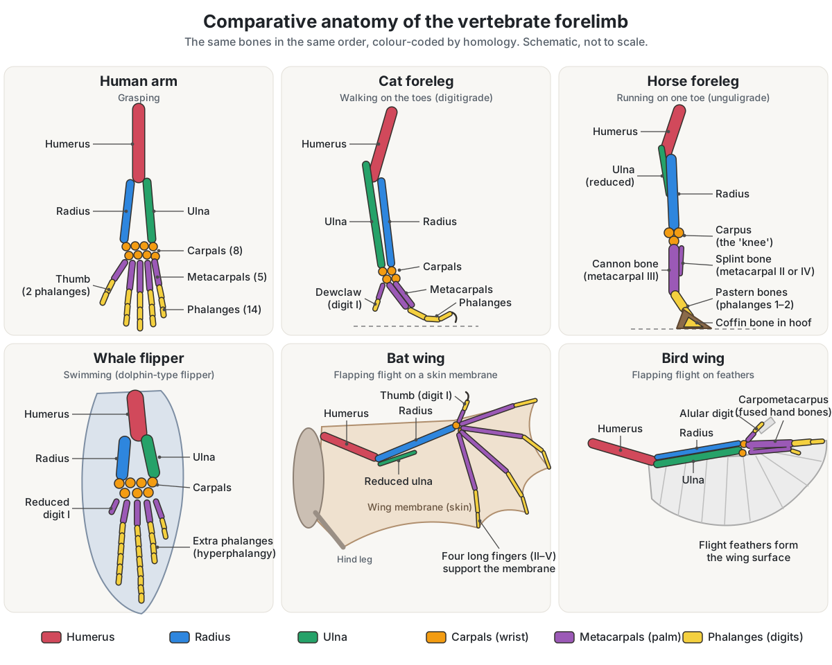
| Forelimb | Job | What changed from the basic plan | 3D model |
|---|---|---|---|
| Human arm | Grasping | Full set: 30 bones, with a two-phalanx thumb | Arm anatomy |
| Cat foreleg | Walking, climbing (digitigrade) | Walks on its digits; five front toes, the first being the dewclaw | Cat skeleton |
| Horse foreleg | Running (unguligrade) | One digit: cannon bone (metacarpal III), splint bones (metacarpals II and IV), reduced ulna | Horse skeleton |
| Whale flipper | Swimming | Paddle with extra phalanges (hyperphalangy) and a reduced first digit | Whale anatomy |
| Bat wing | Flapping flight | Four very long fingers carry a skin membrane; small clawed thumb; thin ulna | Bat skeleton |
| Bird wing | Flapping flight | Hand bones fused into a carpometacarpus; three reduced digits; feathers form the wing | Bird skeleton |
Forelimb homology animal by animal: arm, leg, flipper and wing
Darwin chose the forelimb to show homology in 1859: "What can be more curious than that the hand of a man, formed for grasping, that of a mole for digging, the leg of the horse, the paddle of the porpoise, and the wing of the bat, should all be constructed on the same pattern, and should include the same bones, in the same relative positions?" He credited Geoffroy Saint-Hilaire with the rule behind it: parts may change almost without limit in form and size, but they stay connected in the same order. (Darwin (1859), On the Origin of Species, chapter XIII (Project Gutenberg).)
The human arm shows the full pattern. Each upper limb has 30 bones: the humerus in the upper arm, the radius and ulna in the forearm, 8 carpals in the wrist, 5 metacarpals in the palm and 14 phalanges in the fingers, two of them in the thumb. (OpenStax Anatomy and Physiology 2e, 8.2 Bones of the Upper Limb.)
Running mammals stretch the same bones. Humans are plantigrade and put the whole foot down, cats and dogs are digitigrade and walk on their digits, and horses are unguligrade and walk on the tips of their toes. In fast runners the metacarpals lengthen and become fewer, the ulna is reduced or fused to the radius, and the collarbone is reduced or lost. (Animal Diversity Web, Legs, feet and cursorial locomotion.)
A cat keeps all five front digits. Cats are digitigrade, with five toes on each front foot and four on each hind foot, and the first digit of the front foot, the equivalent of our thumb, is the dewclaw. (Lamberski (2015), Felidae, Fowler's Zoo and Wild Animal Medicine 8 (PMC).)
The horse takes reduction furthest. Its cannon bone is the third metacarpal, followed by one row of phalanges: the long pastern, short pastern and coffin bone inside the hoof. The second and fourth metacarpals survive as thin, vestigial splint bones. (University of Minnesota CVM, Ungulate dissection lab 3: distal forelimb.)
Whales went the other way. The flipper evolved from a five-fingered, weight-bearing limb, but whales add extra finger bones, a condition called hyperphalangy: belugas have extra phalanges on every digit except a reduced first digit. (Gavazzi et al. (2023), comparative embryology of beluga, bowhead and spotted dolphin, J Morphol 284 (PMC).)
In a bat the hand is the wing. Four highly elongated fingers support the wing membrane, while the first digit, homologous to our thumb, stays small and clawed and is used for climbing and walking. The ulna is shortened and thin. (UC Museum of Paleontology, Introduction to the Chiroptera.)
A bird's wing rests mainly on an elongated radius and ulna and the carpometacarpus, made by the reduction and fusion of wrist and hand bones; feathers, not skin, form the flight surface. (UC Museum of Paleontology, Avian flight.)
Which fingers a bird keeps is still argued. The adult wing has three reduced digits, one carrying the alula, a major digit carrying the large flight feathers, and one with a single phalanx. They are numbered either I–II–III or II–III–IV; de Bakker and colleagues' data favour II–III–IV in the chicken. (de Bakker et al. (2021), selection on phalanx development in the evolution of the bird wing, Mol Biol Evol 38 (PMC).)
Pterosaurs, the first vertebrates to evolve true flight, built a third kind of wing: a membrane carried on a greatly elongated fourth finger. (UC Museum of Paleontology, Pterosaur flight.)
Comparing hearts: two, three and four chambers
Vertebrate hearts form a classic comparative series. Fish have a single circuit and a two-chambered heart with one atrium and one ventricle; blood leaving the heart passes through the gill capillaries and then on to the body. Amphibians have a three-chambered heart with two atria and one ventricle, so some oxygen-rich and oxygen-poor blood mixes in the ventricle, and they also exchange gas through the skin in a pulmocutaneous circuit. (OpenStax Biology 2e, 40.1 Overview of the Circulatory System.)
Lizards and snakes also have two atria and one ventricle, but the ventricle is partly divided into three compartments, the cavum arteriosum, cavum venosum and cavum pulmonale, by a muscular ridge and other partial septa. It works as a single pressure pump, yet the two blood streams stay well separated inside it. Crocodiles have a full ventricular septum and four chambers, like birds. (Jensen et al. (2013), development of the hearts of lizards and snakes, PLoS ONE 8: e63651.)
Birds and mammals both have four chambers, two atria and two ventricles, that keep oxygen-rich and oxygen-poor blood completely apart. OpenStax points out that this is convergent evolution: mammals and birds did not inherit the four-chambered heart from a shared four-chambered ancestor. (OpenStax Biology 2e, 29.6 Mammals.)
Complete separation is probably required for the warm-blooded lifestyle of birds and mammals. Insects, crustaceans and most molluscs instead have an open circulatory system, in which hemolymph mixes with the fluid around the tissues, while earthworms, like vertebrates, keep their blood inside vessels. (OpenStax Biology 2e, 40.1 Overview of the Circulatory System.)
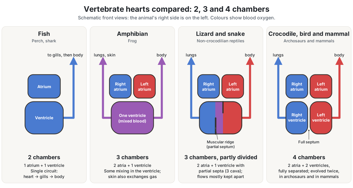
| Animal | Heart | Circulation | 3D model |
|---|---|---|---|
| Fish | 2 chambers: 1 atrium, 1 ventricle | Single: heart → gills → body | Fish, shark |
| Frog | 3 chambers: 2 atria, 1 ventricle | Double, with some mixing; skin also exchanges gas | Frog anatomy |
| Lizard, snake | 2 atria, 1 ventricle partly divided into 3 cava | Double; flows mostly kept apart | Lizard, snake |
| Crocodile | 4 chambers, full ventricular septum | Double, fully separated | — |
| Bird | 4 chambers | Double, fully separated | Bird anatomy |
| Mammal | 4 chambers (evolved separately from birds') | Double, fully separated | Human heart, blood flow |
| Insect, crayfish | Tubular dorsal heart | Open: hemolymph bathes the organs | Insect, crayfish |
Comparing digestive systems: ruminant, hindgut fermenter and carnivore
Diet shapes the gut. Cnidarians and flatworms digest in a gastrovascular cavity with a single opening, an earthworm has a one-way alimentary canal, and among mammals carnivores have a shorter large intestine than herbivores. (OpenStax Biology 2e, 34.1 Digestive Systems.)
Ruminants such as cattle ferment plant food before it reaches the true stomach. Their stomach has four compartments, the rumen, reticulum, omasum and abomasum; the chambers hold microbes that break down cellulose and ferment the food, and the animal regurgitates cud from the reticulum to chew it again. Camels and alpacas are pseudo-ruminants, with a three-chambered stomach and no rumen. (OpenStax Biology 2e, 34.1 Digestive Systems.)
Horses ferment at the other end of the gut. A horse has a simple, one-compartment stomach that makes up only about 10 percent of the volume of its digestive tract, while the cecum and colon make up about 50 percent. The cecum works much like a cow's rumen, a fermentation vat whose microbes digest fibre. (Extension Horses (extension.org), Feeding management of the equine.)
The Merck Veterinary Manual gives the horse's stomach 8–10 L and its cecum 1.2–1.5 m in length and 27–30 L in capacity; horses can neither vomit nor regurgitate. (Merck Veterinary Manual, Overview of colic in horses.)
Rabbits ferment in an enlarged cecum and re-eat the soft cecotropes. Birds have no teeth: food waits in the crop, then passes through the proventriculus and the grinding gizzard. (OpenStax Biology 2e, 34.1 Digestive Systems.)
A carnivore's gut is short and simple. A dog has a large stomach that holds a whole meal eaten at once, a short large intestine of about 40 cm (16 inches) in a medium-sized dog, and a tiny cecum that matters little to carnivores. (Hippocampus Biology (UC Regents and MITE), Vertebrate digestive systems.)
| Animal | Gut type | Key feature | 3D model |
|---|---|---|---|
| Cow | Ruminant (foregut fermenter) | Four-compartment stomach: rumen, reticulum, omasum, abomasum; chews cud | Cow digestive system |
| Horse | Hindgut fermenter | Small simple stomach (~10% of tract); cecum and colon ~50%; cannot vomit | Horse anatomy |
| Rabbit | Hindgut fermenter | Enlarged cecum; re-eats cecotropes | Rabbit anatomy |
| Dog | Carnivore, simple stomach | Large stomach, short large intestine, tiny cecum | Dog anatomy |
| Bird | No teeth | Crop, proventriculus and grinding gizzard | Bird, chicken |
| Earthworm | Tube gut | One-way alimentary canal from mouth to anus | Earthworm anatomy |
Comparing breathing: gills, lungs, air sacs and tracheae
Very small animals need no breathing organs: direct diffusion across the body surface works for organisms less than about 1 mm across, which is how flatworms breathe. Earthworms and amphibians use moist skin as a respiratory surface. Water holds far less oxygen than air, so fish gills are thin, highly branched and folded filaments with a large surface for oxygen to diffuse into the blood. (OpenStax Biology 2e, 39.1 Systems of Gas Exchange.)
Insects carry air straight to their tissues: it enters through spiracles along the thorax and abdomen and travels through a network of small chitin tubes, the tracheal system, to the entire body. (OpenStax Biology 2e, 39.1 Systems of Gas Exchange.)
Aquatic crustaceans breathe with gills, and land-living chelicerates such as spiders use book lungs. (OpenStax Biology 2e, 28.6 Superphylum Ecdysozoa: Arthropods.)
Bird lungs work differently from ours. A mammal breathes in and out of a bronchoalveolar lung, but a bird's lung is essentially fixed in volume and is ventilated by a series of generally nine air sacs that act as bellows. Inhaled air moves through the gas-exchanging parabronchi in the same direction, from back to front, during both inhalation and exhalation, and exchange with the blood is cross-current. One-way airflow has also been found in crocodilians, which have no air sacs outside the lungs, and in monitor lizards and iguanas. (Schachner & Moore (2025), what does it take to make a bird lung?, Phil Trans R Soc B 380 (PMC).)
Air-sac counts vary between species; the chicken has eight. Birds have no diaphragm, and some air sacs extend into bones, where they take the place of marrow and lighten the skeleton. (WikiVet, Avian respiration: anatomy and physiology.)
| Breathing organ | Animals | How it works | 3D model |
|---|---|---|---|
| Body surface | Flatworms | Direct diffusion; works below about 1 mm thickness | — |
| Moist skin | Earthworm, frog | Gas dissolves in the wet skin and diffuses | Earthworm, frog |
| Gills | Fish, crayfish, tadpoles | Thin, folded filaments in water | Fish, crayfish |
| Tracheae | Insects, myriapods | Spiracles open into chitin tubes that reach every tissue | Insect body parts, grasshopper |
| Book lungs | Spiders and other land chelicerates | Internal air-breathing organs | Spider anatomy |
| Alveolar lungs | Mammals | Air breathed in and out; gas exchange only in the alveoli | Human lungs |
| Lungs + air sacs | Birds | Air sacs push air one way through near-rigid lungs (parabronchi) | Bird anatomy |
Comparative embryology: pharyngeal arches and embryonic tails
Embryos show homologies that adults hide. All vertebrate embryos, humans included, have gill slits and a tail early in development; in land vertebrates these disappear, while adult fish and some amphibians keep them. Great ape embryos, humans included, have a tail that is gone by birth. (OpenStax Biology 2e, 18.1 Understanding Evolution.)
The "gill slits" lie between the pharyngeal arches, paired swellings in the embryonic neck that appear in the fourth week of human development. There are six, but the fifth regresses. Their derivatives are listed in the table below. (Kenhub, Pharyngeal arches.)
In fishes the pharyngeal slits are modified into gill supports, and in jawed fishes into jaw supports. In tetrapods they are reworked into parts of the ear and into the tonsils and thymus, and in humans and other great apes the post-anal tail is reduced to a vestigial coccyx. (OpenStax Biology 2e, 29.1 Chordates.)
The middle ear records the change in bone. Reptiles and birds have one middle-ear ossicle, the stapes or columella. Mammals have three, and the two extra ones, the malleus and incus, are homologous to the quadrate and articular, the bones that form the jaw joint in other jawed vertebrates. Karl Reichert proposed this in 1837, and fossils such as Liaoconodon show a transitional stage. (Anthwal, Joshi & Tucker (2013), evolution of the mammalian middle ear and jaw, J Anat 222 (PMC).)
A fish's jaws come from its first and second pharyngeal arches and its gill skeleton from the arches behind them, and on the way to mammals the fish hyomandibula became the stapes. (DeLaurier (2019), evolution and development of the fish jaw skeleton, WIREs Dev Biol (PMC).)
Whale embryos make the same point about legs. No living whale has visible hind limbs, yet beluga, bowhead and spotted-dolphin embryos grow hindlimb buds; in the spotted dolphin they regress fully by Carnegie stage 17, while bowhead embryos keep them for longer. (Gavazzi et al. (2023), comparative embryology of beluga, bowhead and spotted dolphin, J Morphol 284 (PMC).)
| Pharyngeal arch | In fishes | In humans | Nerve |
|---|---|---|---|
| 1st (mandibular) | Upper and lower jaws | Malleus and incus (from Meckel's cartilage), mandible, muscles of chewing | Trigeminal (CN V) |
| 2nd (hyoid) | Hyoid arch; the hyomandibula | Stapes, styloid process, lesser horn of the hyoid, muscles of facial expression | Facial (CN VII) |
| 3rd | Gill arch | Greater horn and lower body of the hyoid | Glossopharyngeal (CN IX) |
| 4th and 6th | Gill arches | Cartilages of the larynx, muscles of the pharynx and larynx | Vagus (CN X) |
See the human ear bones and skull in the ear anatomy and skull bones guides.
How does comparative anatomy support evolution?
If species share common ancestors, they should share similarities inherited from those ancestors, and wherever biologists have looked they have found such homologies. Comparative anatomy supplies several independent kinds: shared bone plans such as the forelimb, structures that appear in embryos and then disappear, and vestigial structures. (Understanding Evolution (UC Berkeley), Lines of evidence: homologies.)
The anatomical pattern matches the rest of the evidence. The universality of DNA points to one common ancestor of all life, and the similarity of DNA sequences reflects how closely groups are related. (OpenStax Biology 2e, 18.1 Understanding Evolution.)
Anatomy can also mislead, because convergent evolution makes unrelated animals look alike. Biologists therefore decide first whether a similarity is homology or analogy, and combine body structure with DNA data: using both is more effective at working out evolutionary relationships than either alone. (OpenStax Biology 2e, 20.2 Determining Evolutionary Relationships.)
History of comparative anatomy: Belon, Cuvier, Owen and Darwin
Pierre Belon (1517–1564), a French traveller, naturalist and diplomat, published L'histoire de la nature des oyseaux in 1555. It set a bird skeleton beside a human skeleton and marked the matching bones with the same letter, and Belon is considered a significant contributor to comparative anatomy for that systematic comparison. (Natural History Society of Northumbria, Treasures of the library: Pierre Belon, L'histoire de la nature des oyseaux (1555).)
Georges Cuvier (1769–1832) created the comparative method of organismal biology. He saw organisms as integrated wholes in which no part could change without upsetting the rest, used living anatomy to reconstruct fossil animals, and firmly established extinction by showing that the fossil mammoths of Europe and Siberia differed from both living elephants. (UC Museum of Paleontology, Georges Cuvier (1769–1832).)
Richard Owen (1804–1892) defined homology in 1843 as "the same organ in different animals under every variety of form and function", pointing to the common plan of a bat's wing, a seal's flipper, a cat's paw and a human hand. He explained the plan as an archetype, an idea in the Divine mind rather than an ancestor, and was not receptive to Darwin's theory when it appeared in 1859. (UC Museum of Paleontology, Richard Owen (1804–1892).)
Charles Darwin's On the Origin of Species (1859) gave the same facts a new explanation. He called morphology the most interesting department of natural history, "its very soul", and treated the unity of type, embryonic resemblances and rudimentary organs as the marks of descent from common ancestors. (Darwin (1859), On the Origin of Species, chapter XIII (Project Gutenberg).)
Explore comparative anatomy in 3D
Open two models side by side to make your own comparisons:
- Human armthe full set of bones
- Cat skeletona digitigrade foreleg with a dewclaw
- Dog skeletona second digitigrade carnivore
- Horse skeletonone toe, the cannon bone and the splint bones
- Whale anatomyflippers and vestigial pelvic bones
- Bat skeletona hand turned into a wing
- Bird skeletonthe carpometacarpus, keel and furcula
- Pterodactyl skeletona wing on the fourth finger
- Snake skeletona python's vestigial pelvis and spurs
- Fish anatomygills and a two-chambered heart
- Frog anatomya three-chambered heart and skin breathing
- Bird anatomyair sacs, crop and gizzard
Frequently asked questions
What is comparative anatomy in simple terms?
Comparative anatomy is the study of how the bodies of different animals are alike and how they differ. By comparing bones, organs and embryos, anatomists work out which features were inherited from a shared ancestor, which evolved separately for the same job, and which are shrunken leftovers. Those comparisons are used to classify animals and are one of the main lines of evidence for evolution.
What are the three types of structures in comparative anatomy?
The three types are homologous, analogous and vestigial structures. Homologous structures come from the same ancestral structure, such as the human arm and the bat wing. Analogous structures do the same job but evolved separately, such as bird and insect wings. Vestigial structures are reduced remnants that have lost their original function, such as whale pelvic bones and horse splint bones.
What is the difference between homologous and analogous structures?
Homologous structures share an origin: they develop from the same embryonic tissue and were inherited from a common ancestor, even if they now do different jobs. Analogous structures share a function but not an origin; they evolved independently under similar demands, by convergent evolution. A bat's wing and a bird's wing are both: the arm bones are homologous, but the wings themselves are analogous.
How does comparative anatomy provide evidence for evolution?
Common ancestry predicts shared, inherited similarities, and comparative anatomy finds them everywhere: the same forelimb bones in arms, flippers, legs and wings, pharyngeal arches and tails in every vertebrate embryo, and vestigial structures such as whale pelvic bones. These anatomical groupings agree with DNA comparisons and with the geographic spread of species.
What is an example of comparative anatomy?
The vertebrate forelimb is the classic example. A human arm, a cat's foreleg, a horse's foreleg, a whale's flipper, a bat's wing and a bird's wing all contain a humerus, a radius and ulna, wrist bones, hand bones and finger bones in the same order. Their shapes and numbers differ because the limbs grasp, run, swim and fly.
Who is the father of comparative anatomy?
The title is given to different people. Pierre Belon's 1555 comparison of a bird and a human skeleton, with matching bones lettered alike, is one of the earliest. Georges Cuvier created the comparative method of organismal biology and established extinction, and Richard Owen defined homology in 1843. Darwin then explained those homologies as inheritance from common ancestors.
Are vestigial structures useless?
Not always. Vestigial means a structure has lost its original function, not that it has no use. Whale pelvic bones no longer support legs, but they anchor the muscles of the penis and are shaped by sexual selection. The human appendix holds lymphoid tissue and may shelter helpful gut bacteria. Others, such as horse splint bones, are simply reduced remnants.
Why do birds and mammals both have four-chambered hearts?
Both groups are warm-blooded, and completely separating oxygen-rich from oxygen-poor blood is probably needed for that lifestyle. The two hearts evolved independently, so they are an example of convergent evolution rather than homology. Crocodiles, which group with birds as archosaurs, also have four chambers, while lizards and snakes have a single, partly divided ventricle.
References
- "Anatomy, Comparative." The Gale Encyclopedia of Science. Gale, via Encyclopedia.com.
- OpenStax. Biology 2e, section 18.1: Understanding Evolution. Rice University.
- OpenStax. Biology 2e, section 20.2: Determining Evolutionary Relationships.
- OpenStax. Biology 2e, section 28.6: Superphylum Ecdysozoa: Arthropods.
- OpenStax. Biology 2e, section 29.1: Chordates.
- OpenStax. Biology 2e, section 29.4: Reptiles.
- OpenStax. Biology 2e, section 29.6: Mammals.
- OpenStax. Biology 2e, section 34.1: Digestive Systems.
- OpenStax. Biology 2e, section 39.1: Systems of Gas Exchange.
- OpenStax. Biology 2e, section 40.1: Overview of the Circulatory System.
- OpenStax. Anatomy and Physiology 2e, section 8.2: Bones of the Upper Limb.
- Understanding Evolution, University of California Museum of Paleontology. Homologies and analogies.
- Understanding Evolution, University of California Museum of Paleontology. Lines of evidence: homologies.
- University of California Museum of Paleontology. Georges Cuvier (1769–1832).
- University of California Museum of Paleontology. Richard Owen (1804–1892).
- University of California Museum of Paleontology. Introduction to the Chiroptera.
- University of California Museum of Paleontology. Vertebrate flight: avian flight.
- University of California Museum of Paleontology. Vertebrate flight: pterosaur flight.
- Natural History Society of Northumbria. Treasures of the library: Pierre Belon, L'histoire de la nature des oyseaux, 1555.
- Wellcome Collection. The skeleton of a bird for comparison with that of a man, from Belon (1555).
- Darwin, C. On the Origin of Species, 1st edition. John Murray, 1859. Chapter XIII. Project Gutenberg.
- Animal Diversity Web, University of Michigan. Legs, feet and cursorial locomotion.
- Animal Diversity Web, University of Michigan. Chiroptera.
- Lamberski, N. Felidae. In: Miller & Fowler (eds), Fowler's Zoo and Wild Animal Medicine, vol. 8. Elsevier, 2015.
- VCA Animal Hospitals. 10 facts about cats.
- Cox, Clarkson, Brown & Fletcher. Ungulate dissection lab 3: distal forelimb. University of Minnesota College of Veterinary Medicine.
- Gavazzi, L. et al. Comparative embryology of Delphinapterus leucas, Balaena mysticetus and Stenella attenuata. Journal of Morphology 284 (2023): e21543.
- de Bakker, M. A. G. et al. Selection on phalanx development in the evolution of the bird wing. Molecular Biology and Evolution 38 (2021): 4222–4237.
- Jensen, B. et al. Development of the hearts of lizards and snakes and perspectives to cardiac evolution. PLoS ONE 8 (2013): e63651.
- Schachner, E. R. & Moore, A. J. Unidirectional airflow, air sacs or the horizontal septum: what does it take to make a bird lung? Philosophical Transactions of the Royal Society B 380 (2025).
- WikiVet. Avian respiration: anatomy and physiology.
- Extension Horses. Feeding management of the equine.
- Moore, R. M. Overview of colic in horses. Merck Veterinary Manual.
- Hippocampus Biology (Regents of the University of California and Monterey Institute for Technology and Education). Vertebrate digestive systems.
- Kenhub. Pharyngeal arches.
- Anthwal, N., Joshi, L. & Tucker, A. S. Evolution of the mammalian middle ear and jaw: adaptations and novel structures. Journal of Anatomy 222 (2013): 147–160.
- DeLaurier, A. Evolution and development of the fish jaw skeleton. WIREs Developmental Biology 8 (2019): e337.
- Dines, J. P. et al. Sexual selection targets cetacean pelvic bones. Evolution 68 (2014): 3296–3306.
- Collard, M. K. et al. The cecal appendix is correlated with greater maximal longevity in mammals. Journal of Anatomy 239 (2021): 1157–1169.
- Yoshida, M. & Ogura, A. Genetic mechanisms involved in the evolution of the cephalopod camera eye. BMC Evolutionary Biology 11 (2011): 180.
- Bhattacharjee, S. et al. On the importance of integrating comparative anatomy and One Health perspectives in anatomy education. Journal of Anatomy 240 (2022): 429–446.
More interactive anatomy models
- What is anatomy?What anatomy studies, its branches, how anatomists name and describe the body, and how to learn it.
- Anatomy and physiologyHow structure and function fit together, the levels of organisation and the organ systems, with study tips.
- Animal anatomyEvery animal on the site by group, with what each body plan teaches.
- Anatomy quizFree anatomy quizzes and games: find-the-structure quizzes on 3D models and labelling practice.
- Human anatomy1,000+ bones, muscles, organs, vessels and nerves from real anatomical data.
- Octopus anatomyThree hearts, a ring-shaped brain, eight arms and a beak, in 3D.