

Neuron anatomy at a glance
The anatomy of a neuron follows its job: it is an excitable cell built to carry information in one direction. Signals arrive on the dendrites, spread across the cell body, are turned into action potentials at the start of the axon, and travel down the axon to its terminals, which pass the message on at synapses. (OpenStax Anatomy and Physiology 2e, 12.2 Nervous tissue.)
The model is a vertebrate spinal motor neuron, the cell that makes skeletal muscles contract. Its cell body sits in the ventral (anterior) horn of the spinal cord's grey matter, and its axon leaves through the ventral root to run in a peripheral nerve to a muscle. (OpenStax Anatomy and Physiology 2e, 13.2 The central nervous system; OpenStax Biology 2e, 35.4 The peripheral nervous system.) It is the classic textbook neuron because it shows every part clearly: a large multipolar cell body, many dendrites, one long myelinated axon and terminal branches on muscle fibres.
Neurons share the organelles of other cells. What sets them apart are their dendrites and axons, specialised for receiving and sending electrical signals, and the connections they make with other neurons and with muscle. (OpenStax Biology 2e, 35.1 Neurons and glial cells.) The human brain alone holds about 86 billion neurons. (Herculano-Houzel (2009): The human brain in numbers, Frontiers in Human Neuroscience 3:31; OpenStax Biology 2e, 35.1 Neurons and glial cells.)
Labeled diagram of a neuron
A labeled neuron diagram shows, from the input end to the output end: dendrites with their spines, the cell body with its nucleus, nucleolus and Nissl bodies, the axon hillock and initial segment, the axon wrapped in myelin sheaths separated by nodes of Ranvier, a side branch (collateral), and the terminal branches ending in synaptic boutons.
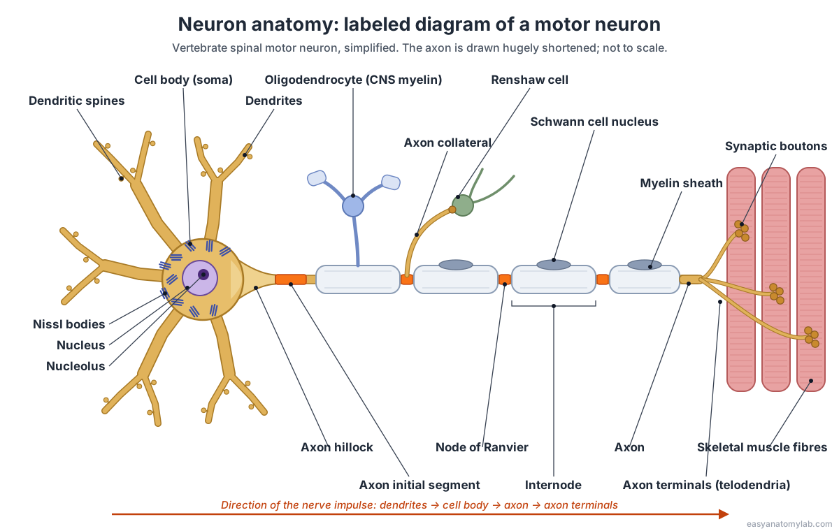
In the 3D model, switch on All labels to name every part at once, or click any part to zoom to it and read what it does. Drag the Myelin sheath slider down to see the bare axon underneath.
Parts of a neuron and their functions
Every neuron has three main parts, dendrites, a cell body and an axon, plus specialised zones along the axon. This table gives each part, where it is, and its job.
| Part | Where it is | Function |
|---|---|---|
| Dendrites | Branches around the cell body | Receive signals from other neurons at synapses |
| Dendritic spines | Small bumps on dendrites | Sites of most excitatory synapses; add surface area |
| Cell body (soma) | Centre of the neuron | Holds the nucleus; makes proteins; sums incoming signals |
| Nucleus and nucleolus | Inside the cell body | Store DNA; make ribosome subunits |
| Nissl bodies | Cell body and proximal dendrites | Rough ER and ribosomes that make proteins |
| Axon hillock | Where the cell body narrows into the axon | Joins soma to axon; part of the trigger zone |
| Initial segment | First, bare stretch of the axon | Usually starts the action potential |
| Axon | From the initial segment to the terminals | Conducts signals away from the cell body |
| Axon collateral | Side branch of the axon | Sends a copy of the signal to another target |
| Myelin sheath | Wrapped around the axon in segments | Insulates the axon and speeds conduction |
| Nodes of Ranvier | Gaps between myelin segments | Regenerate the signal, which jumps node to node |
| Axon terminals and boutons | End of the axon | Release neurotransmitter onto the next cell |
The cell body: nucleus, Nissl bodies and organelles
The cell body (soma or perikaryon) is the neuron's metabolic centre. It contains the nucleus, endoplasmic reticulum, Golgi apparatus, mitochondria and other organelles common to most cells, and it makes almost all of the neuron's proteins. (OpenStax Biology 2e, 35.1 Neurons and glial cells.)
A large spinal motor neuron has a large, central nucleus surrounded by dark-staining Nissl bodies. These are stacks of rough endoplasmic reticulum with ribosomes; they extend into the dendrites but stop at the pale, Nissl-free axon hillock. (Clermont, Lalli & Bencsath-Makkai, Light-Microscopic Histology Atlas (McGill), 4.3 Neuron; Clermont, Lalli & Bencsath-Makkai, Light-Microscopic Histology Atlas (McGill), 4.1 Large pyramidal neuron; Kenhub: Parts of a neuron.) Because the axon has no rough ER of its own, proteins made in the cell body must be carried down it by axonal transport. Fast transport moves vesicles at about 250–400 mm a day, slow transport of cytoskeletal proteins only about 1 mm a day; a review gives 50–400 mm and under 8 mm a day. (BioNumbers 113074: fast and slow axonal transport rates in mammals (Brady et al., Basic Neurochemistry); Schenone et al. (2025): Basic pathological mechanisms in peripheral nerve diseases, International Journal of Molecular Sciences.) A neuron has over a thousand mitochondria to power all this. (OpenStax Anatomy and Physiology 2e, 3.2 The cytoplasm and cellular organelles.)
Dendrites and dendritic spines
Dendrites are the receiving branches of a neuron. They are usually short, tapering and highly branched, and other neurons' axon terminals contact them at synapses. Excitatory inputs commonly land on dendrites, inhibitory inputs nearer the axon hillock. (OpenStax Anatomy and Physiology 2e, 12.2 Nervous tissue; OpenStax Anatomy and Physiology 2e, 12.5 Communication between neurons.) A single cerebellar Purkinje cell can receive contacts from up to about 200,000 other neurons. (OpenStax Biology 2e, 35.1 Neurons and glial cells.)
Dendritic spines are tiny protrusions that add surface area for synapses and receive input from excitatory axons. (OpenStax Biology 2e, 35.1 Neurons and glial cells; Rochefort & Konnerth (2012): Dendritic spines: from structure to in vivo function, EMBO Reports 13:699.) Spinal motor neurons have them too: in mice their spines peak around two weeks after birth and then thin out, leaving mostly short "stubby" spines on the outer dendrites, with an occasional mushroom-shaped one. (Kanjhan, Noakes & Bellingham (2016): Emerging roles of filopodia and dendritic spines in motoneuron plasticity, Neural Plasticity 2016:3423267.)
The axon: hillock, initial segment, collaterals and terminals
The axon is the neuron's single output cable. It starts at the axon hillock, a cone where the cell body tapers, continues as the initial segment, and runs to the terminals. The hillock and initial segment together form the trigger zone where action potentials begin; the initial segment has a high density of voltage-gated sodium channels. (OpenStax Anatomy and Physiology 2e, 12.2 Nervous tissue; OpenStax Anatomy and Physiology 2e, 12.4 The action potential.) A protein scaffold of ankyrin-G and βIV-spectrin anchors these channels and also acts as a filter that keeps the axon's contents separate from the dendrites'. (Jones & Svitkina (2016): Axon initial segment cytoskeleton: architecture, development, and role in neuron polarity, Neural Plasticity 2016:6808293.) How the action potential itself rises and falls is covered on our action potential diagram page.
A human motor axon can be about a metre long, from the base of the spine to the toes, yet only about 10 µm wide. (OpenStax Biology 2e, 35.1 Neurons and glial cells; Milo & Phillips, Cell Biology by the Numbers: How big is a human cell?.) Axons in general are 1–20 µm in diameter. (OpenStax Anatomy and Physiology 2e, 12.2 Nervous tissue.)
Axons can branch. A motor axon gives off a recurrent collateral inside the spinal cord that turns back and excites Renshaw cells, small interneurons that then inhibit the same motor neurons, a negative feedback loop called recurrent inhibition. The collateral releases acetylcholine onto nicotinic receptors, and one impulse in one motor axon can make a Renshaw cell fire. (Alvarez, Dewey, McMillin & Fyffe (1999): Distribution of cholinergic contacts on Renshaw cells in the rat spinal cord, Journal of Physiology 515:787; Enjin et al. (2017): Developmental disruption of recurrent inhibitory feedback in the Renshaw cell–motor neuron circuit, Journal of Neuroscience 37:5634.) In the muscle, the axon branches profusely so that one neuron supplies many muscle fibres; each branch ends in swollen boutons 1–5 µm across. (Slater (2017): The structure of human neuromuscular junctions: some unanswered molecular questions, International Journal of Molecular Sciences 18:2183.)
Myelin sheath and nodes of Ranvier
The myelin sheath is a fatty insulating wrap made of many layers of glial cell membrane. Schwann cells make it in the peripheral nervous system, each wrapping one segment of one axon; oligodendrocytes make it in the brain and spinal cord, each wrapping segments of several axons. (OpenStax Anatomy and Physiology 2e, 12.2 Nervous tissue; OpenStax Biology 2e, 35.1 Neurons and glial cells.) So the motor axon in the model is myelinated by an oligodendrocyte while it is still inside the spinal cord, and by Schwann cells once it leaves through the ventral root.
Between the myelin segments (internodes) are nodes of Ranvier, gaps about 1 µm long where the axon membrane is exposed and packed with voltage-gated channels; the signal is regenerated at each node and jumps from node to node. (OpenStax Biology 2e, 35.2 How neurons communicate.) Internodes are about 1–2 mm long, 100–1,000 times the axon's diameter. (OpenStax Anatomy and Physiology 2e, 12.2 Nervous tissue.) A review of Schwann cell sheaths gives maximal internodes of about 1 mm in mice and 2.4 mm in cows, and minimum lengths of 0.15–0.3 mm in all species. (Tricaud (2018): Myelinating Schwann cell polarity and mechanically-driven myelin sheath elongation, Frontiers in Cellular Neuroscience 11:414.)
Myelin and thickness together set the speed. The fastest axons, the thick myelinated Aα fibres that include alpha motor axons, are about 12–20 µm across and conduct at about 100 m/s. (e-Learning for Healthcare, e-Safe Anaesthesia: Nerve classification.) Measured in cats, alpha motor axons conducted at 71–91 m/s and the thinner gamma motor axons at 15–48 m/s. (Westbury (1982): A comparison of the structures of α- and γ-spinal motoneurones of the cat, Journal of Physiology 325:79.) Across all nerves, speeds range from about 100 m/s down to less than 0.1 m/s. (Byrne, Neuroscience Online (UTHealth), ch. 3: Propagation of the action potential.) When myelin is damaged, as in multiple sclerosis, current leaks out and conduction slows. (OpenStax Biology 2e, 35.2 How neurons communicate.)
The synapse: where a motor neuron meets muscle
A synapse is the junction where an axon terminal passes its signal to another cell. A motor neuron's synapse with a skeletal muscle fibre is called the neuromuscular junction, and it is the magnified close-up in the 3D model. (OpenStax Anatomy and Physiology 2e, 10.2 Skeletal muscle; OpenStax Anatomy and Physiology 2e, 12.5 Communication between neurons.)
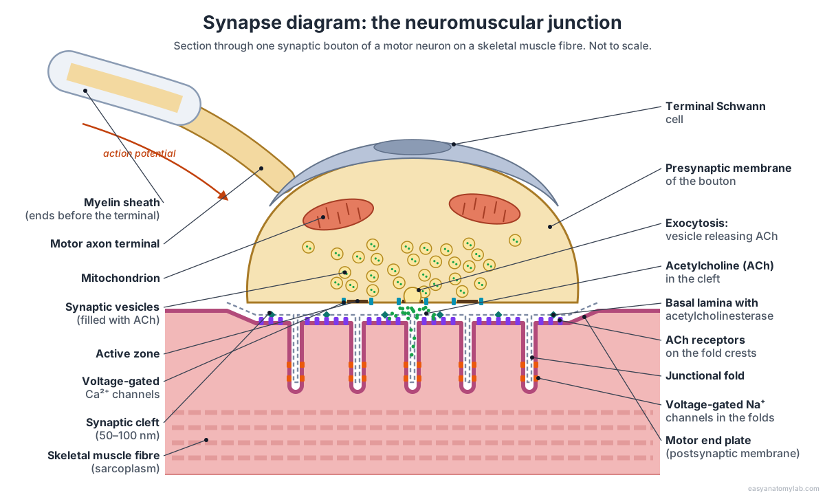
- An action potential reaches the bouton and opens voltage-gated calcium channels; calcium entry makes synaptic vesicles fuse with the membrane. (OpenStax Anatomy and Physiology 2e, 12.5 Communication between neurons.)
- Each vesicle, about 50 nm across, releases a packet of about 10,000 acetylcholine molecules at an active zone. (Slater (2017): The structure of human neuromuscular junctions: some unanswered molecular questions, International Journal of Molecular Sciences 18:2183.)
- Acetylcholine crosses a cleft 50–100 nm wide, with a basal lamina running through its middle; a synapse between two neurons has a cleft of only about 20–30 nm and no basal lamina. (Zou & Pan (2022): Post-synaptic specialization of the neuromuscular junction: junctional folds formation, function, and disorders, Cell & Bioscience 12:93; Slater (2017): The structure of human neuromuscular junctions: some unanswered molecular questions, International Journal of Molecular Sciences 18:2183.)
- It binds acetylcholine receptors packed at about 10,000 per µm² on the crests of the junctional folds, which open and depolarise the muscle membrane. Sodium channels deep in the folds then start the muscle action potential. (Zou & Pan (2022): Post-synaptic specialization of the neuromuscular junction: junctional folds formation, function, and disorders, Cell & Bioscience 12:93; Slater (2017): The structure of human neuromuscular junctions: some unanswered molecular questions, International Journal of Molecular Sciences 18:2183.)
- Acetylcholinesterase in the cleft breaks acetylcholine down so it cannot act again. (OpenStax Anatomy and Physiology 2e, 10.2 Skeletal muscle.)
What happens next inside the muscle fibre is shown on the sarcomere diagram page.
Types of neurons by structure
Neurons are classified by structure according to how many processes leave the cell body: multipolar (one axon and two or more dendrites), bipolar (one axon and one dendrite), unipolar or pseudounipolar (a single process that splits in two) and anaxonic (no process recognisable as an axon). (OpenStax Anatomy and Physiology 2e, 12.2 Nervous tissue; OpenStax Biology 2e, 35.1 Neurons and glial cells.)
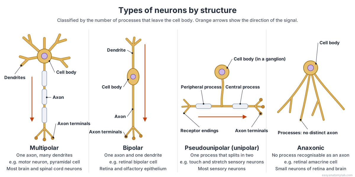
| Type | Shape | Examples and where found |
|---|---|---|
| Multipolar | One axon, many dendrites | Most neurons of the brain and spinal cord: motor neurons, pyramidal cells, Purkinje cells |
| Bipolar | One axon and one dendrite at opposite ends | Rare: retinal bipolar cells and olfactory epithelium |
| Pseudounipolar | One process that splits into a peripheral and a central branch | Most sensory neurons; cell bodies in dorsal root and cranial ganglia |
| Unipolar (true) | One process, no dendrites | Invertebrates only, for example insect neurons |
| Anaxonic | Many processes, none recognisable as an axon | Retinal amacrine cells; small brain neurons |
Textbooks disagree slightly on names. One calls human pseudounipolar sensory neurons "unipolar" and notes that true unipolar cells occur only in invertebrates; it also says "anaxonic" is a misnomer, because these cells do have axons that simply cannot be told apart from dendrites under the light microscope, so they count as multipolar. (OpenStax Anatomy and Physiology 2e, 12.2 Nervous tissue.) Another lists amacrine cells of the retina as neurons with no axon at all. (OpenStax Biology 2e, 35.1 Neurons and glial cells.)
Types of neurons by function: sensory, motor and interneurons
By function there are three types of neurons. Sensory (afferent) neurons carry information from receptors toward the central nervous system; motor (efferent) neurons carry commands out to muscles and glands; and interneurons sit between them in the brain and spinal cord, relaying signals and forming circuits. (Queensland Brain Institute (University of Queensland): Types of neurons; OpenStax Biology 2e, 35.4 The peripheral nervous system.)
| Type | Direction | Usual structure | Cell body | Example |
|---|---|---|---|---|
| Sensory (afferent) | Receptor → CNS | Pseudounipolar | Dorsal root or cranial ganglion | Touch and stretch receptors in skin and muscle |
| Motor (efferent) | CNS → muscle or gland | Multipolar | Ventral grey matter (ventral horn) of the spinal cord | Alpha motor neuron (this model) |
| Interneuron | Within the CNS | Multipolar | Brain and spinal cord | Renshaw cell (in the model) |
Sensory neuron cell bodies are grouped in the dorsal root ganglia; motor neuron cell bodies lie in the ventral grey matter and project through the ventral root. (OpenStax Biology 2e, 35.4 The peripheral nervous system.) See how these fit into the cord on our spinal cord anatomy guide, and how neurons are organised into the brain on the brain anatomy and cranial nerves guides.
Glial cells: the neuron's support team
Glial cells support neurons, maintain their surroundings and make myelin. There are six main types, four in the central nervous system and two in the peripheral nervous system. (OpenStax Anatomy and Physiology 2e, 12.2 Nervous tissue; OpenStax Biology 2e, 35.1 Neurons and glial cells.)
| Glial cell | Where | Main job |
|---|---|---|
| Astrocytes | CNS | Regulate ions and chemicals around neurons, support synapses, help form the blood–brain barrier |
| Oligodendrocytes | CNS | Myelinate segments of several axons each |
| Microglia | CNS | Resident immune cells: remove dead cells and microbes |
| Ependymal cells | CNS | Line the ventricles and central canal; help make and move cerebrospinal fluid |
| Schwann cells | PNS | Myelinate one segment of one axon each (terminal Schwann cells cap nerve endings) |
| Satellite cells | PNS | Surround cell bodies in ganglia and support them |
How many glia are there? Some textbooks still say the brain has about ten times as many glial cells as neurons. (OpenStax Biology 2e, 35.1 Neurons and glial cells.) Direct counts disagree: the adult human brain has about 86 billion neurons and 85 billion other cells, and a review of 150 years of counting found that histological data always supported a ratio of about one glial cell per neuron. (Herculano-Houzel (2009): The human brain in numbers, Frontiers in Human Neuroscience 3:31; von Bartheld, Bahney & Herculano-Houzel (2016): The search for true numbers of neurons and glial cells in the human brain, Journal of Comparative Neurology 524:3865; OpenStax Psychology 2e, 3.2 Cells of the nervous system.) For how neurons and glia make up nervous tissue, see our tissue types guide.
Neuron anatomy in numbers
Sizes and speeds vary widely between neurons. These are the values for the motor neuron modelled here.
| Measure | Value | Sources |
|---|---|---|
| Axon length | Up to about 1 m (spinal cord to toes) | OpenStax Biology 2e 35.1; Kenhub |
| Axon diameter | 1–20 µm in general; Aα motor axons about 12–20 µm | OpenStax A&P 2e 12.2; e-LfH |
| Internode length | About 1–2 mm (0.15–2.4 mm across species) | OpenStax A&P 2e 12.2; Tricaud 2018 |
| Node of Ranvier | About 1 µm long | OpenStax Biology 2e 35.2 |
| Conduction speed (alpha motor axon) | About 100 m/s; 71–91 m/s measured in cats | e-LfH; Westbury 1982 |
| Synaptic bouton | 1–5 µm across | Slater 2017 |
| Synaptic vesicle | About 50 nm; about 10,000 ACh molecules | Slater 2017 |
| Neuromuscular cleft | 50–100 nm (CNS synapse 20–30 nm) | Zou & Pan 2022; Slater 2017 |
| Neurons in the human brain | About 86 billion | Herculano-Houzel 2009; OpenStax Biology 2e 35.1 |
Every structure in the 3D neuron model
This is the text version of the 3D model: each labeled structure, grouped by region, with what it does. Use it as a study list for a neuron labeling worksheet.
Dendrites & inputs
- Dendrites
- Tapering, branched processes that spread out from the cell body like the branches of a tree. Receive signals from other neurons at synapses and carry the resulting small (graded) electrical changes toward the cell body.
- Dendritic spines
- Tiny knob- or mushroom-shaped bumps on the dendrites, each typically the site of one excitatory synapse. Add surface area for synapses and give each input its own small compartment that can change in strength.
- Synapses from other neurons (Presynaptic boutons on the dendrites and soma)
- The swollen endings of other neurons' axons, pressed against this neuron's dendrites and cell body. Deliver excitatory and inhibitory inputs. The motor neuron adds them up, and fires only when the sum at the axon's trigger zone reaches threshold.
Cell body
- Cell body (soma) (Soma, perikaryon)
- The rounded, organelle-filled centre of the neuron, from which the dendrites and the axon grow. Keeps the cell alive: it holds the nucleus and makes the proteins and membranes that the dendrites and axon need. It also sums the inputs arriving from the dendrites.
- Nucleus
- A large, round, pale nucleus in the middle of the cell body. Holds the DNA and controls which proteins the neuron makes.
- Nucleolus
- A dark, dense body inside the nucleus. Makes ribosomal RNA and assembles ribosome subunits, which the neuron needs in large numbers to make protein.
- Nissl bodies (Nissl substance, chromatophilic substance)
- Clumps of rough endoplasmic reticulum and free ribosomes that stain dark blue with basic dyes. Make proteins: the large amounts of membrane proteins, enzymes and transmitter-handling proteins a big neuron needs.
- Golgi apparatus
- Stacks of flattened membrane sacs near the nucleus. Modifies, sorts and packages proteins and lipids from the rough ER into vesicles bound for the membrane, the dendrites or the axon.
- Mitochondria
- Small, bean-shaped organelles that make ATP. Supply ATP for the sodium-potassium pumps, transport and transmitter recycling, which make neurons some of the most energy-hungry cells.
Axon
- Axon hillock
- A cone-shaped region where the cell body narrows into the axon. Joins the cell body to the axon. Together with the initial segment it forms the trigger zone that decides whether the neuron fires.
- Axon initial segment (Initial segment, AIS)
- The first, unmyelinated stretch of the axon, between the hillock and the start of the myelin. Usually where the action potential starts: it is packed with voltage-gated sodium channels held in place by a scaffold of ankyrin-G and βIV-spectrin.
- Axon (Nerve fibre)
- A single long, thin process that carries signals away from the cell body. Conducts action potentials from the trigger zone to the axon terminals, and carries proteins and organelles both ways by axonal transport.
- Axon collateral (Recurrent collateral)
- A side branch of the axon. In a motor neuron the main collateral turns back into the spinal cord grey matter. Lets one neuron signal to more than one target. The motor axon's recurrent collateral excites Renshaw cells, which then inhibit the motor neuron (recurrent inhibition).
- Nodes of Ranvier (Myelin sheath gaps)
- Short, bare gaps in the myelin sheath between neighbouring internodes. Hold clusters of voltage-gated sodium channels, so the action potential is regenerated at each node and 'jumps' from node to node (saltatory conduction).
- Axon terminals (telodendria) (Terminal arborization, terminal branches)
- The fine branches at the end of the axon, one for each muscle fibre the neuron controls. Distribute the signal to many target cells at once: here, the muscle fibres of one motor unit.
- Synaptic boutons (Terminal boutons, synaptic end bulbs, synaptic knobs)
- Small swellings at the tips of the terminal branches, where neurotransmitter is released. Store neurotransmitter in vesicles and release it when an action potential arrives.
Myelin & glia
- Myelin sheath
- A thick, fatty wrapping of many layers of glial cell membrane around the axon. Insulates the axon so current does not leak out, which makes conduction much faster and saves energy.
- Schwann cells (Neurolemmocytes; their outer layer is the neurilemma)
- The glial cells that myelinate peripheral axons. Each one wraps a single internode of a single axon. Build and maintain the peripheral myelin sheath and support axon regrowth after injury.
- Oligodendrocyte
- A CNS glial cell whose processes each wrap a segment of a different axon in myelin. Myelinates the part of the motor axon that is still inside the spinal cord.
Target cells
- Renshaw cell
- A small inhibitory interneuron in the ventral horn of the spinal cord. Excited by the motor neuron's collateral, it releases glycine and/or GABA back onto the motor neuron, a negative-feedback loop called recurrent inhibition.
- Skeletal muscle fibres
- The long, striated muscle cells that this motor neuron controls. Three short pieces are drawn. Contract when acetylcholine from the axon terminal triggers an action potential in their membrane. One motor neuron and all the fibres it supplies form a motor unit.
Synapse close-up
- Presynaptic terminal (Synaptic bouton, cut open)
- One bouton of the motor nerve terminal, magnified and cut in half. Converts the electrical signal into a chemical one: an arriving action potential opens calcium channels and triggers release of acetylcholine.
- Synaptic vesicles
- Tiny membrane bubbles filled with the neurotransmitter acetylcholine (ACh). Store transmitter and release it by exocytosis: the vesicle fuses with the presynaptic membrane and empties into the cleft.
- Active zones
- Dense strips of protein on the inside of the presynaptic membrane. The release sites: they dock vesicles next to voltage-gated calcium channels so release is fast and precise.
- Mitochondrion (in the terminal)
- A mitochondrion inside the nerve terminal. Supplies ATP for refilling vesicles and pumping ions after each burst of release.
- Acetylcholine (ACh) (Neurotransmitter)
- The neurotransmitter of motor neurons, shown as green dots released into the cleft. Diffuses across the cleft and binds ACh receptors on the muscle fibre. It is then broken down by acetylcholinesterase so it cannot keep acting.
- Synaptic cleft and basal lamina
- The narrow gap between nerve and muscle, with a sheet of extracellular matrix (basal lamina) running through it. The space ACh must cross. The basal lamina anchors acetylcholinesterase and molecules that keep the synapse aligned.
- Motor end plate (junctional folds) (Postsynaptic membrane)
- The specialised patch of muscle-fibre membrane (sarcolemma) under the terminal, folded into deep junctional folds. Carries the receptors that turn ACh into an electrical signal; the folds increase its area and help start the muscle action potential.
- Acetylcholine receptors (Nicotinic AChRs)
- Ligand-gated ion channels packed on the crests of the junctional folds. Open when ACh binds, letting positive ions into the muscle fibre to depolarise it (the end-plate potential).
- Voltage-gated sodium channels
- Sodium channels concentrated deep in the junctional folds. When the end-plate potential reaches threshold they open and start the muscle action potential.
- Terminal Schwann cell (Perisynaptic Schwann cell)
- A non-myelinating Schwann cell that caps the nerve terminal. Covers and supports the terminal; unlike the Schwann cells along the axon, it makes no myelin.
- Muscle fibre (close-up) (Sarcoplasm)
- The magnified muscle fibre under the terminal. Receives the signal; the action potential that starts at the end plate spreads over the fibre and triggers contraction.
Frequently asked questions
What are the main parts of a neuron?
A neuron has three main parts: dendrites, which receive signals; the cell body (soma), which holds the nucleus and makes the cell's proteins; and the axon, which carries signals away to other cells. Many axons are wrapped in a myelin sheath interrupted by nodes of Ranvier, and they end in axon terminals with synaptic boutons that release neurotransmitter.
What is the difference between an axon and a dendrite?
Dendrites carry signals toward the cell body; the axon carries them away. A neuron usually has many short, tapering, branched dendrites but only one axon, which keeps an even width and can be very long. Dendrites contain Nissl bodies (rough ER) and receive synapses; the axon has no Nissl bodies, starts at the axon hillock and may be myelinated.
What does the myelin sheath do?
The myelin sheath insulates the axon so electrical current does not leak out. The action potential is then regenerated only at the nodes of Ranvier and jumps from node to node, which is much faster than in an unmyelinated axon. Schwann cells make myelin in the peripheral nervous system and oligodendrocytes in the brain and spinal cord.
What are the four structural types of neurons?
By the number of processes, neurons are multipolar (one axon and many dendrites, such as motor neurons), bipolar (one axon and one dendrite, in the retina and olfactory epithelium), unipolar or pseudounipolar (one process that splits in two, as in most sensory neurons), and anaxonic (no process recognisable as an axon, such as retinal amacrine cells).
What are the three functional types of neurons?
Sensory (afferent) neurons carry information from receptors toward the brain and spinal cord. Motor (efferent) neurons carry commands out to muscles and glands. Interneurons sit between sensory and motor neurons inside the brain and spinal cord, relaying signals and forming circuits with each other. The motor neuron on this page is efferent; the Renshaw cell beside it is an interneuron.
How long is the longest neuron?
In humans the longest axons run from the lower spinal cord to the toes, as in the motor neurons of the foot muscles: about a metre long, yet only around 10 to 20 micrometres wide. Thick, myelinated motor axons like these carry signals at up to about 100 metres per second.
How many neurons are in the human brain?
About 86 billion. A 2009 count found 86 billion neurons and 85 billion non-neuronal cells in the adult male human brain. The old idea that glia outnumber neurons ten to one is wrong: careful counts give roughly one glial cell per neuron overall.
What is the difference between a neuron and a nerve?
A neuron is a single cell. A nerve is a bundle of axons from many neurons in the peripheral nervous system; inside the brain and spinal cord the same kind of bundle is called a tract. The cell bodies of those axons lie elsewhere, in the brain, the spinal cord or a ganglion. So a nerve is made of neurons, but it is not one neuron.
References
- OpenStax Anatomy and Physiology 2e, 12.1 Basic structure and function of the nervous system
- OpenStax Anatomy and Physiology 2e, 12.2 Nervous tissue
- OpenStax Anatomy and Physiology 2e, 12.4 The action potential
- OpenStax Anatomy and Physiology 2e, 12.5 Communication between neurons
- OpenStax Anatomy and Physiology 2e, 13.2 The central nervous system
- OpenStax Anatomy and Physiology 2e, 10.2 Skeletal muscle
- OpenStax Anatomy and Physiology 2e, 3.2 The cytoplasm and cellular organelles
- OpenStax Biology 2e, 35.1 Neurons and glial cells
- OpenStax Biology 2e, 35.2 How neurons communicate
- OpenStax Biology 2e, 35.4 The peripheral nervous system
- OpenStax Psychology 2e, 3.2 Cells of the nervous system
- Queensland Brain Institute (University of Queensland): Types of neurons
- Kenhub: Parts of a neuron
- Clermont, Lalli & Bencsath-Makkai, Light-Microscopic Histology Atlas (McGill), 4.3 Neuron
- Clermont, Lalli & Bencsath-Makkai, Light-Microscopic Histology Atlas (McGill), 4.1 Large pyramidal neuron
- Byrne, Neuroscience Online (UTHealth), ch. 3: Propagation of the action potential
- e-Learning for Healthcare, e-Safe Anaesthesia: Nerve classification
- Westbury (1982): A comparison of the structures of α- and γ-spinal motoneurones of the cat, Journal of Physiology 325:79
- Herculano-Houzel (2009): The human brain in numbers, Frontiers in Human Neuroscience 3:31
- von Bartheld, Bahney & Herculano-Houzel (2016): The search for true numbers of neurons and glial cells in the human brain, Journal of Comparative Neurology 524:3865
- Kanjhan, Noakes & Bellingham (2016): Emerging roles of filopodia and dendritic spines in motoneuron plasticity, Neural Plasticity 2016:3423267
- Rochefort & Konnerth (2012): Dendritic spines: from structure to in vivo function, EMBO Reports 13:699
- Jones & Svitkina (2016): Axon initial segment cytoskeleton: architecture, development, and role in neuron polarity, Neural Plasticity 2016:6808293
- Alvarez, Dewey, McMillin & Fyffe (1999): Distribution of cholinergic contacts on Renshaw cells in the rat spinal cord, Journal of Physiology 515:787
- Enjin et al. (2017): Developmental disruption of recurrent inhibitory feedback in the Renshaw cell–motor neuron circuit, Journal of Neuroscience 37:5634
- Slater (2017): The structure of human neuromuscular junctions: some unanswered molecular questions, International Journal of Molecular Sciences 18:2183
- Zou & Pan (2022): Post-synaptic specialization of the neuromuscular junction: junctional folds formation, function, and disorders, Cell & Bioscience 12:93
- Tricaud (2018): Myelinating Schwann cell polarity and mechanically-driven myelin sheath elongation, Frontiers in Cellular Neuroscience 11:414
- Schenone et al. (2025): Basic pathological mechanisms in peripheral nerve diseases, International Journal of Molecular Sciences
- BioNumbers 113074: fast and slow axonal transport rates in mammals (Brady et al., Basic Neurochemistry)
- Milo & Phillips, Cell Biology by the Numbers: How big is a human cell?
More interactive anatomy models
- Cellular respiration diagramGlycolysis, Krebs cycle and electron transport chain, with a 3D mitochondrion.
- Cell anatomyAn animal cell in 3D: nucleus, mitochondria, endoplasmic reticulum, Golgi and more.
- Human anatomy1,000+ bones, muscles, organs, vessels and nerves from real anatomical data.
- Octopus anatomyThree hearts, a ring-shaped brain, eight arms and a beak, in 3D.
- Squid anatomyPen, eight arms, two tentacles, giant axons and three hearts of the longfin squid.
- Squid dissectionStep-by-step guided squid dissection with every structure labeled.