

Anatomy definition and meaning
In science, anatomy is the study of the structure of organisms and their parts. The U.S. National Library of Medicine's subject headings file it as a branch of biology, and Dictionary.com's scientific definition is the study of the shape and structure of organisms and their parts. Human anatomy, in the words of the OpenStax textbook, is the scientific study of the body's structures. (NLM Medical Subject Headings (MeSH), Anatomy.)
Anatomy is not only about people. The American Association for Anatomy describes it as the study of the structure of living things, whether animal, human or plant, from microscopic cells and molecules up to whole organisms as large as whales. Its definition of an anatomist even includes people who study extinct species such as dinosaurs and Neanderthals. (American Association for Anatomy, What Is Anatomy?.)
The word has everyday meanings too. 'Anatomy' can mean the structure itself (the anatomy of the knee), a dissection, a skeleton, informally the human body, or any detailed analysis or minute examination of something. The plural is anatomies. (Dictionary.com, anatomy.)
What does anatomy mean? Where the word comes from
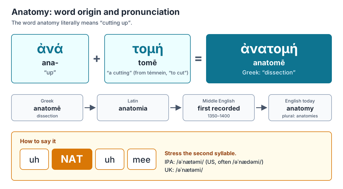
Anatomy literally means 'cutting up'. English borrowed it in the Middle English period, first recorded between 1350 and 1400, from Latin anatomia, which came from Greek anatomḗ, 'dissection'. The Greek word joins ana-, 'up', with tomḗ, 'a cutting', a noun from the verb témnein, 'to cut'. (Dictionary.com, anatomy.)
The name fits how the subject began. Anatomy was first learned by looking at the outside of the body and at the wounds of soldiers and other injured people; later, physicians dissected the bodies of the dead to see inside. Dissection, taking an organism apart to find out its internal structure, is still a core method today. (OpenStax Anatomy & Physiology 2e, 1.1 Overview of Anatomy and Physiology.)
How to pronounce anatomy
Anatomy is pronounced uh-NAT-uh-mee, with the stress on the second syllable. Dictionary.com gives the IPA as /əˈnætəmi/ in American English and /əˈnætəmɪ/ in British English. (Dictionary.com, anatomy.)
Vocabulary.com's American pronunciation softens the t towards a d, /əˈnædəmi/, as in most everyday American speech; its British pronunciation keeps a clear t, /əˈnætəmi/. Either way the first and third syllables are a weak 'uh'. (Vocabulary.com, anatomy.)
Branches of anatomy
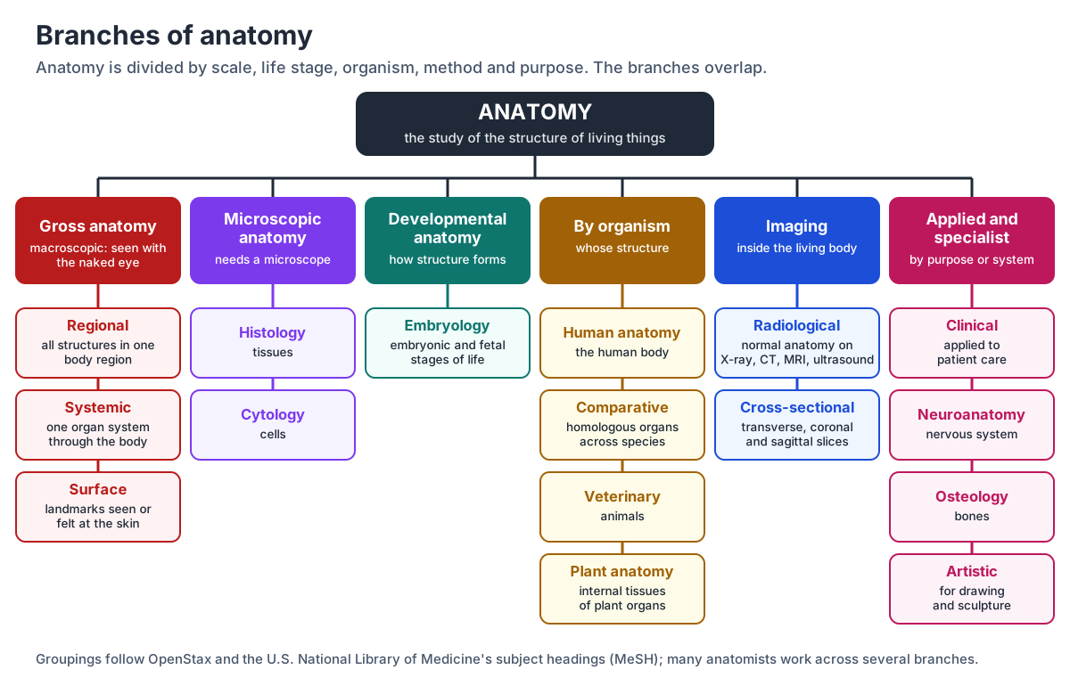
Anatomy is divided first by scale. Gross (macroscopic) anatomy studies structures large enough to see without magnification; microscopic anatomy studies structures that can only be seen with a microscope or other magnifying device. The prefixes say it: macro- means large and micro- means small. (OpenStax Anatomy & Physiology 2e, 1.1 Overview of Anatomy and Physiology.)
Other branches are defined by time (how structure develops), by species (comparing animals, or studying domestic animals or plants), by method (imaging) and by purpose (medicine or art). The National Library of Medicine's subject headings list artistic, comparative, cross-sectional, regional and veterinary anatomy, cell biology, embryology, histology, neuroanatomy and osteology as narrower fields of anatomy. (NLM Medical Subject Headings (MeSH), Anatomy.)
| Branch | What it studies | How it is studied |
|---|---|---|
| Gross (macroscopic) | Structures visible to the naked eye: organs, bones, muscles, vessels | Dissection, models, imaging |
| Regional | Every structure in one region, such as the abdomen | Dissection region by region |
| Systemic | One organ system through the whole body, such as all skeletal muscles | Dissection and models, system by system |
| Surface | Landmarks identified from the surface of the body | Observation, without dissection |
| Microscopic | Structures too small to see unaided | Microscopes |
| Histology | Tissues: epithelial, connective, muscle and nervous | Stained tissue slides |
| Cytology | The structure and function of cells | Microscopy |
| Developmental / embryology | How structure forms in the embryonic and fetal stages | Microscopy, imaging |
| Comparative | The same (homologous) organs across species | Dissection of different animals |
| Veterinary | The anatomical structures of animals | Dissection, imaging |
| Plant | Internal tissues and structures of plant organs | Thin sections under a microscope |
| Radiological / cross-sectional | Normal anatomy on images, in transverse, coronal and sagittal slices | X-ray, CT, MRI, ultrasound |
| Clinical | Anatomy applied to diagnosis, surgery and patient care | Clinical cases, imaging, cadavers |
| Neuroanatomy | The anatomy of the nervous system | Dissection, imaging, microscopy |
| Osteology | The structure and function of bones | Skeletons, imaging |
| Artistic | Body structure for drawing, painting and sculpture | Observation and drawing |
What is gross anatomy?
Gross anatomy, also called macroscopic anatomy, is the study of body structures that can be seen with the naked eye, such as the internal organs and external features. 'Gross' here means large, not unpleasant. It is usually approached in one of three ways: regionally, systemically or from the surface. (LibreTexts Medicine, 1.1A Defining Anatomy (A and P for STEM Educators, James Madison University).)
Regional anatomy studies everything in one region of the body at once, such as the abdomen, and how its structures relate to each other. Systemic anatomy follows one body system through the whole body; a systemic study of the muscular system, for example, covers all of the skeletal muscles. (OpenStax Anatomy & Physiology 2e, 1.1 Overview of Anatomy and Physiology.)
Surface (superficial) anatomy is the study of anatomical landmarks that can be identified by observing the surface of the body, without dissection. (LibreTexts Medicine, 1.1A Defining Anatomy (A and P for STEM Educators, James Madison University).)
| Gross anatomy | Microscopic anatomy | |
|---|---|---|
| Also called | Macroscopic anatomy | Histology and cytology |
| Scale | Seen with the naked eye | Seen only with a microscope |
| Subdivisions | Regional, systemic, surface | Histology (tissues), cytology (cells) |
| Main methods | Dissection, models, X-ray, CT, MRI, ultrasound | Thin, stained slides under a microscope |
| Typical question | Which muscles cross the knee, and where do they attach? | Which tissue lines the small intestine? |
For gross anatomy in practice, explore the 3D human anatomy model, then study it by system in the guides to the skeletal system, muscular system, circulatory system, digestive system or by region in the guides to the head, chest, abdomen, arm, leg.
Microscopic anatomy: histology and cytology
Microscopic anatomy studies structures too small to see without magnification. Its two main parts are cytology, the study of the structure and function of cells, and histology, the study of how tissues are organised and built. (LibreTexts Medicine, 1.1A Defining Anatomy (A and P for STEM Educators, James Madison University).)
Histology is organised around four primary tissue types: epithelial tissue, which covers surfaces and forms glands; connective tissue, which binds and supports; muscle tissue, which contracts; and nervous tissue, which carries electrochemical signals. OpenStax defines histology as the microscopic study of tissue appearance, organisation and function. (OpenStax Anatomy & Physiology 2e, 4.1 Types of Tissues.)
See microscopic anatomy in action in the guides to the four tissue types, the layers of the skin, the parts of a cell and the sarcomere, the repeating unit of muscle.
Developmental anatomy and embryology
Developmental anatomy follows how structure forms and changes as an organism develops. Its best-known part is embryology, defined in MeSH as the study of the development of an organism during the embryonic and fetal stages of life. (NLM MeSH, Embryology.)
Embryology traces the body's tissues back to the embryo's three germ layers, cell lineages named by their position: ectoderm (outer), mesoderm (middle) and endoderm (inner). (OpenStax Anatomy & Physiology 2e, 4.1 Types of Tissues.)
Comparative, veterinary and plant anatomy
Comparative anatomy is the comparative study of animal structure with regard to homologous organs or parts: it asks how the same organ differs between species. Veterinary anatomy is the study of the anatomical structures of animals. (NLM MeSH, Anatomy, Comparative.)
Plant anatomy studies the internal tissues and structures of plant organs: the arrangement of cells and tissues inside roots, stems, leaves and flowers. A classic plant anatomy exercise compares cross-sections of dicot and monocot roots, stems and leaves. (Bidlack, Plant Anatomy (SERC, Carleton College).)
Radiological, clinical and neuroanatomy
Radiological anatomy is normal anatomy as it appears on medical images. Kenhub puts the difference neatly: radiology helps you understand what is abnormal, radiological anatomy helps you understand what is normal, and it sharpens your sense of the spatial relationships inside the body. (Kenhub, Radiological anatomy: a must-have tool for every medical student.)
Its close relative, cross-sectional anatomy, describes the body from three-dimensional imaging as a series of transverse, coronal and sagittal slices. It is essential for interpreting ultrasound, MRI and CT. (NLM MeSH, Anatomy, Cross-Sectional.)
Clinical anatomy applies anatomical knowledge to patient care. A 2025 consensus statement from the American Association of Clinical Anatomists defines it as an integrative discipline that applies anatomy to clinical practice, research and education with the goal of improving patient care, and says it is defined by that integration rather than by holding clinical credentials. (Iwanaga et al., What Is Clinical Anatomy? A Consensus Statement From the American Association of Clinical Anatomists, Clinical Anatomy 39:72 (2026).)
Neuroanatomy is the study of the anatomy of the nervous system as a specialty, and osteology is the branch that deals with the structure and function of bones. (NLM MeSH, Neuroanatomy.)
Artistic anatomy studies the structure of organisms for drawing, painting, sculpture and illustration. (NLM MeSH, Anatomy, Artistic.)
Compare body plans across species on the comparative anatomy and animal anatomy pages, or go straight to a 3D model such as the dog, frog or fetal pig dissection. For plants, start with plant anatomy, the leaf cross-section and flower anatomy. Neuroanatomy starts with the brain, spinal cord, cranial nerves and the neuron.
How anatomy is studied: dissection, microscopes, imaging and models
Dissection, the process of taking an organism apart to find its internal structure, remains central to learning gross anatomy. Students also learn from anatomical models, skeletons, textbooks, diagrams, photographs, lectures and tutorials. Over the centuries the field has moved from examining animals and cadavers by dissection to non-invasive imaging and radiology. (LibreTexts Medicine, 1.1A Defining Anatomy (A and P for STEM Educators, James Madison University).)
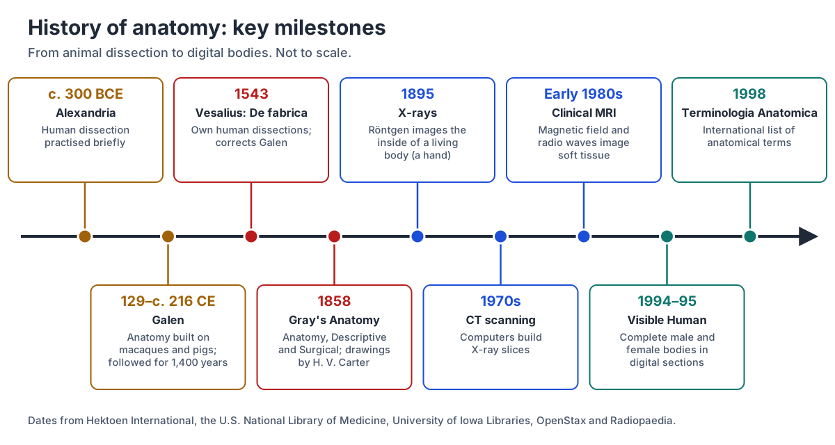
Imaging made it possible to see inside the living body. Wilhelm Röntgen made the first durable X-ray image of a living person's internal parts, his wife's hand, in 1895. Computed tomography (CT) followed in the 1970s, clinical MRI scanners by the early 1980s, and ultrasound was developed in the 1940s and 1950s. PET scans add function: they track short-lived radioactive tracers to show metabolism and blood flow rather than a static picture. (OpenStax Anatomy & Physiology 2e, 1.7 Medical Imaging.)
Digital anatomy builds on those images. The U.S. National Library of Medicine's Visible Human Project released a complete male body in 1994 (1,871 cross-sections at 1 mm intervals, with CT and MRI) and a female body in 1995 (5,189 anatomical images at 0.33 mm intervals). Three-dimensional atlases built from such data let you rotate and dissect a body on screen. (U.S. National Library of Medicine, The Visible Human Project.)
The 3D models on this site use that approach. The human body is built from BodyParts3D, a database from the Database Center for Life Science in Japan that represents anatomical concepts as segments of a three-dimensional whole-body model of an adult man, released under a CC BY licence. (BodyParts3D, Database Center for Life Science (CC BY 4.0).)
| Method | In use since | Shows best | Limits |
|---|---|---|---|
| X-ray (radiography) | 1895 | Bones, teeth and fractures; soft tissue looks grey | Radiation can damage cells |
| CT (computed tomography) | 1970s | Cross-sectional slices with fine soft-tissue detail, such as the brain and abdominal organs | Radiation dose many times that of a standard X-ray |
| MRI | Clinical scanners by the early 1980s | Precise images, especially for finding tumours; functional MRI shows brain activity | Costly, noisy, enclosed tube; not for some metal implants |
| PET | First scanner 1961; tracers about 15 years later | Function: metabolism and blood flow, not just shape | Needs short-lived radioactive tracers |
| Ultrasound | Developed in the 1940s–1950s | Heart function, blood flow, the gallbladder and fetal growth, in real time | Operator-dependent; cannot see through bone or gas |
The interactive heart, 3D human body and dissection guides such as the sheep heart dissection on this site let you combine the methods: rotate a model, peel back layers, then compare with a real specimen.
The language of anatomy
Anatomists describe the body with a precise vocabulary. Most terms come from ancient Greek and Latin; because those languages are no longer spoken, the meanings of their words stay fixed. Every description assumes the anatomical position: standing upright, facing forward, feet at shoulder width and parallel, toes forward, arms at the sides and palms facing forward. (OpenStax Anatomy & Physiology 2e, 1.6 Anatomical Terminology.)
From that position come the directional terms (anterior and posterior, superior and inferior, medial and lateral, proximal and distal, superficial and deep) and the three body planes: the sagittal plane divides the body into right and left, the frontal (coronal) plane into front and back, and the transverse plane into upper and lower parts. The abdomen is mapped either into four quadrants or into nine regions. (OpenStax Anatomy & Physiology 2e, 1.6 Anatomical Terminology.)
The global standard for anatomical names is Terminologia Anatomica, released in 1998 to replace the older Nomina Anatomica; the Federative International Programme for Anatomical Terminology (FIPAT) published a second edition in 2019. (Radiopaedia, Terminologia Anatomica.)
Terminologia Anatomica lists more than 7,500 gross anatomical structures of the human body. Every Latin term has an English equivalent and a unique code number, and an index of eponyms (structures named after people) leads to the correct descriptive term. (Kenhub's fact box dates the first edition to 1999; its own text and Radiopaedia say 1998.) (Kenhub, Terminologia Anatomica.)
Learn the vocabulary step by step in the guides to anatomical terms, directional terms, anatomical planes, body regions.
A short history of anatomy
Human dissection was rare in the Greek and Roman world because of social and cultural constraints; it was practised only briefly, in Hellenistic Alexandria around 300 BCE. Galen of Pergamon (129 to about 216 CE) therefore built his anatomy mostly on rhesus and Barbary macaques, using pigs when monkeys were not available. His books were accepted as standard and rarely questioned for more than fourteen centuries. (Štrkalj, Galen, macaques, and the growth of the discipline of human anatomy (Hektoen International).)
Andreas Vesalius (1514–1564), professor of anatomy at Padua from 1537, changed that. He performed his own careful dissections and in 1543 published De fabrica, the Seven Books on the Fabric of the Human Body (Basel: Johannes Oporinus), confirming and refuting many of Galen's anatomical and physiological teachings. Its woodcut illustrations, by artists now known only as the workshop of Titian, shaped anatomical drawing for centuries. (U.S. National Library of Medicine, Historical Anatomies on the Web: Vesalius.)
Unlike his own teachers, Vesalius insisted on dissecting with his own hands and encouraged his students to do the same. He was 28 when the Fabrica appeared, and he contradicted more than 200 of Galen's teachings, among them the claim that the human jaw consists of two bones. (Gunderman, The man who revolutionized our knowledge of the human body (The Conversation).)
Henry Gray (1825–1861) published Anatomy, Descriptive and Surgical in London in 1858 (John W. Parker): 750 pages, with drawings by H. V. Carter. (University of Iowa Libraries, Heirs of Hippocrates: Henry Gray, Anatomy, Descriptive and Surgical (1858).)
Known since as Gray's Anatomy, the book has been revised many times and remains a standard text, with detailed descriptions of the parts of the human body. (Vocabulary.com, anatomy.)
Anatomy vs physiology
Anatomy studies structure; physiology studies function. Human physiology is the scientific study of the chemistry and physics of the body's structures and of how they work together to support life. The two depend on each other: form is closely related to function in all living things, so it is hard to learn anatomy without the physiology it supports. (OpenStax Anatomy & Physiology 2e, 1.1 Overview of Anatomy and Physiology.)
For the full comparison table, worked structure-and-function examples, the levels of organisation, the organ systems and homeostasis, read anatomy and physiology.
Why study anatomy? Careers and uses
Anatomy underpins every health profession. Anatomists teach future doctors, nurses, physical therapists, dentists and veterinarians, mostly in medical, dental and veterinary schools and large undergraduate programmes. They work at universities, research institutions and in private industry, and their cell and molecular research has improved the understanding of cleft palate, congenital heart defects, neurological disorders and cancer. (American Association for Anatomy, What Is Anatomy?.)
Anatomy is used well beyond medicine. The American Association for Anatomy lists palaeontologists among the careers that rely on it, along with biomedical engineers, who design prosthetics and pacemakers. (American Association for Anatomy, What Is Anatomy?.)
Start learning anatomy
Begin with the body as a whole: rotate the 3D human anatomy model, turn systems on and off, and click any structure to read about it. Then learn the organs of the body and work through one system at a time. Test yourself with the anatomy quiz or the label-it-yourself heart diagram on the heart blood flow page. Self-testing and spacing your study over several days work better than rereading.
Anatomy: frequently asked questions
What is anatomy in simple words?
Anatomy is the study of what living things are made of and how their parts are arranged: bones, muscles, organs, tissues and cells, and where each sits in relation to the others. It applies to humans, animals and plants. The word comes from Greek anatomē, 'dissection', because the subject began with cutting bodies open to look inside.
What does anatomy mean in Greek?
Anatomy comes from Greek anatomē, 'dissection', made of ana- ('up') and tomē ('a cutting'), from the verb temnein, 'to cut'. So the literal meaning is 'cutting up'. The word passed into Latin as anatomia and into English in the late Middle Ages, first recorded between 1350 and 1400.
How do you pronounce anatomy?
Say uh-NAT-uh-mee, with the stress on the second syllable: /əˈnætəmi/. In American English the t is often softened so it sounds closer to uh-NAD-uh-mee (/əˈnædəmi/). The first and third syllables are both a short, unstressed 'uh'. The plural is anatomies, and someone who studies anatomy is an anatomist.
What are the main branches of anatomy?
The two main branches are gross (macroscopic) anatomy, which studies structures visible to the naked eye, and microscopic anatomy, which covers histology (tissues) and cytology (cells). Gross anatomy is studied regionally, systemically or from the surface. Other branches include developmental anatomy and embryology, comparative, veterinary and plant anatomy, radiological and clinical anatomy, and neuroanatomy.
What is gross anatomy?
Gross anatomy is the study of body structures large enough to see without a microscope, such as bones, muscles and organs. It is also called macroscopic anatomy; 'gross' means large. It is learned by dissection, models and imaging, and is organised either by region (everything in the chest, say) or by system (all the bones, all the muscles).
What is the difference between anatomy and physiology?
Anatomy is about structure: what a part is, its shape and where it lies. Physiology is about function: how that part works, down to its chemistry and physics. The heart's four chambers and valves are anatomy; how pressure changes open and close those valves to pump blood is physiology. The two are usually taught together because form and function are linked.
Who is the father of anatomy?
The title usually goes to Andreas Vesalius, whose 1543 De fabrica, based on his own human dissections, is described as marking the birth of modern anatomy. It corrected many errors in the anatomy of Galen, which had been built largely on monkeys and pigs and accepted for over 1,400 years. Galen and the anatomists of ancient Alexandria came much earlier.
Is anatomy hard to learn?
Anatomy is less about difficult ideas than about volume: there are thousands of named structures, and the official list, Terminologia Anatomica, covers more than 7,500. It gets easier once you learn the anatomical position, the directional terms and the Greek and Latin word roots, and when you study in 3D and test yourself instead of rereading.
References
- OpenStax Anatomy & Physiology 2e, 1.1 Overview of Anatomy and Physiology
- OpenStax Anatomy & Physiology 2e, 1.6 Anatomical Terminology
- OpenStax Anatomy & Physiology 2e, 1.7 Medical Imaging
- OpenStax Anatomy & Physiology 2e, 4.1 Types of Tissues
- LibreTexts Medicine, 1.1A Defining Anatomy (A and P for STEM Educators, James Madison University)
- Dictionary.com, anatomy
- Vocabulary.com, anatomy
- NLM Medical Subject Headings (MeSH), Anatomy
- NLM MeSH, Histology
- NLM MeSH, Embryology
- NLM MeSH, Anatomy, Comparative
- NLM MeSH, Anatomy, Veterinary
- NLM MeSH, Neuroanatomy
- NLM MeSH, Anatomy, Cross-Sectional
- NLM MeSH, Anatomy, Artistic
- Iwanaga et al., What Is Clinical Anatomy? A Consensus Statement From the American Association of Clinical Anatomists, Clinical Anatomy 39:72 (2026)
- Kenhub, Radiological anatomy: a must-have tool for every medical student
- Bidlack, Plant Anatomy (SERC, Carleton College)
- American Association for Anatomy, What Is Anatomy?
- Štrkalj, Galen, macaques, and the growth of the discipline of human anatomy (Hektoen International)
- Gunderman, The man who revolutionized our knowledge of the human body (The Conversation)
- U.S. National Library of Medicine, Historical Anatomies on the Web: Vesalius
- University of Iowa Libraries, Heirs of Hippocrates: Henry Gray, Anatomy, Descriptive and Surgical (1858)
- U.S. National Library of Medicine, The Visible Human Project
- Radiopaedia, Terminologia Anatomica
- Kenhub, Terminologia Anatomica
- BodyParts3D, Database Center for Life Science (CC BY 4.0)
More interactive anatomy models
- Anatomy and physiologyHow structure and function fit together, the levels of organisation and the organ systems, with study tips.
- Comparative anatomyHomologous, analogous and vestigial structures compared across vertebrates, with diagrams.
- Animal anatomyEvery animal on the site by group, with what each body plan teaches.
- Anatomy quizFree anatomy quizzes and games: find-the-structure quizzes on 3D models and labelling practice.
- Human anatomy1,000+ bones, muscles, organs, vessels and nerves from real anatomical data.
- Octopus anatomyThree hearts, a ring-shaped brain, eight arms and a beak, in 3D.