

What is plant anatomy?
Plant anatomy is the branch of botany that studies how a plant is built inside: the development, pattern and significance of its cells, tissues and organs, and the microscope methods used to see them (University of Guelph). A standard textbook works organ by organ, through the stem, root, leaf, flower, pollen, seed and fruit, with tools from the hand lens to the electron microscope, and calls the subject fundamental to classifying plants and essential to physiology, ecology and evolutionary biology (Rudall, Anatomy of Flowering Plants).
| Field | What it studies | Example question |
|---|---|---|
| Plant anatomy | Internal structure: cells, tissues and organs, mostly in thin sections | What layers do you see in a leaf cross section? |
| Plant morphology | External form: the shape and arrangement of the plant's parts | Are the leaves simple or compound, opposite or alternate? |
| Plant physiology | How the plant works: moving water, minerals and sugars, and responding to light and gravity | How does water reach the top of a tree? |
The split between anatomy (internal) and morphology (external form) follows university botany courses (University of Rwanda); the topics of physiology follow OpenStax (OpenStax ch. 30). In practice the three overlap: you need the anatomy of the xylem to explain how water moves.
Parts of a plant: the shoot system and the root system
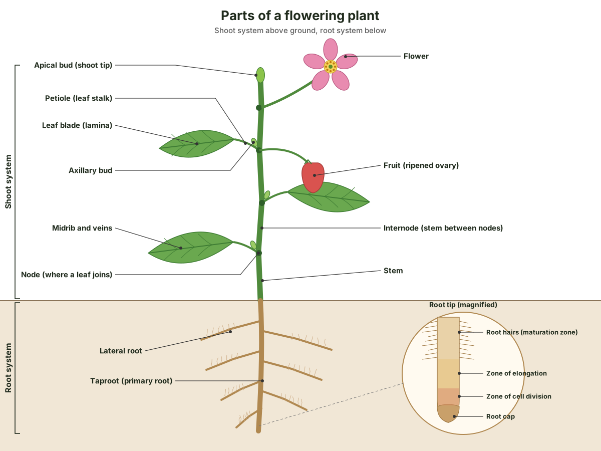
A flowering plant has two organ systems. The shoot system, usually above ground, holds the vegetative organs (stems and leaves) and the reproductive ones (flowers and fruits). The root system, usually underground, anchors the plant and absorbs water and minerals (OpenStax 30.1). Every plant body is built of stems, roots and leaves (OpenStax ch. 30).
| Organ | Main tissues | Main job | Learn more |
|---|---|---|---|
| Root | Epidermis with root hairs, cortex, endodermis, central vascular cylinder | Anchors, absorbs water and minerals, stores food | Roots, below |
| Stem | Epidermis, cortex, pith, vascular bundles | Supports leaves and flowers; links roots and leaves | Stems, below |
| Leaf | Epidermis with stomata, mesophyll, veins | Photosynthesis and gas exchange | Leaf cross section |
| Flower | Sepals, petals, stamens, carpels | Sexual reproduction | Flower anatomy |
| Fruit | Ovary wall (pericarp) | Protects and disperses the seeds | Fruits, below |
| Seed | Seed coat, embryo, cotyledons, endosperm | Carries the embryo of the next plant | Seed anatomy |
Plant cells: what makes them different
Plant cells have three things most animal cells lack: a rigid cell wall of cellulose, chloroplasts and other plastids, and a large central vacuole. Most plant cells, in turn, have no centrosome and no lysosomes (OpenStax 4.3). For a full 3D tour of the organelles, see cell anatomy.
| Structure | What it is | What it does |
|---|---|---|
| Cell wall | A rigid covering outside the cell membrane, mainly cellulose, a polysaccharide made of glucose units | Protects the cell, supports it and gives it its shape |
| Middle lamella | The layer between neighbouring cells, part of it made by each cell; rich in pectin, but lignified in wood | Glues cells together so they act as one tissue |
| Plasmodesmata | Channels through the walls of two neighbouring cells | Let materials pass straight from one cell's cytoplasm to the next |
| Central vacuole | A very large, fluid-filled sac | Regulates the cell's water, keeps up pressure against the wall, and lets the cell grow cheaply |
| Chloroplasts | Plastids with an outer and an inner membrane, stacked thylakoids (grana) and stroma, and their own DNA | Photosynthesis |
| Other plastids | Other members of the plastid family | Store pigments |
Table sources: OpenStax on the wall, vacuole and plastids (OpenStax 4.3) and on plasmodesmata, which play the part that gap junctions play between animal cells (OpenStax 4.6); the Wood Handbook on the middle lamella (Wood Handbook, ch. 3). The vacuole is why a growing plant cell can become much larger without making much new cytoplasm.
A wall is built in layers. Each cell first lays down a thin primary wall, with cellulose microfibrils running in random directions. Inside it, many cells, including those of wood, add a secondary wall in three layers (S1, S2 and S3), each with its microfibrils at a different angle. Every layer combines cellulose microfibrils, hemicelluloses and a matrix: mostly pectin in primary walls and lignin in secondary walls (Wood Handbook, ch. 3). That chemical difference is what stains tell apart on a slide (see how plant anatomy is studied, below): the lignified walls of sclerenchyma and xylem take a different stain from the thin walls of parenchyma. For the inside of a chloroplast, see chloroplast structure.
Meristems: where plants grow
Plants grow from meristems, regions of continuously dividing cells. Meristem cells are undifferentiated or only partly differentiated; the cells they leave behind become the permanent tissues, which no longer divide (OpenStax 30.1).
| Meristem | Where | What it does |
|---|---|---|
| Apical meristems | Tips of shoots and roots | Make the plant longer (primary growth) |
| Lateral meristems: vascular cambium and cork cambium | A cylinder inside the stem and root, and in the bark | Make woody plants thicker (secondary growth): new wood, phloem and cork |
| Intercalary meristems | Base of leaf blades and at nodes, in monocots only | Let grass leaves grow back from the base after mowing |
Table from OpenStax (OpenStax 30.1) (OpenStax 30.2). The vascular cambium is the cylinder of meristem that makes the secondary xylem and phloem of shrubs and trees (Bellairs 3.1). To see secondary growth in 3D, from the cambium to the growth rings, explore the tree anatomy model of an oak trunk.
The three tissue systems: dermal, ground and vascular
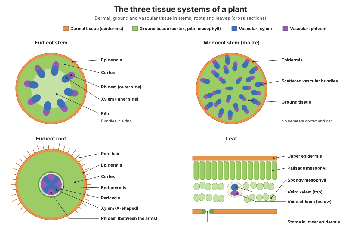
All the tissues of a plant belong to three tissue systems: dermal, ground and vascular (Bellairs 2.1). Dermal tissue covers and protects the plant and controls gas exchange; vascular tissue transports water, minerals and sugars; and ground tissue is where photosynthesis happens, supports the vascular tissue and stores water and sugars (OpenStax 30.1).
| Tissue system | Tissues and cells | Job | Where |
|---|---|---|---|
| Dermal | Epidermis, with a waxy cuticle, stomata and guard cells, hairs (trichomes) and, on roots, root hairs; in woody stems, cork | Protection; controls water loss and gas exchange; absorption in roots | The outer layer of every organ |
| Ground | Parenchyma, collenchyma, sclerenchyma | Photosynthesis, storage, support | Cortex and pith of stems and roots; mesophyll of leaves |
| Vascular | Xylem (tracheids, vessel elements, xylem parenchyma); phloem (sieve elements, companion cells, phloem parenchyma, fibres) | Water and minerals up (xylem); sugars to the rest of the plant (phloem) | Bundles in stems and leaves (veins); a central cylinder in roots |
Simple tissues: parenchyma, collenchyma and sclerenchyma
A simple tissue is made of similar cells; a complex tissue, such as xylem or phloem, is made of several cell types (OpenStax 30.1).
- Parenchyma: the most common plant cells, with thin cellulose walls. They carry out photosynthesis, store starch and help heal wounds (OpenStax 30.2). Parenchyma with large air spaces, called aerenchyma, keeps water plants afloat and aerated (Bellairs 2.2).
- Collenchyma: elongated, living cells with unevenly thickened walls that support the plant, found below the epidermis (OpenStax 30.2); celery is the classic example (Bellairs 2.2).
- Sclerenchyma: support cells with lignin-thickened walls, many of them dead at maturity (OpenStax 30.2). Long, thin fibres and variously shaped sclereids, such as the gritty stone cells of a pear (Bellairs 2.2).
Xylem and phloem
Xylem carries water and minerals from the roots; its water-conducting cells, tracheids and vessel elements, are dead at maturity, and vessel elements join end to end through perforation plates. Phloem carries sugars and amino acids in sieve-tube elements, which are alive and supported by companion cells (OpenStax 30.2). In stems the two form vascular bundles; in roots they form the vascular cylinder, or stele (OpenStax 30.1).
See the three tissue systems in 3D
A leaf shows all three systems at once. In this 3D block of a lilac leaf, the epidermis and cuticle are dermal tissue, the palisade and spongy mesophyll are ground tissue, and the veins are vascular tissue. Hide a system with the buttons at the bottom of the viewer, or open the full leaf cross section guide.
Drag to rotate · Scroll or pinch to zoom · Right-drag to pan · Click a part
Structures
Click a part on the model or pick one below.
Plant organs: root, stem, leaf, flower, fruit and seed
An organ is a group of tissues working together. These are the six organs of a flowering plant, with a short answer for each and a link to the full guide where one exists.
Root anatomy
A root has an outer epidermis, a cortex, and a central stele of xylem and phloem. The innermost layer of the cortex, the endodermis, is found only in roots: a waxy band of suberin in its walls, the Casparian strip, makes water and minerals pass through living membranes before they reach the xylem (OpenStax 30.3) (Bellairs 3.3). Just inside it, the pericycle can give rise to lateral roots.
The root tip is covered by a root cap, behind which lie the zones of cell division, elongation and maturation; root hairs, extensions of epidermal cells, start in the maturation zone and increase the surface for absorption. In eudicot roots the xylem and phloem alternate in an X shape; in monocot roots the vascular tissue forms a ring around a pith. Eudicots usually have a taproot, monocots a fibrous root system, and taproots such as carrot and beet are modified to store food (OpenStax 30.3). Most plants also partner with fungi in their roots (mycorrhizae), trading sugars for phosphate and other minerals (OpenStax 31.3).
Stem anatomy
A stem carries leaves at nodes, separated by internodes. Inside, a single-layered epidermis covers a cortex of ground tissue, the vascular tissue, and a central pith. In eudicot stems the vascular bundles form a ring; in monocot stems they are scattered through the ground tissue (OpenStax 30.2) (Bellairs 3.1).
Herbaceous stems mostly grow in length only. Woody stems also grow in girth: the vascular cambium adds wood inside and phloem outside, and the cork cambium makes bark, with lenticels for gas exchange (OpenStax 30.2). The tree anatomy page follows these layers through an oak trunk, from bark to heartwood.
Leaf anatomy
A leaf has a flat blade (lamina), often a stalk (petiole), and a midrib with smaller veins; leaves are simple or compound and arranged alternately, oppositely or in whorls. Inside, a waxy cuticle and epidermis cover the palisade and spongy mesophyll, veins carry xylem and phloem, and pairs of guard cells open and close the stomata (OpenStax 30.4). See the leaf cross section for every layer, the stomata diagram for how the pores work, and chloroplast structure for where photosynthesis happens.
Flower anatomy
Flowers are modified leaves arranged around a receptacle: a whorl of sepals (the calyx), then petals (the corolla), then the stamens, each a filament topped by an anther, and in the centre one or more carpels, each with a stigma, style and ovary (OpenStax 26.3). Explore every part on a 3D lily in flower anatomy.
Fruit anatomy
Botanically, a fruit is a ripened ovary. Its wall, the pericarp, has three layers: the exocarp (skin), mesocarp (middle) and endocarp (inner part). Fruits can be dry or fleshy, and they are classed by how they form (OpenStax 32.2):
| Fruit type | Forms from | Examples |
|---|---|---|
| Simple | One carpel or fused carpels of one flower | Nuts, beans |
| Aggregate | Several separate carpels of one flower | Raspberry |
| Multiple | A whole cluster of flowers | Pineapple |
| Accessory (false) | Mainly another part of the flower, such as the receptacle | Strawberry, apple |
Seed anatomy
A typical seed has a seed coat, cotyledons, endosperm and a single embryo, whose axis has three parts: the plumule (future shoot), the radicle (future root) and the hypocotyl between them. Endosperm forms after double fertilization, when one sperm fertilizes the egg and the other fuses with two polar nuclei (OpenStax 32.2). Compare a bean and a corn grain in 3D in seed anatomy.
Secondary growth: how stems and roots get thicker
Secondary growth is the thickening of stems and roots by the two lateral meristems. The vascular cambium, a thin cylinder of dividing cells between the bark and the wood, makes secondary xylem (wood) to the inside and secondary phloem to the outside; the cork cambium makes cork, whose walls contain waxy, water-repelling suberin. Herbaceous plants mostly show primary growth only (OpenStax 30.2), and monocot stems rarely form wood (EBSCO).
| Primary growth | Secondary growth | |
|---|---|---|
| Made by | Apical meristems at shoot and root tips | Lateral meristems: vascular cambium and cork cambium |
| Direction | Length | Girth (thickness) |
| Tissues it makes | Epidermis, cortex, pith, primary xylem and phloem | Secondary xylem (wood), secondary phloem, and cork (periderm) |
| Found in | All flowering plants | Woody eudicots and conifers; rare in monocots |
| What you see | A young green shoot | Bark outside, growth rings of wood inside |
As the cambium adds wood each year, the young stem's tissues are rearranged. The secondary xylem gives shrubs and trees extra support and extra water-conducting tissue, new secondary phloem replaces the primary phloem (Bellairs 2.5), and the expanding wood squeezes the pith into the centre (Bellairs 3.1). Wood made early in the season has large cells; later wood is denser, and the contrast between them shows as annual rings (OpenStax 30.2). The cambium also grows in circumference by dividing sideways, and its two kinds of starting cells make the vertical cells of the wood and the horizontal rays (Wood Handbook, ch. 3).
Wood does three jobs for the plant: it conducts water from the roots to the leaves, holds the plant body up, and stores food and other chemicals (Wood Handbook, ch. 3). Everything outside the vascular cambium is bark: the inner bark (phloem) and the outer bark (cork). To follow all of these layers through a real trunk, from bark to heartwood, explore the tree anatomy model of an oak.
Monocot vs dicot anatomy
Flowering plants are split mainly into monocots, with one seed leaf, and eudicots (true dicots), with two. The difference runs through every organ, so it is the first thing to check on a slide (OpenStax 26.3).
| Feature | Monocots | Dicots (eudicots) |
|---|---|---|
| Cotyledons in the seed | One | Two |
| Leaf veins | Parallel | Net-like (reticulate) |
| Vascular bundles in the stem | Scattered | In a ring |
| Root system | Fibrous, no main root | Usually a taproot from the embryonic radicle |
| Vascular tissue in the root | A ring around a central pith | X-shaped xylem with phloem between the arms |
| Flower parts | In threes or sixes | In fours or fives |
| Pollen | One furrow or pore | Three furrows or pores |
| Wood (secondary growth) | Rarely | Common in trees and shrubs |
| Examples | Grasses, lilies, orchids, palms | Roses, sunflowers, mints, most trees |
| Number of species | About 65,000 | About 165,000 (two-thirds of flowering plants) |
Sources: OpenStax for cotyledons, veins, stems, roots, flowers, pollen and examples (OpenStax 26.3) (OpenStax 30.3); EBSCO for wood and species numbers (EBSCO). OpenStax's statement that eudicots make up two-thirds of flowering plants matches EBSCO's counts. A few early-branching groups, such as water lilies and the magnoliids, fit neither column neatly (OpenStax 26.3).
How plant anatomy is studied: sections, stains and microscopes
Plant anatomists cut thin slices (sections) of an organ, stain them so that different walls show in different colours, and examine them under a microscope. Because a single slice is flat, wood scientists cut in three planes: a transverse section across the stem, a radial section from the centre outward along its length, and a tangential section at right angles to the radius; only together do they show the structure in three dimensions (Wood Handbook, ch. 3).
You can make sections by hand. Hold the leaf or stem between two halves of a carrot, potato or elder pith, keep the tissue, blade and fingers wet, and cut with one smooth stroke of a razor blade, never sawing. Cut many slices, float them in water, and mount the thinnest, almost transparent ones in a drop of water (White, Prometheus protocols). Laboratory slides are often cut from tissue embedded in paraffin, or with a vibratome (University of Vigo atlas).
| Stain | Colours | What it shows |
|---|---|---|
| Safranin | Red | Lignified (secondary) walls, such as xylem and sclerenchyma, and walls with cutin or suberin |
| Fast green or Alcian blue | Green or blue | Non-lignified (primary) walls, such as those of parenchyma |
Safranin and fast green compete, so the more green a wall takes up, the less red it shows (University of Vigo atlas). Stains are needed because most cell parts are colourless, but staining kills the cells. A light microscope can show living cells; an electron microscope magnifies far more but only works on specially prepared, dead specimens. The scanning electron microscope shows the surface of a specimen, and the transmission electron microscope shows the structures inside its cells (OpenStax 4.1). Plant anatomy uses the whole range, from the hand lens to the electron microscope (Rudall, Anatomy of Flowering Plants). To see what a stained leaf section looks like, compare the leaf cross section.
Explore plant anatomy in 3D
- Flower anatomy: petals, sepals, stamens, pistil, ovary and ovules of a lily.
- Leaf cross section: every tissue layer of a lilac leaf, plus monocot, C4 and pine leaves.
- Stomata diagram: guard cells and how the pore opens and closes.
- Chloroplast structure: envelope, stroma, thylakoids and grana.
- Seed anatomy: bean and corn seeds, seed coat, cotyledons and embryo.
- Tree anatomy: bark, cambium, sapwood, heartwood and growth rings of an oak.
- Cell anatomy: the organelles of a cell in 3D.
Frequently asked questions
What is plant anatomy?
Plant anatomy is the branch of botany that studies the internal structure of plants: how cells are built into tissues, and tissues into organs such as roots, stems and leaves. It is usually studied in thin sections under a microscope. It differs from plant morphology, which describes outward form, and from plant physiology, which studies how plants work.
What are the three tissue systems of a plant?
Dermal, ground and vascular. Dermal tissue (the epidermis, or bark in woody plants) covers and protects the plant. Ground tissue (parenchyma, collenchyma and sclerenchyma) carries out photosynthesis, stores water and sugars, and supports the plant. Vascular tissue (xylem and phloem) carries water, minerals and sugars around the plant.
What are the main parts of a plant?
A flowering plant has a shoot system, usually above ground, and a root system below. The vegetative organs are the root, stem and leaf. The reproductive structures are the flower, which forms fruit and seeds: the fruit is the ripened ovary, and the seeds inside it hold the embryo of the next plant.
What is the difference between xylem and phloem?
Xylem carries water and dissolved minerals up from the roots; its conducting cells, tracheids and vessel elements, are dead when they work. Phloem carries sugars and other organic compounds from the leaves to the rest of the plant; its sieve-tube elements are alive and supported by companion cells.
What are meristems in plants?
Meristems are regions of cells that keep dividing for the life of the plant. Apical meristems at the tips of shoots and roots make the plant longer (primary growth). Lateral meristems, the vascular cambium and cork cambium, make woody plants thicker (secondary growth). Intercalary meristems, found only in monocots, let grass leaves regrow from the base.
What is the difference between monocots and dicots?
Monocots have one seed leaf (cotyledon), parallel leaf veins, scattered vascular bundles in the stem, a fibrous root system and flower parts in threes. Dicots (eudicots) have two cotyledons, net-veined leaves, bundles in a ring, usually a taproot and flower parts in fours or fives. Monocot stems rarely form wood; many dicot trees and shrubs thicken every year.
What are the three types of ground tissue?
Parenchyma, collenchyma and sclerenchyma. Parenchyma cells have thin walls and do photosynthesis, storage and wound healing. Collenchyma cells are alive, with unevenly thickened walls, and support stems and leaves, as in celery. Sclerenchyma cells have thick, lignified walls, are often dead at maturity and form fibres and stone cells.
References
- University of Guelph, Department of Molecular and Cellular Biology (2021). BOT*3410 Plant Anatomy, course outline.
- Rudall, P. J. (2020). Anatomy of Flowering Plants: An Introduction to Plant Structure and Development, 4th edition. Cambridge University Press (front matter).
- University of Rwanda. BIO 102: Plant diversity, structure and function (course outline).
- Clark, M. A., Douglas, M. & Choi, J. (2018). Biology 2e, chapter 30 introduction: Plant Form and Physiology. OpenStax.
- Clark, M. A., Douglas, M. & Choi, J. (2018). Biology 2e, 30.1 The Plant Body. OpenStax.
- Clark, M. A., Douglas, M. & Choi, J. (2018). Biology 2e, 30.2 Stems. OpenStax.
- Clark, M. A., Douglas, M. & Choi, J. (2018). Biology 2e, 30.3 Roots. OpenStax.
- Clark, M. A., Douglas, M. & Choi, J. (2018). Biology 2e, 30.4 Leaves. OpenStax.
- Clark, M. A., Douglas, M. & Choi, J. (2018). Biology 2e, 26.3 Angiosperms. OpenStax.
- Clark, M. A., Douglas, M. & Choi, J. (2018). Biology 2e, 32.2 Pollination and Fertilization. OpenStax.
- Clark, M. A., Douglas, M. & Choi, J. (2018). Biology 2e, 4.3 Eukaryotic Cells. OpenStax.
- Clark, M. A., Douglas, M. & Choi, J. (2018). Biology 2e, 31.3 Nutritional Adaptations of Plants. OpenStax.
- Bellairs, S. Plant Anatomy and Physiology, 2.1 Development of a plant. Biology LibreTexts.
- Bellairs, S. Plant Anatomy and Physiology, 2.2 Ground tissues. Biology LibreTexts.
- Bellairs, S. Plant Anatomy and Physiology, 2.5 Secondary tissues. Biology LibreTexts.
- Bellairs, S. Plant Anatomy and Physiology, 3.1 Stems. Biology LibreTexts.
- Bellairs, S. Plant Anatomy and Physiology, 3.3 Roots. Biology LibreTexts.
- Clark, M. A., Douglas, M. & Choi, J. (2018). Biology 2e, 4.6 Connections between Cells and Cellular Activities. OpenStax.
- Clark, M. A., Douglas, M. & Choi, J. (2018). Biology 2e, 4.1 Studying Cells. OpenStax.
- Wiedenhoeft, A. (2010). Structure and function of wood. Wood Handbook, ch. 3, FPL-GTR-190. USDA Forest Service, Forest Products Laboratory.
- Atlas of Plant and Animal Histology, Department of Functional Biology and Health Sciences, University of Vigo. Safranin with Alcian blue or fast green.
- White, R. Using other plant tissues as support tissue to make hand sections. Prometheus protocols.
- EBSCO Research Starters. Monocots vs. dicots.
More interactive anatomy models
- Flower anatomyParts of a flower in 3D: petals, sepals, stamens, pistil, ovary and ovules of a lily.
- Leaf cross sectionLabeled leaf cross section: cuticle, epidermis, palisade and spongy mesophyll, veins.
- Stomata diagramGuard cells, stomatal pore and how stomata open and close, in 3D.
- Seed anatomyBean and corn seed anatomy: seed coat, cotyledons, endosperm and embryo.
- Chloroplast structureEnvelope, stroma, thylakoids, grana and lumen of a chloroplast, in 3D.
- Tree anatomyA tree trunk in 3D: bark, phloem, cambium, sapwood, heartwood and growth rings.