

Cell anatomy at a glance
An animal cell has three main parts: the plasma membrane around it, the cytoplasm inside it (the jelly-like cytosol plus the organelles), and the nucleus, which holds the DNA. Every cell, from a bacterium to a neuron, shares four components: a plasma membrane, cytoplasm, DNA and ribosomes. (OpenStax Biology 2e, 4.2 Prokaryotic cells; OpenStax Anatomy and Physiology 2e, 3.2 The cytoplasm and cellular organelles.)
Cellular anatomy differs most between prokaryotes and eukaryotes. Animal cells are eukaryotic: they keep their DNA in a nucleus and divide their cytoplasm into membrane-bound organelles, each with its own job. Eukaryotic cells are typically 10–100 µm across, compared with 0.1–5 µm for bacteria. (OpenStax Biology 2e, 4.2 Prokaryotic cells.) Most mammalian cells have a volume of about 1,000–10,000 µm³. (Milo & Phillips, Cell Biology by the Numbers: How many cells are there in an organism?.) The human body is made of about 30 trillion human cells by a 2016 estimate, or 37 trillion by an earlier one, of more than 200 types. (Sender, Fuchs & Milo (2016): Revised estimates for the number of human and bacteria cells in the body, PLOS Biology 14:e1002533; Milo & Phillips, Cell Biology by the Numbers: How big is a human cell?.)
The model shows a generalized animal cell: real cells specialise. A neuron grows long dendrites and an axon (see our neuron anatomy model), a muscle fibre fills with contractile filaments, and a red blood cell loses its nucleus altogether. How cells group into tissues is covered in our tissue types guide.
Labeled diagram of an animal cell
A labeled animal cell diagram shows the plasma membrane, cytoplasm and nucleus (with nuclear envelope, pores, nucleolus and chromatin), and the organelles of the cytoplasm: rough and smooth endoplasmic reticulum, ribosomes, Golgi apparatus, vesicles, lysosomes, peroxisomes, mitochondria, the centrosome and the cytoskeleton.
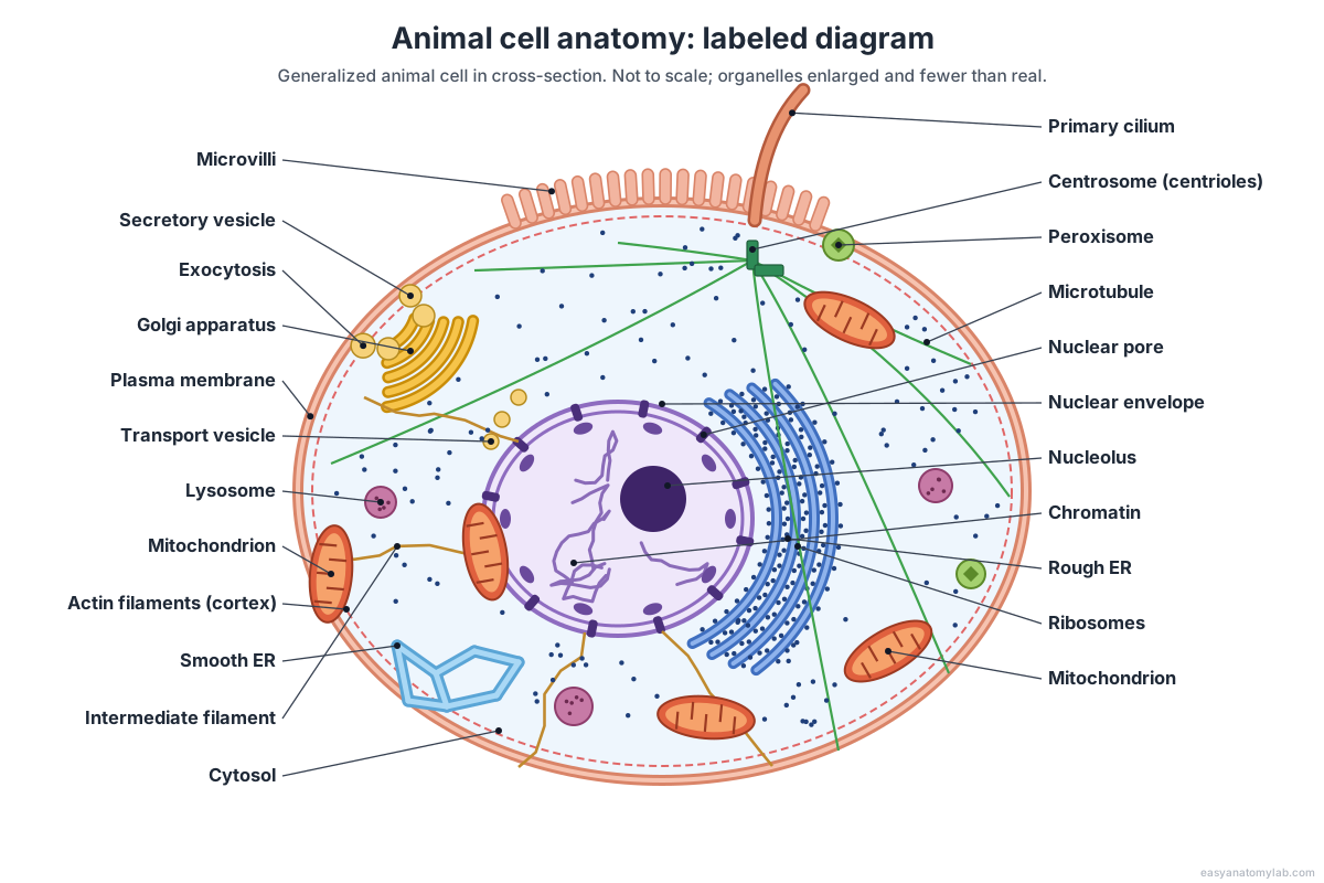
In the 3D model, turn on All labels to name everything at once, or click a part to zoom to it. Slide the Plasma membrane control down to see the whole cell interior from any angle.
Parts of a cell and their functions
Each organelle has a main job. This table lists the parts of an animal cell, what they look like and what they do.
| Organelle | Structure | Function |
|---|---|---|
| Plasma membrane | Phospholipid bilayer with proteins and cholesterol | Controls what enters and leaves; cell signalling and recognition |
| Cytosol | Watery gel, 70–80% water | Medium for many chemical reactions |
| Nucleus | Double-membrane envelope with pores | Holds the DNA; controls the cell |
| Nucleolus | Dense, dark-staining body in the nucleus | Makes ribosomal RNA and ribosome subunits |
| Ribosomes | RNA–protein particles (80S), no membrane | Make proteins |
| Rough ER | Sheets of membrane sacs studded with ribosomes | Makes and folds membrane and secreted proteins |
| Smooth ER | Network of tubes, no ribosomes | Makes lipids and steroids; detoxifies; stores Ca²⁺ |
| Golgi apparatus | Stack of flattened sacs (cis and trans faces) | Modifies, sorts and packages proteins and lipids |
| Vesicles | Small membrane bubbles | Carry cargo between organelles and to the surface |
| Lysosomes | Acidic sacs of digestive enzymes | Break down worn-out parts and engulfed material |
| Peroxisomes | Small sacs with oxidising enzymes and catalase | Break down fatty acids; detoxify; destroy H₂O₂ |
| Mitochondria | Double membrane; inner one folded into cristae | Make ATP by aerobic respiration |
| Centrosome | Two centrioles of 9 microtubule triplets | Organises microtubules and the spindle |
| Cytoskeleton | Microtubules, actin filaments, intermediate filaments | Shape, support, movement and transport |
| Microvilli and cilia | Finger-like folds; hair-like projections | Absorption; sensing (primary cilium) or movement (motile cilia) |
The nucleus: the cell's control centre
The nucleus is usually the largest and most prominent organelle. It is wrapped in a nuclear envelope of two lipid bilayers with a thin space between them, and holds the chromatin and the nucleolus in a gel-like nucleoplasm. (OpenStax Anatomy and Physiology 2e, 3.3 The nucleus and DNA replication; OpenStax Biology 2e, 4.3 Eukaryotic cells.) Typical nuclei are 2–10 µm across. (Milo & Phillips, Cell Biology by the Numbers: How big are nuclei?.)
Traffic in and out passes through nuclear pores, protein complexes about 120 nm across with eightfold symmetry; each human pore weighs about 110 MDa, and molecules smaller than about 40 kDa can diffuse through it freely. (Dultz, Wojtynek, Medalia & Onischenko (2022): The nuclear pore complex: birth, life, and death of a cellular behemoth, Cells 11:1456; Rush et al. (2023): Unveiling the complexity: assessing models describing the structure and function of the nuclear pore complex, Frontiers in Cell and Developmental Biology 11:1245939.) Inside, DNA wraps around histone proteins as nucleosomes, about 147 base pairs per histone core, forming the threads of chromatin; a human cell carries 46 chromosomes and almost 22,000 genes. (OpenStax Anatomy and Physiology 2e, 3.3 The nucleus and DNA replication; Milo & Phillips, Cell Biology by the Numbers: How big are nuclei?; OpenStax Biology 2e, 4.3 Eukaryotic cells.) The nucleolus builds ribosome subunits, which leave through the pores. Just inside the envelope, a mesh of intermediate filaments called the nuclear lamina shapes the nucleus. (OpenStax Microbiology, 3.4 Unique characteristics of eukaryotic cells; Gruenbaum & Aebi (2014): Intermediate filaments: a dynamic network that controls cell mechanics, F1000Prime Reports 6:54.)
The endomembrane system: ER, Golgi apparatus, vesicles and lysosomes
The endomembrane system is a group of membranes and organelles that work together to make, modify, package and ship proteins and lipids. It includes the nuclear envelope, endoplasmic reticulum, Golgi apparatus, vesicles, lysosomes and, by interaction, the plasma membrane; mitochondria are not part of it. (OpenStax Biology 2e, 4.4 The endomembrane system and proteins.)
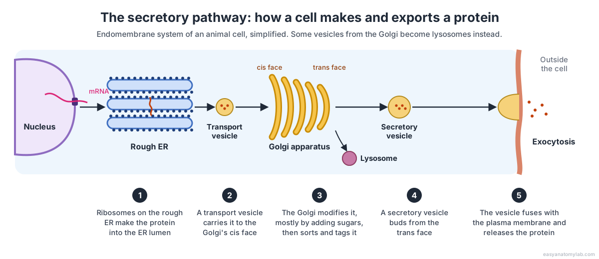
- Ribosomes on the rough ER insert a new protein into its lumen, where it is folded and may gain sugar chains. (OpenStax Biology 2e, 4.4 The endomembrane system and proteins.)
- Transport vesicles bud from the ER and fuse with the cis face of the Golgi apparatus. (OpenStax Biology 2e, 4.4 The endomembrane system and proteins; OpenStax Microbiology, 3.4 Unique characteristics of eukaryotic cells.)
- The Golgi modifies the protein, most often by adding short sugar chains, then sorts and tags it. (OpenStax Biology 2e, 4.4 The endomembrane system and proteins.)
- Secretory vesicles bud from the trans face and fuse with the plasma membrane, releasing their contents (exocytosis); the Golgi also produces lysosomes. (OpenStax Biology 2e, 4.4 The endomembrane system and proteins; OpenStax Anatomy and Physiology 2e, 3.2 The cytoplasm and cellular organelles.)
The ER is big. In liver and pancreas cells it can hold up to 60% of all the cell's membrane, and in a pancreatic acinar cell its surface area is 10–20 times that of the plasma membrane. (Milo & Phillips, Cell Biology by the Numbers: How big is the endoplasmic reticulum of cells?.) The smooth ER makes lipids and steroid hormones, detoxifies drugs and poisons, and stores calcium; in muscle it is called the sarcoplasmic reticulum. (OpenStax Biology 2e, 4.4 The endomembrane system and proteins; OpenStax Anatomy and Physiology 2e, 3.2 The cytoplasm and cellular organelles.) Lysosomes are the recycling centre: their enzymes work at about pH 4.5–5, far more acidic than the cytosol at about pH 7.2, and digest worn-out organelles (autophagy) and engulfed bacteria. (Dharmasivam & Kaya (2025): Lysosome as a chemical reactor; Weiss (2012): Cross-talk between TRPML1 channel, lipids and lysosomal storage diseases, Communicative & Integrative Biology 5:111; OpenStax Anatomy and Physiology 2e, 3.2 The cytoplasm and cellular organelles.)
Mitochondria and peroxisomes
Mitochondria are the powerhouses of the cell: double-membraned organelles that make most of its ATP by aerobic respiration. The inner membrane folds into cristae around a matrix that holds the mitochondrion's own DNA and 70S ribosomes. (OpenStax Microbiology, 3.4 Unique characteristics of eukaryotic cells; OpenStax Biology 2e, 4.3 Eukaryotic cells.) A textbook mitochondrion is about 2 µm long and 1 µm wide, and a mammalian cell holds roughly 1,000–10,000 of them. (Milo & Phillips, Cell Biology by the Numbers: How big are mitochondria?.) Numbers vary with energy demand: a neuron has over a thousand, a bone cell a couple of hundred, and muscle cells are packed with them. (OpenStax Anatomy and Physiology 2e, 3.2 The cytoplasm and cellular organelles.) Their bacteria-like DNA and ribosomes are the main evidence that mitochondria descend from bacteria taken up by an ancestral cell (endosymbiotic theory). (OpenStax Microbiology, 3.4 Unique characteristics of eukaryotic cells.) How they make ATP is shown on our cellular respiration diagram.
Peroxisomes are small single-membrane sacs whose enzymes break down fatty acids and toxins such as alcohol, producing hydrogen peroxide that the enzyme catalase then splits into water and oxygen. Liver cells have especially many. (OpenStax Anatomy and Physiology 2e, 3.2 The cytoplasm and cellular organelles; OpenStax Microbiology, 3.4 Unique characteristics of eukaryotic cells; OpenStax Biology 2e, 4.3 Eukaryotic cells.)
The cytoskeleton, centrosome, cilia and microvilli
The cytoskeleton is a network of protein fibres that gives the cell its shape, holds organelles in place and moves things around. It has three kinds of fibre, which differ in thickness and job. (OpenStax Biology 2e, 4.5 The cytoskeleton; OpenStax Anatomy and Physiology 2e, 3.2 The cytoplasm and cellular organelles.)
| Fibre | Diameter | Made of | Main jobs |
|---|---|---|---|
| Actin filaments (microfilaments) | About 6–7 nm | Actin | Cell shape and movement; with myosin, contraction and cell division |
| Intermediate filaments | About 8–10 nm | Keratins and others (70+ genes) | Bear tension; anchor the nucleus and organelles |
| Microtubules | About 23–25 nm, hollow | α- and β-tubulin | Resist compression; tracks for vesicles; spindle, cilia, centrioles |
Sources differ slightly on the diameters: OpenStax Biology gives 7 nm for actin and 25 nm for microtubules, OpenStax Microbiology gives 6 nm and 23 nm, and Cell Biology by the Numbers gives 6 nm and 25 nm. (OpenStax Biology 2e, 4.5 The cytoskeleton; OpenStax Microbiology, 3.4 Unique characteristics of eukaryotic cells; Milo & Phillips, Cell Biology by the Numbers: How big are the cell's filaments?.) Intermediate filaments are 8–10 nm or about 10 nm. (OpenStax Biology 2e, 4.5 The cytoskeleton; Gruenbaum & Aebi (2014): Intermediate filaments: a dynamic network that controls cell mechanics, F1000Prime Reports 6:54.)
Microtubules grow out from the centrosome, a pair of centrioles at right angles, each built from nine triplets of microtubules. (OpenStax Biology 2e, 4.3 Eukaryotic cells; OpenStax Microbiology, 3.4 Unique characteristics of eukaryotic cells.) One centriole can dock under the membrane and become the basal body of a primary cilium, a single non-moving 9 + 0 "antenna" found on most vertebrate cells; motile cilia, such as those that sweep mucus from the airways, have a 9 + 2 pattern. (Bai et al. (2022): Primary cilium in kidney development, function and disease, Frontiers in Endocrinology 13:952055; OpenStax Anatomy and Physiology 2e, 3.2 The cytoplasm and cellular organelles.) Microvilli, finger-like folds of the membrane, increase the surface area of absorptive cells such as those lining the small intestine. (OpenStax Biology 2e, 4.3 Eukaryotic cells.)
Membrane-bound vs non-membrane organelles
Organelles can be sorted by their membranes. The nucleus and mitochondria are wrapped in two membranes; the ER, Golgi apparatus, vesicles, lysosomes and peroxisomes in one; and ribosomes, centrioles and the cytoskeleton in none, so they sit directly in the cytosol. (OpenStax Biology 2e, 4.3 Eukaryotic cells; OpenStax Microbiology, 3.4 Unique characteristics of eukaryotic cells; Pearson Channels, Anatomy & Physiology study guide: Cellular anatomy, structure and function of the cell.)
| Membranes | Organelles |
|---|---|
| Two (double membrane) | Nucleus (nuclear envelope), mitochondria |
| One | Rough and smooth ER, Golgi apparatus, vesicles, lysosomes, peroxisomes |
| None (non-membranous) | Ribosomes, centrosome and centrioles, cytoskeleton |
Animal cell vs plant cell
Animal and plant cells are both eukaryotic and share a nucleus, plasma membrane, cytoplasm, mitochondria, ER, Golgi apparatus and ribosomes. Only animal cells have centrosomes and lysosomes; only plant cells have a cellulose cell wall, chloroplasts and a large central vacuole. (OpenStax Biology 2e, 4.3 Eukaryotic cells.)
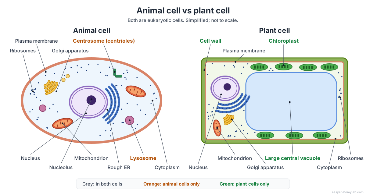
| Feature | Animal cell | Plant cell |
|---|---|---|
| Cell wall | None; an extracellular matrix instead | Yes, mainly cellulose |
| Chloroplasts | No | Yes, in green tissues |
| Large central vacuole | No | Yes, can fill most of the cell |
| Centrosome and centrioles | Yes | Most plant cells lack them |
| Lysosomes | Yes | Most plant cells lack them |
| Shared | Nucleus, plasma membrane, mitochondria, ER, Golgi, ribosomes, peroxisomes, cytoskeleton | Same |
Look inside a chloroplast on our chloroplast structure model, and see plant cells in place in the leaf cross-section.
Prokaryotic vs eukaryotic cells
Prokaryotic cells (bacteria and archaea) have no nucleus and no membrane-bound organelles; their DNA lies in a region called the nucleoid. Eukaryotic cells, including animal and plant cells, have both. (OpenStax Biology 2e, 4.2 Prokaryotic cells.)
| Feature | Prokaryotic cell | Eukaryotic animal cell |
|---|---|---|
| Size | 0.1–5 µm | 10–100 µm |
| Nucleus | None (nucleoid) | Yes, double membrane |
| Membrane-bound organelles | None | ER, Golgi, mitochondria, lysosomes and more |
| Ribosomes | 70S | 80S in the cytosol; 70S inside mitochondria |
| Cell wall | Usually peptidoglycan | None |
Sources: OpenStax Biology 2e and Microbiology. (OpenStax Biology 2e, 4.2 Prokaryotic cells; OpenStax Microbiology, 3.4 Unique characteristics of eukaryotic cells.) Explore a bacterium in 3D on our bacterial cell diagram.
Why are cells so small? Surface area to volume ratio
Cells stay small because of their surface area-to-volume ratio. Everything a cell takes in or gets rid of must cross its plasma membrane, but as a cell grows its volume rises faster than its surface area. The ratio falls, and past a certain size the membrane can no longer pass enough material by diffusion to serve the whole cell. (OpenStax Biology 2e, 4.2 Prokaryotic cells.)
A worked example with spherical cells shows why. Surface area is 4πr² and volume is 4/3πr³, so the ratio is 3 divided by the radius: each tenfold increase in width gives 100 times the surface but 1,000 times the volume to supply. The same rule helps explain the size gap between the two great kinds of cell: prokaryotic cells are only 0.1–5 µm across, while eukaryotic cells, at 10–100 µm, divide their interior into membrane-bound organelles. (OpenStax Biology 2e, 4.2 Prokaryotic cells.)
| Cell diameter | Surface area | Volume | Surface area : volume |
|---|---|---|---|
| 2 µm (bacterium-sized) | 13 µm² | 4.2 µm³ | 3 per µm |
| 20 µm (typical animal cell) | 1,260 µm² | 4,190 µm³ | 0.3 per µm |
| 200 µm | 126,000 µm² | 4,190,000 µm³ | 0.03 per µm |
The 20 µm cell's volume of about 4,000 µm³ sits in the usual 1,000–10,000 µm³ range for mammalian cells. (Milo & Phillips, Cell Biology by the Numbers: How many cells are there in an organism?.) Cells beat the limit in the ways the 3D model shows: they divide, fold their membrane into microvilli, grow thin or long, and build internal membranes as organelles. (OpenStax Biology 2e, 4.2 Prokaryotic cells.) In a pancreatic cell the ER alone has 10–20 times the area of the plasma membrane, and a neuron can be over a metre long yet only about 10 µm wide. (Milo & Phillips, Cell Biology by the Numbers: How big is the endoplasmic reticulum of cells?; Milo & Phillips, Cell Biology by the Numbers: How big is a human cell?.)
Cell anatomy in numbers
| Measure | Value | Sources |
|---|---|---|
| Eukaryotic cell diameter | 10–100 µm | OpenStax Biology 2e 4.2 |
| Mammalian cell volume | About 1,000–10,000 µm³ | Cell Biology by the Numbers |
| Cells in the human body | About 30 trillion (2016); 37 trillion (earlier estimate) | Sender et al. 2016; Cell Biology by the Numbers |
| Nucleus diameter | About 2–10 µm | Cell Biology by the Numbers |
| Nuclear pore | About 120 nm across, about 110 MDa | Dultz et al. 2022; Rush et al. 2023 |
| Mitochondrion | About 2 µm × 1 µm; 1,000–10,000 per cell | Cell Biology by the Numbers; OpenStax A&P 2e 3.2 |
| Lysosome pH | About 4.5–5 (cytosol about 7.2) | Dharmasivam & Kaya 2025; Weiss 2012 |
| Microtubule / actin / intermediate filament | 23–25 nm / 6–7 nm / 8–10 nm | OpenStax Biology 2e and Microbiology; Cell Biology by the Numbers |
Every structure in the 3D cell model
The text version of the 3D model: every labeled part of the cell, grouped by system, with its function. Use it as a study list for a cell labeling worksheet.
Membrane & surface
- Plasma membrane (Cell membrane)
- The thin, flexible boundary of the cell: a double layer of phospholipids with proteins embedded in it. Controls what enters and leaves the cell, receives signals, and holds the cell's identity tags.
- Microvilli
- Many tiny finger-like folds of the plasma membrane on the cell's upper (apical) surface. Increase the surface area of the membrane for absorption.
- Primary cilium
- A single hair-like projection that acts as the cell's antenna. Detects signals from outside the cell and passes them on to signalling pathways inside it.
Nucleus
- Nuclear envelope (Nuclear membrane)
- Two membranes (inner and outer) that wrap the nucleus, with a thin space between them. Separates the DNA from the cytoplasm and, through its pores, controls what moves between them.
- Nuclear pores (Nuclear pore complexes)
- Large ring-shaped protein channels through the nuclear envelope. Let RNA and ribosome subunits out of the nucleus and let selected proteins in.
- Nucleoplasm
- The gel-like fluid inside the nucleus. Holds the chromatin and nucleolus and the building blocks of nucleic acids.
- Chromatin
- The cell's DNA wound around histone proteins, spread through the nucleus as loose threads. Stores the genes. It condenses into visible chromosomes only when the cell divides.
- Nucleolus
- A dense, dark-staining body inside the nucleus. Makes ribosomal RNA and assembles it with proteins into ribosome subunits, which leave through the nuclear pores.
ER, Golgi & vesicles
- Rough endoplasmic reticulum (Rough ER, RER)
- Stacks of flattened membrane sacs (cisternae) studded with ribosomes, wrapped around the nucleus. Makes proteins that go into membranes or out of the cell, folds them and adds sugar chains, then sends them on in vesicles.
- Smooth endoplasmic reticulum (Smooth ER, SER)
- A network of branching membrane tubes with no ribosomes. Makes lipids and steroid hormones, breaks down some drugs and poisons, and stores calcium ions.
- Golgi apparatus (Golgi body, Golgi complex)
- A stack of flattened, curved membrane sacs (cisternae) that looks like a pile of plates. Receives proteins and lipids from the ER, modifies them (mostly by adding sugars), sorts them and ships them out in vesicles.
- Vesicles (Transport and secretory vesicles)
- Small membrane bubbles that carry cargo between organelles and to the cell surface. Move proteins and lipids around the cell; secretory vesicles fuse with the plasma membrane to release their contents (exocytosis).
- Lysosomes
- Small, round sacs of digestive enzymes. Break down worn-out organelles (autophagy), engulfed bacteria and large molecules so their parts can be reused.
Other organelles
- Mitochondria (Singular: mitochondrion)
- Bean-shaped organelles with two membranes; the inner one is folded into cristae. Each is drawn cut open. Make most of the cell's ATP by aerobic cellular respiration, using oxygen.
- Peroxisomes
- Small, round organelles with a single membrane, full of oxidising enzymes. Break down fatty acids and amino acids, detoxify poisons such as alcohol, and destroy the hydrogen peroxide they make.
- Ribosomes
- Tiny particles of RNA and protein, either free in the cytosol or bound to the rough ER. Translate messenger RNA into protein. Free ribosomes make proteins for the cytosol; bound ones make proteins for membranes and export.
- Centrosome and centrioles
- A pair of short cylinders (centrioles) at right angles to each other, the cell's main microtubule-organising centre. Organises the microtubules and helps build the spindle that separates chromosomes when the cell divides. A centriole can also anchor a cilium.
Cytoskeleton
- Microtubules
- Hollow tubes of the protein tubulin, the thickest fibres of the cytoskeleton. Resist compression, act as tracks for vesicles and organelles, and form the spindle, cilia and centrioles.
- Actin filaments (microfilaments)
- The thinnest cytoskeletal fibres, made of two twisted strands of actin. Give the cell its shape, let it change shape and move, and, with myosin, pinch the cell in two during division.
- Intermediate filaments
- Rope-like fibres, thicker than actin but thinner than microtubules. Bear tension, hold the nucleus and organelles in place, and make the cell tough.
Cytosol
- Cytosol (Cytoplasmic fluid)
- The jelly-like fluid that fills the cell around the organelles. Provides the fluid medium for many of the cell's chemical reactions, including protein synthesis on free ribosomes.
Frequently asked questions
What are the main parts of a cell?
Every animal cell has three main parts: the plasma membrane around it, the cytoplasm (the jelly-like cytosol plus the organelles) inside it, and the nucleus, which holds the DNA. The organelles include the endoplasmic reticulum, Golgi apparatus, mitochondria, lysosomes, peroxisomes, ribosomes, centrosome and cytoskeleton.
What is the largest organelle in an animal cell?
The nucleus is usually the largest and most prominent organelle, typically 2–10 µm across. Measured by membrane area, though, the endoplasmic reticulum is often bigger: in liver and pancreas cells it holds up to 60% of all the cell's membrane.
Which organelle is called the powerhouse of the cell?
The mitochondrion. It makes most of the cell's ATP by aerobic respiration on its folded inner membrane (the cristae). A mammalian cell holds roughly 1,000 to 10,000 of them; muscle cells are packed with mitochondria, while a bone cell has only a couple of hundred.
What is the difference between cytoplasm and cytosol?
Cytosol is just the jelly-like fluid inside the cell, mostly water with dissolved ions, sugars and proteins. Cytoplasm is everything between the plasma membrane and the nucleus: the cytosol plus all the organelles and the cytoskeleton suspended in it. Cytoplasm is about 70–80% water.
What does an animal cell have that a plant cell does not?
Animal cells have centrosomes with centrioles and lysosomes, which most plant cells lack. Plant cells instead have a cellulose cell wall, chloroplasts and a large central vacuole, none of which animal cells have. Both have a nucleus, mitochondria, ER, Golgi apparatus, ribosomes and a plasma membrane.
How many cells are in the human body?
About 30 to 37 trillion. A 2016 analysis estimated 30 trillion human cells in a 70 kg adult, most of them red blood cells; an earlier widely quoted estimate is 37 trillion. They belong to more than 200 cell types, which vary a lot in size and shape.
What does the Golgi apparatus do?
The Golgi apparatus is the cell's packaging and shipping centre. Vesicles from the rough ER deliver new proteins and lipids to its cis face; it modifies them, mainly by adding sugar chains, sorts them, and sends them from its trans face in vesicles to the plasma membrane, to lysosomes or out of the cell.
Do all human cells have a nucleus?
No. Mature red blood cells eject their nucleus to make room for haemoglobin, so they have none. Most cells have one nucleus, and skeletal muscle fibres have many. Where it is present, the nucleus is usually the largest organelle and holds the cell's 46 chromosomes, which carry almost 22,000 genes.
References
- OpenStax Biology 2e, 4.2 Prokaryotic cells
- OpenStax Biology 2e, 4.3 Eukaryotic cells
- OpenStax Biology 2e, 4.4 The endomembrane system and proteins
- OpenStax Biology 2e, 4.5 The cytoskeleton
- OpenStax Anatomy and Physiology 2e, 3.2 The cytoplasm and cellular organelles
- OpenStax Anatomy and Physiology 2e, 3.3 The nucleus and DNA replication
- OpenStax Microbiology, 3.4 Unique characteristics of eukaryotic cells
- Milo & Phillips, Cell Biology by the Numbers: How big is a human cell?
- Milo & Phillips, Cell Biology by the Numbers: How big are nuclei?
- Milo & Phillips, Cell Biology by the Numbers: How big are mitochondria?
- Milo & Phillips, Cell Biology by the Numbers: How big is the endoplasmic reticulum of cells?
- Milo & Phillips, Cell Biology by the Numbers: How big are the cell's filaments?
- Milo & Phillips, Cell Biology by the Numbers: How many cells are there in an organism?
- Sender, Fuchs & Milo (2016): Revised estimates for the number of human and bacteria cells in the body, PLOS Biology 14:e1002533
- Dultz, Wojtynek, Medalia & Onischenko (2022): The nuclear pore complex: birth, life, and death of a cellular behemoth, Cells 11:1456
- Rush et al. (2023): Unveiling the complexity: assessing models describing the structure and function of the nuclear pore complex, Frontiers in Cell and Developmental Biology 11:1245939
- Gruenbaum & Aebi (2014): Intermediate filaments: a dynamic network that controls cell mechanics, F1000Prime Reports 6:54
- Bai et al. (2022): Primary cilium in kidney development, function and disease, Frontiers in Endocrinology 13:952055
- Dharmasivam & Kaya (2025): Lysosome as a chemical reactor
- Pearson Channels, Anatomy & Physiology study guide: Cellular anatomy, structure and function of the cell
- Weiss (2012): Cross-talk between TRPML1 channel, lipids and lysosomal storage diseases, Communicative & Integrative Biology 5:111
More interactive anatomy models
- Cellular respiration diagramGlycolysis, Krebs cycle and electron transport chain, with a 3D mitochondrion.
- Neuron anatomyThe parts of a neuron in 3D: dendrites, cell body, axon, myelin and synapse.
- Human anatomy1,000+ bones, muscles, organs, vessels and nerves from real anatomical data.
- Octopus anatomyThree hearts, a ring-shaped brain, eight arms and a beak, in 3D.
- Squid anatomyPen, eight arms, two tentacles, giant axons and three hearts of the longfin squid.
- Squid dissectionStep-by-step guided squid dissection with every structure labeled.