

Deer anatomy at a glance
The white-tailed deer is a member of the deer family, the Cervidae, which in North America also includes elk, moose, caribou and mule deer. The Pennsylvania Game Commission sums up the family: cervids are split-hoofed mammals with no incisor teeth in the front of the upper jaw, and they are ruminants, with a four-chambered stomach, that chew a cud. Adult bucks grow and shed a set of antlers every year.[3]
Size varies more than most people expect. The figures below come from wildlife agencies and university accounts. Texas deer are much smaller than northern ones (The Mammals of Texas gives 30–70 kg for males), and a buck may weigh 25–30% more at the start of the rut than at its end.[2, 3]
| Measurement | Typical values | Source |
|---|---|---|
| Shoulder height | 80–100 cm; average Pennsylvania buck 32–34 in (81–86 cm); bucks 60–105 cm range-wide | Animal Diversity Web; Pennsylvania Game Commission; Handbook of the Mammals of the World[1, 3, 5] |
| Chest girth | About 38 in (97 cm) for a 157 lb deer and 39 in (99 cm) for 169 lb, measured just behind the front legs | Pennsylvania Game Commission weight chart[4] |
| Head and body length | 150–200 cm; about 70 in (178 cm) nose to tail base in Pennsylvania | Animal Diversity Web; Pennsylvania Game Commission[1, 3] |
| Weight | 57–137 kg range-wide; Missouri males 155–195 lb, females 110–145 lb; Texas Hill Country males about 50–59 kg | Animal Diversity Web; Missouri Department of Conservation; Luna & Weckerly 2013[1, 6, 42] |
| Tail | 10–28 cm; about 11 in of vertebrae | Animal Diversity Web; Pennsylvania Game Commission[1, 3] |
| Teeth | 32: I 0/3, C 0/1, P 3/3, M 3/3 | The Mammals of Texas[2] |
| Speed and jumping | Up to 40 mph in bursts, 25 mph sustained; clears 9 ft high or 25 ft wide | Pennsylvania Game Commission[3] |
Directions used on this page: cranial means towards the head, caudal towards the tail, dorsal the back and ventral the belly. Left and right are the deer's own. The model opens on the deer's left side, the view used in most diagrams and the broadside view hunters see.
Parts of a deer: external anatomy
The outside of a white-tailed deer is brown above and white below. The coat is reddish brown in summer and greyish brown in winter; the belly, throat, a band behind the black nose, rings round the eyes, the insides of the ears and the underside of the tail stay white all year. When it flees, a whitetail raises its tail and shows the white underside as a flag.[3, 6, 1]
| Part | Where it is | What to know |
|---|---|---|
| Antlers and pedicles | Top of the skull | Bone, grown and cast every year from two permanent pedicles[36, 1] |
| Ears | Either side of the poll | Large, around half the length of the face; smaller than a mule deer's[5, 2] |
| Eyes | Sides of the head | Especially good at detecting motion[3] |
| Nose | Tip of the narrow snout | Narrow snout with a dark nose and a white band behind it[5, 14] |
| Tail | Over the rump | Brown above, white below, raised as an alarm flag[3, 5] |
| Hooves | Two per foot (toes III and IV) | Leave a heart-shaped, pointed track[6] |
| Dewclaws | Back of each foot | Toes II and V; print two dots when the deer bounds[6, 39] |
| Scent glands | Face, legs and feet | Preorbital, forehead, tarsal, metatarsal and interdigital[3, 34] |
Where is a deer's heart? The vital organs
A deer's heart lies low in the chest, just above the breastbone and inside the rib cage, with most of it directly above the front leg rather than behind it. The lungs surround the heart and fill the chest from the belly side up to the spine. Together the heart and lungs form the vital area, behind the shoulder and about a third of the way up the body.[24, 25, 9]
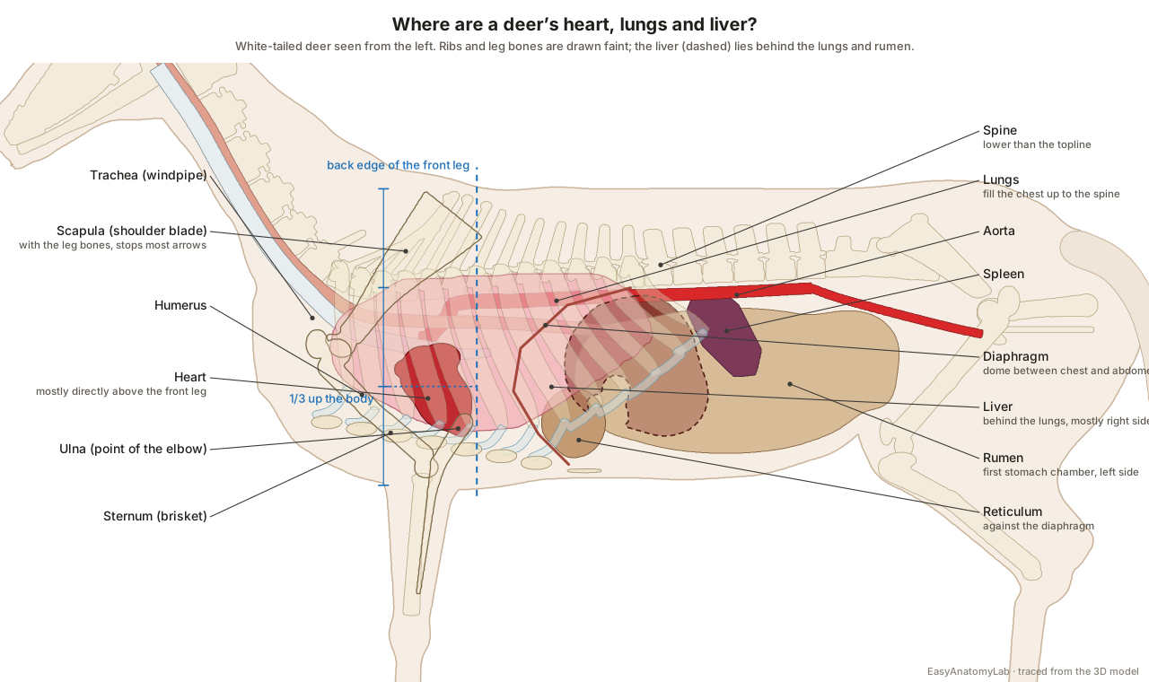
Texas Parks and Wildlife's hunter-education course describes the broadside view as the largest target for the heart and lungs: look at the front leg facing you, follow a line up its back edge and come up one third of the way into the body. The National Deer Association's anatomy diagrams make three points that surprise many hunters. Most of the heart sits directly above the leg, not behind it. The backbone does not run along the top of the body; in the chest it sits low and dips between the lungs. And there is no empty space between the lungs and the spine: the lungs fill the arch of the ribs all the way up. A high shot can pass above the backbone through muscle with no vital organs.[9, 24, 25]
| Organ | Position in a broadside deer | Notes |
|---|---|---|
| Heart | Low in the chest, mostly directly above the front leg | Apex formed by the left ventricle[24, 20] |
| Lungs | Fill the chest from the floor up to the spine, behind the shoulder | The largest part of the vital area[25, 9] |
| Liver | Just behind the lungs and diaphragm, under the last ribs, mostly on the right | Tucked between the lungs and the rumen[27, 19] |
| Diaphragm | Thin dome of muscle from the inside of the last ribs | Separates the chest from the abdomen[28] |
| Rumen and intestines | Behind the diaphragm, filling the abdomen | A shot here is a "gut shot"[9] |
| Aorta and venae cavae | Along the underside of the spine and through the chest | The great vessels of the mediastinum[20] |
| Shoulder blade and leg bones | In front of and below the vital area | Will stop most arrows[26] |
For this reason the National Deer Association recommends only the heart–lung area as a target, ideally with the deer broadside or slightly quartering away, and warns bowhunters off the shoulder blade and leg bones beside it. The liver is tucked behind the lungs, so a shot slightly too far back often hits it: lethal, but the deer may take longer to die. Further back still is the rumen. Select Heart, Lungs or Liver in the viewer to see each one in place with the skin removed.[26, 32, 27]
Deer internal organs
Inside a deer, the chest holds the heart and lungs, and the diaphragm separates them from the abdomen. The left side of the abdomen is filled by the rumen and reticulum; the liver, omasum, abomasum and intestines lie to the right. The kidneys are wrapped in fat that biologists weigh to judge a deer's condition; the bladder and the end of the large intestine lie in the pelvis, reached when hunters cut through the pelvic bone; and the tenderloins run under the spine between the last ribs and the pelvis.[19, 30, 28, 29]
The University of Minnesota's ruminant dissection guide puts it plainly: the ruminoreticulum occupies the entire left side of the abdominal cavity, while the liver, omasum and abomasum lie to the right of its cranial part, with the liver most dorsal, between the diaphragm and the omasum. Turn the model to the right side to see the intestines, which the rumen hides from the left.[19]
How many stomachs does a deer have?
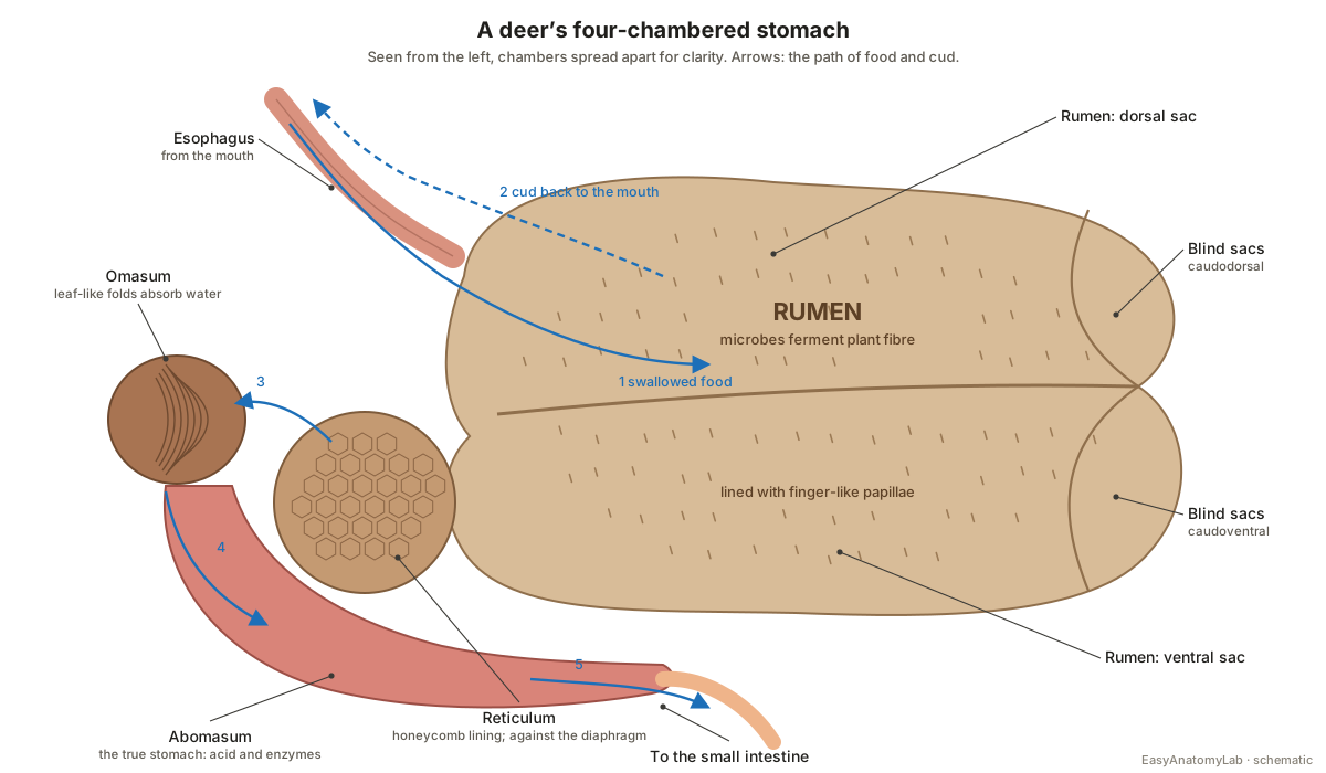
A deer has one stomach with four chambers: the rumen, reticulum, omasum and abomasum. Food is swallowed after little chewing, fermented by microbes in the rumen and reticulum, brought back up as cud and chewed again, then passed on through the omasum to the abomasum, the true glandular stomach, and into the small intestine.[16, 18]
| Chamber | Nickname | Job |
|---|---|---|
| Rumen | Paunch | Largest chamber; microbial fermentation of plant fibre; divided into dorsal, ventral and two blind sacs[18, 16] |
| Reticulum | Honeycomb | Sorts particles; traps heavy objects; lies against the diaphragm, near the heart[16, 19] |
| Omasum | Manyplies | Leaf-like folds absorb water and nutrients[16] |
| Abomasum | True stomach | Acid and enzymes (such as pepsin) digest the food; in one farmed deer it was blocked by about 5 kg of sand[16, 38] |
Whitetails are browsers, which biologists call concentrate selectors: they pick the most digestible leaves, buds, fruits and nuts rather than mature grass. Mississippi State University's Deer Lab explains that their relatively smaller and less complex digestive tract needs higher-quality food than cattle, elk or moose eat; deer have starved with their stomachs full of poor forage. Their saliva neutralises tannins, which lets them eat amounts of acorns that would kill a cow. Rumen size is not fixed either: in Texas, rumen-reticulum capacity changed from year to year with rainfall and forage quality.[18, 14, 42] For a full account of how ruminant digestion works, see our cow digestive system page.
Do deer have a gallbladder?
No. White-tailed deer have no gallbladder, as Penn State's deer research team and the Pennsylvania Game Commission point out; in their quiz, 80% of people got this wrong. Bile from the liver flows straight into the duodenum. A study of the marsh deer of South America likewise found that the missing gallbladder is the main difference between its liver and other ruminants' livers, which do have one.[21, 22, 23]
Deer skeleton
The deer skeleton is light and built for speed, with long cannon bones and slim legs. It has a long skull carrying the antler pedicles, a neck of seven cervical vertebrae, thirteen pairs of ribs, six loin vertebrae, a sacrum, the tail vertebrae and four long, slender legs.[36, 40, 41]
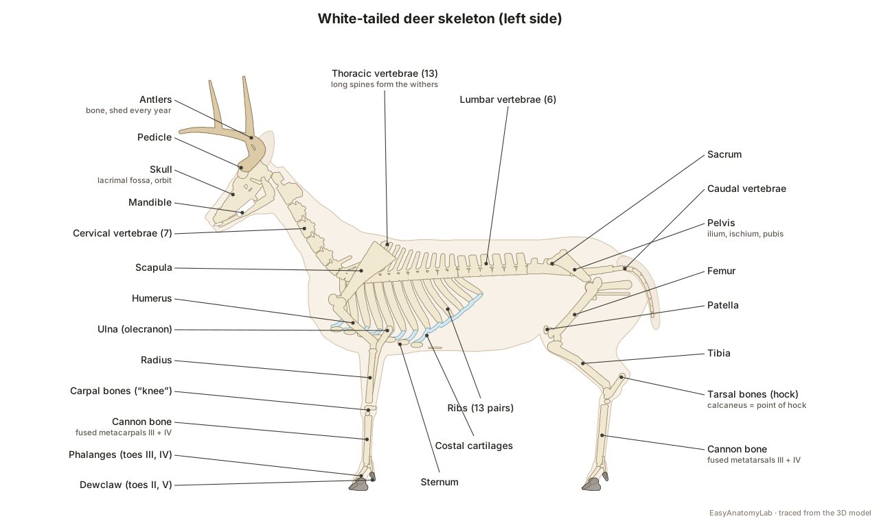
Several features mark it as a deer. The skull has a lacrimal fossa, the pit that holds the preorbital gland, a preorbital vacuity (a gap in the bones in front of each eye) and two lacrimal foramina, which taxonomists list among the diagnostic characters of the family. There are no upper incisors or canines. Each cannon bone is the fused third and fourth metapodial, and the deer stands on two toes. White-tailed deer belong to the telemetacarpal deer, in which only the lower ends of the side metacarpals survive, as splinters of bone by the dewclaws.[35, 3, 39]
Vertebral counts: we found no open-access count made on white-tailed deer, so the model follows two other deer species studied in detail, the spotted deer (13 thoracic, 6 lumbar, 4 sacral and 12 caudal vertebrae) and the Calamian deer (7 cervical, 13 thoracic, 6 lumbar and 5 sacral).[40, 41]
Deer teeth and aging by the jaw
A white-tailed deer has 32 teeth: on each side, three incisors and one incisor-shaped canine in the lower jaw only, three premolars and three molars above and below. A hard dental pad replaces the upper front teeth. Wildlife biologists age deer from the replacement and wear of the lower cheek teeth: fawns have five or fewer, yearlings have all six with a three-cusped milk premolar that is then replaced by a two-cusped permanent one, and wear exposes more dark dentine every year. The teeth usually wear out in the eighth or ninth year.[2, 16, 8, 31, 10, 7]
Antler anatomy: pedicle, burr, beam and tines
Antlers are bone. Each grows from a pedicle, a permanent outgrowth of the frontal bone, and starts at the burr (coronet), the rough ring at its base. The main beam sweeps back, out and forward, and the tines rise from its top, the first being the brow tine. In whitetails all major points come off the main beam, and the brow tines are more prominent than in mule deer.[36, 11, 2]
| Stage | Timing in white-tailed deer | What happens |
|---|---|---|
| Growth in velvet | From April or May (Missouri: April to August) | Grows from the tip under velvet, a hairy skin full of blood vessels[1, 6, 3] |
| Hardening | Mid-July to mid-August | Rising testosterone mineralises the antler from base to tip[3] |
| Velvet shed | August or September | Velvet dies and is rubbed off on vegetation, sometimes within 24 hours[1, 11] |
| Casting | January to March (Texas: late January–February) | Bone-dissolving cells cut it free at the pedicle[1, 7, 11] |
Growth is fast. Mississippi State's Deer Lab measured up to 3/4 inch a week in yearlings and 1.5 inches a week in adults at peak growth. The Pennsylvania Game Commission calls antler the fastest-growing tissue known, with the capacity to grow an inch or more a day, and a 2023 review gives up to 2 cm a day for antlers in general; those daily figures are not specific to white-tailed deer. A buck's first antlers are 10–25% of his eventual maximum, which takes about six years to reach, and calcium and phosphorus for them are borrowed from the ribs and other bones. Antlers are mainly weapons for contests between bucks, not defences against predators.[11, 3, 37, 13, 12]
Deer muscles: backstrap and tenderloin
The muscles hunters know best are the backstraps and the tenderloins. The backstrap is the longissimus dorsi, the largest of the erector spinae muscles, lying on either side of the spine on top of the ribs from the shoulder blades to the hindquarters. The tenderloins are the psoas muscles inside the abdomen, under the spine between the last ribs and the pelvis, close to the intestines and rectum.[29, 17, 28] To see the superficial muscles, hide External body and set the skin slider to 100%: the triceps behind the shoulder, the latissimus over the ribs and the biceps femoris and semitendinosus of the hindquarter appear.
Deer scent glands
White-tailed deer communicate by smell through several skin glands, and their secretions become strongest in the rut.[1, 3]
| Gland | Location | Use |
|---|---|---|
| Preorbital | Below the inner corner of each eye | Marks the overhanging branch above a scrape[3] |
| Forehead | Skin of the forehead | Rubbed on trees and branches; thicker skin in bucks[6, 34] |
| Tarsal | Inside of each hock (hair tuft) | Scent of excitement or fear; left on a carcass, it does not taint the meat[3, 33] |
| Metatarsal | Outside of each hind leg, hock to hoof | Small and round in whitetails, larger in mule deer[2, 34] |
| Interdigital | Between the hooves of each foot | A scent trail other deer can follow[3] |
Male vs female deer anatomy
Bucks and does differ mainly in antlers and reproductive organs. Bucks grow antlers, which only rarely appear on does, and their testes in the scrotum make the testosterone that times both the rut and the antler cycle. Does have two ovaries, a uterus with two horns and an udder between the hind legs. Gestation lasts about 200 days (given as six and a half to seven months), and adult does usually have twins.[3, 2, 6, 1, 7]
| Buck | Doe | |
|---|---|---|
| Antlers | Grown and cast every year | Rarely present |
| Gonads | Testes in the scrotum | Ovaries near the pelvic inlet |
| Other organs | Penis in a sheath under the belly | Uterus with two horns, vagina, udder |
| Breeding | Can breed from velvet shed to antler cast[15] | In heat for about a day, again about 28 days later if not bred[1] |
Switch the viewer between Male and Female to see each set of organs in place.
White-tailed deer vs mule deer anatomy
The two North American Odocoileus species share the same organs and skeleton but differ in a few external features. White-tailed deer have relatively short ears, a broad tail that is white underneath, antlers whose points all rise from the main beam with prominent brow tines, and small round metatarsal glands. Mule deer antlers fork (branch in two) and their metatarsal glands and preorbital gland sacs are larger.[2, 11, 34]
Deer anatomy labeled: every structure in the model
This is the text version of the 3D model: all 90 labeled structures, grouped by body system. Click any name in the panel beside the model to find it in 3D.
External body
- Skin and coat (Hide, pelage)
- The hide and its hair: reddish brown in summer, greyish brown in winter, with white on the belly, throat, eye rings, nose band, insides of the ears and legs and under the tail. Insulation, camouflage and signalling; the white underside of the raised tail is a visible alarm flag.
- Nose and muzzle (Rhinarium)
- The black, moist, hairless nose pad at the tip of the narrow snout, with a white band behind it. Smell is a deer's main early-warning sense; a narrow snout and long tongue let whitetails pick out particular plant parts.
- Ears (Pinnae)
- Two large, mobile, oval ears, white inside, held up and out behind the antlers; in the model about 15 cm long, roughly half the length of the head. Catch and locate sound; whitetails rely on keen hearing and smell to detect danger.
- Eyes
- Large dark eyes set on the sides of the head, each framed by a ring of white hair. A wide field of view for spotting predators; the eyes are especially good motion detectors.
- Antlers · males only
- Branched bones grown from the pedicles of the frontal bones: a main beam on each side with tines (points) rising from its top, the first being the brow tine. Mainly weapons and displays in contests between bucks for dominance; antler size may also signal a buck's quality to does.
- Tail
- A broad, flat tail, brown above and pure white below, held down at rest and raised as a 'flag' when the deer runs. Signalling: the raised white underside warns other deer and helps fawns follow their mother.
- Hooves (Cloven hoof, digits III and IV)
- Each foot stands on two pointed hooves, the horny claws of the third and fourth toes, which leave a heart-shaped track. Carry the body's weight; deer are built to run in short bursts and jump.
- Dewclaws (Digits II and V)
- Two small horny claws at the back of each foot, above the hooves: the reduced second and fifth toes. Touch the ground only on soft ground or when the deer bounds, leaving two dots behind each track.
- Scrotum · males only
- The pouch of skin between the hind legs that holds the testes. Holds the testes outside the body cavity.
- Udder (Mammary glands) · females only
- The mammary glands between the hind legs, with teats, that feed the fawns. Produces milk; fawns are nursed for 8 to 10 weeks.
Scent glands
- Preorbital gland (Lacrimal or tear-duct gland)
- A small pocket of skin in front of the inner corner of each eye, lying in a pit of the skull (the lacrimal fossa). Bucks rub it on the overhanging 'licking branch' above a scrape to leave their scent.
- Forehead glands (Sudoriferous glands of the forehead)
- Scent-producing skin glands across the forehead between the eyes and the antler bases. Bucks rub them on trees and branches when making rubs and scrapes.
- Tarsal glands
- Large tufts of hair over scent glands on the inside of each hind leg at the hock. Release scents that convey excitement or fear; their odour is strongest during the rut.
- Metatarsal glands
- A small, round gland ringed with white hair on the outside of each hind leg between the hock and the hoof. Releases scent conveying excitement or fear.
- Interdigital glands
- A gland between the two hooves of each foot that opens into the cleft between the toes. Leaves a scent trail with every step, which lets deer follow each other by smell.
Skeleton
- Skull (Cranium and facial bones)
- A long, narrow skull with complete bony rings round the orbits, a pit and a gap in front of each orbit, and no teeth at the front of the upper jaw. Protects the brain and eyes, carries the antler pedicles and anchors the jaws and chewing muscles.
- Pedicles
- Two short, permanent outgrowths of the frontal bones, covered with ordinary scalp skin, from which the antlers grow. The base of the antlers; the antler is cast at the junction with the pedicle and regrows from it.
- Mandible (Lower jaw)
- The two halves of the lower jaw, joined at the chin, carrying the lower incisors and the cheek teeth. Chewing and cud chewing (rumination).
- Cervical vertebrae (Neck vertebrae (C1–C7), including the atlas and axis)
- The seven vertebrae of the long neck: the ring-shaped atlas, the long axis with its peg (dens), and five more. Support and move the head; the neck vertebrae curve down to the base of the neck, well below the crest.
- Thoracic vertebrae (Back vertebrae (T1–T13))
- The thoracic vertebrae, each carrying a pair of ribs; the long spines of the first few form the withers. Carry the ribs and anchor the back muscles.
- Lumbar vertebrae (Loin vertebrae (L1–L6))
- Six loin vertebrae with long, flat transverse processes that stick out sideways. Support the abdomen; the backstraps lie above the transverse processes and the tenderloins below them.
- Sacrum
- Fused vertebrae that join the spine to the pelvis. Transfers the thrust of the hind legs to the spine.
- Caudal vertebrae (Tail vertebrae)
- A chain of small tail vertebrae, simple rods towards the tip. Support and move the tail.
- Ribs
- Thirteen pairs of curved, flattened ribs forming the rib cage round the heart and lungs. Protect the heart, lungs and liver and move with the diaphragm in breathing.
- Costal cartilages
- Bars of cartilage that continue the ribs to the breastbone; those of the last ribs join one another to form the costal arch. Make the chest wall flexible for breathing.
- Sternum (Breastbone, brisket)
- The breastbone along the floor of the chest, made of several segments (sternebrae). Anchors the ribs' cartilages and the chest muscles; the heart's apex lies just above it.
- Scapula (Shoulder blade)
- A long triangular shoulder blade with a ridge (spine) on its outer face, lying flat on the chest wall. Anchors the shoulder muscles and forms the shoulder joint with the humerus.
- Humerus (Upper arm bone)
- The upper bone of the front leg, from the shoulder joint down and back to the elbow. Lever for the shoulder and elbow muscles.
- Radius (Forearm bone)
- The main bone of the forearm, carrying the weight from the elbow to the carpus. Weight-bearing bone of the forearm.
- Ulna
- A slender bone behind the radius whose upper end, the olecranon, forms the point of the elbow. Lever for the triceps, which straightens the elbow.
- Carpal bones (Carpus, the 'knee' of the front leg)
- Two rows of small bones between the forearm and the cannon bone. A flexible joint equivalent to the human wrist.
- Cannon bones (Metacarpus and metatarsus III–IV)
- The long lower-leg bones, each formed by the fusion of the third and fourth metacarpals (front) or metatarsals (hind). Long, light levers that give the deer its speed and jumping power.
- Phalanges (Toe bones and sesamoids)
- The three bones of each of the two main toes, the last one inside the hoof, with small sesamoid bones behind the fetlock and the tiny bones of the dewclaws. Carry the hooves; deer walk on the tips of their third and fourth toes.
- Pelvis (Hip bones: ilium, ischium and pubis)
- The two hip bones, joined to the sacrum above and to each other below, forming the bony ring of the pelvic canal. Carries the hip joints and protects the bladder, rectum and reproductive tract.
- Femur (Thigh bone)
- The thigh bone, from the hip joint down and forward to the stifle. Lever for the powerful thigh muscles.
- Patella (Kneecap)
- The kneecap at the front of the stifle, the true knee of the hind leg. Lets the thigh muscles straighten the stifle.
- Tibia (Shin bone)
- The main bone of the lower hind leg, from the stifle down and back to the hock. Weight-bearing bone of the gaskin.
- Tarsal bones (Hock, including the calcaneus)
- The bones of the hock, the ankle of the hind leg; the calcaneus forms its backward-pointing point. The calf muscles pull on the calcaneus to straighten the hock and push the deer forward.
Muscles
- Masseter
- The large cheek muscle. Closes the jaws for chewing and cud chewing.
- Brachiocephalicus
- A long strap muscle down the side of the neck from the head to the upper arm. Pulls the front leg forward and turns the head and neck.
- Sternocephalicus
- A strap muscle along the underside of the neck from the breastbone to the head. Lowers the head and flexes the neck.
- Trapezius
- A thin triangular sheet from the top of the neck and withers to the shoulder blade's spine. Lifts and swings the shoulder blade.
- Deltoideus
- The shoulder muscle over the outside of the shoulder joint. Flexes the shoulder joint.
- Triceps brachii
- The thick muscle mass filling the angle between the shoulder blade and the elbow. Straightens the elbow, the main engine of the front leg.
- Latissimus dorsi
- A broad fan-shaped muscle from the back to the upper arm. Pulls the front leg backwards.
- Pectoral muscles (Brisket)
- The chest muscles under the breastbone between the front legs. Hold the front legs against the body and swing them.
- Longissimus dorsi (Backstrap, loin)
- The long muscle on each side of the spine, resting on the ribs and loin vertebrae: the 'backstrap' of venison. Extends and stiffens the back.
- External abdominal oblique
- The outer sheet of the belly wall, from the ribs to the flank and belly line. Supports the rumen and intestines and flexes the trunk.
- Gluteal muscles
- The muscles of the croup, from the hip bones and sacrum to the thigh bone. Extend the hip to drive the deer forward.
- Biceps femoris
- The large muscle on the outside of the thigh. Extends the hip and helps flex the stifle.
- Semitendinosus
- One of the hamstring muscles along the back of the thigh. Extends the hip and hock in running and jumping.
- Gastrocnemius
- The calf muscle, ending in the tendon on the point of the hock. Straightens the hock to push off.
- Forearm extensors (Extensor carpi radialis and digital extensors)
- Muscles on the front of the forearm whose tendons run down over the 'knee' to the foot. Straighten the carpus and toes.
- Psoas muscles (Tenderloins)
- Muscles under the loin vertebrae inside the abdominal cavity: the 'tenderloins' of venison. Flex the hip and stabilise the loin.
Digestive (four-chambered stomach)
- Lower incisors (Incisors and incisor-like canines)
- Eight chisel-shaped front teeth in the lower jaw: three incisors and one incisor-shaped canine on each side. Bite and snip leaves and twigs against the dental pad.
- Cheek teeth (Premolars and molars)
- Three premolars and three molars in each row, upper and lower, with ridged grinding surfaces. Grind plant food, again and again during rumination.
- Dental pad
- A tough, hard pad of gum in place of upper front teeth. The lower incisors cut against it.
- Tongue
- A long, mobile tongue. Picks and moves food; with the narrow snout it lets deer select particular plant parts.
- Salivary glands (Parotid gland shown)
- Glands that pour saliva into the mouth; the parotid lies below the ear behind the jaw. Saliva buffers the rumen and, in deer, helps neutralise plant tannins.
- Esophagus (Gullet)
- The muscular tube from the throat down the neck, through the chest above the heart, to the rumen. Carries swallowed food down and, in rumination, brings the cud back up.
- Rumen (Paunch, first stomach chamber)
- The largest stomach chamber: a fermentation vat of microbes, divided into sacs, filling the left side of the abdomen. Microbes break down plant fibre into volatile fatty acids the deer absorbs for energy.
- Reticulum (Honeycomb, second chamber)
- A small chamber at the front of the rumen with a honeycomb lining. Works with the rumen as one unit, sorting particles and trapping heavy objects.
- Omasum (Manyplies, third chamber)
- A round chamber packed with leaf-like folds, to the right of the reticulum. Absorbs water and nutrients before food reaches the true stomach.
- Abomasum (True stomach, fourth chamber)
- The glandular 'true stomach', lying along the floor of the abdomen on the right. Digests food with acid and enzymes, like a human stomach, before it enters the small intestine.
- Small intestine (Duodenum, jejunum and ileum)
- The long, coiled tube where most nutrients are absorbed; the duodenum leaves the abomasum and the jejunum hangs in loops on the right side. Digestion and absorption of nutrients.
- Caecum (Cecum)
- A blind-ended pouch where the small intestine meets the colon. Receives what is left after the small intestine; in ruminants it does far less than in horses.
- Colon and rectum (Large intestine, including the spiral colon)
- The large intestine: a flat spiral coil on the right side, then the descending colon under the spine, the rectum and the anus. Absorbs water and forms the pellets.
- Liver
- The large dark-red organ against the diaphragm, mostly on the right, just behind the lungs. There is no gallbladder. Processes absorbed nutrients, makes bile and stores energy.
- Pancreas
- A small gland along the duodenum. Makes digestive juices that enter the duodenum.
Respiratory
- Larynx (Voice box)
- The cartilage box at the top of the windpipe, behind the jaw. Guards the airway and makes calls such as bleats and grunts.
- Trachea (Windpipe)
- The ringed air tube down the underside of the neck into the chest, where it divides into the bronchi. Carries air to the lungs.
- Lungs
- Two large lungs filling the chest from the spine down round the heart. Gas exchange.
- Diaphragm
- The thin dome-shaped wall of muscle between the chest and the abdomen. Its contractions, with the ribs, draw air into the lungs.
Heart & vessels
- Heart
- A four-chambered heart low in the chest, its point (apex) just above the breastbone. Pumps blood through the lungs and the body.
- Aorta
- The main artery: it arches up from the heart and runs back under the spine to the pelvis. Carries oxygen-rich blood to the body.
- Pulmonary trunk (Pulmonary arteries)
- The vessel from the right ventricle that divides to both lungs. Carries oxygen-poor blood to the lungs.
- Caudal vena cava
- The great vein returning blood from the hindquarters and abdomen through the liver to the heart. Returns blood to the heart.
- Jugular veins (With the cranial vena cava)
- Large veins down each side of the neck in the jugular groove, joining the cranial vena cava at the chest. Drain blood from the head.
- Carotid arteries
- Arteries running up the neck beside the windpipe to the head. Supply the brain and head with blood.
- Spleen
- A flat, oval, purplish organ on the left side of the rumen. Filters blood and stores blood cells; part of the immune system.
Urinary
- Kidneys
- Two bean-shaped kidneys high in the abdomen under the loin, wrapped in fat. Filter blood and make urine.
- Ureters
- Thin tubes from each kidney to the bladder. Carry urine to the bladder.
- Urinary bladder
- The bladder at the entrance to the pelvis. Stores urine.
Reproductive
- Testes (With the ductus deferens) · males only
- The two testes in the scrotum, with the sperm ducts that run up into the pelvis. Make sperm and testosterone, which also times the antler cycle.
- Penis · males only
- The penis runs from the pelvis forward under the belly inside the sheath (prepuce). Mating and urination.
- Ovaries · females only
- Two small ovaries near the entrance to the pelvis. Release eggs and make the hormones that time estrus.
- Uterus (With the cervix) · females only
- A uterus with two horns, where the fawns develop. Carries the fawns through pregnancy.
- Vagina and vulva · females only
- The birth canal from the cervix to the vulva below the tail. Mating and birth.
Brain & spinal cord
- Brain
- The brain in the braincase: cerebrum, cerebellum and brainstem. Controls the body; the pineal gland at its base measures day length and times the rut.
- Spinal cord
- The cord of nerves running from the brain through the vertebral canal. Carries signals between the brain and the body.
Frequently asked questions
Where is a deer's heart located?
Low in the chest, just above the breastbone and medial to the elbow. In a deer standing broadside, most of the heart lies directly above the front leg rather than behind it. The lungs surround it and fill the chest up to the spine, so the heart–lung area forms the vital zone behind the shoulder, about a third of the way up the body.
Do deer have a gallbladder?
No. White-tailed deer have no gallbladder; Penn State's deer researchers and the Pennsylvania Game Commission both point this out. Bile from the liver flows straight into the small intestine instead of being stored. A study of the marsh deer of South America also found the missing gallbladder to be the main way its liver differs from other ruminants'.
How many stomachs does a deer have?
One stomach with four chambers: the rumen, reticulum, omasum and abomasum. Food is fermented by microbes in the rumen and reticulum, the cud is brought back up and chewed again, and the omasum and abomasum, the true glandular stomach, finish the job. Whitetails are browsers, or concentrate selectors, with a relatively small, fast digestive tract.
Are deer antlers bone?
Yes. Antlers are bone grown from two permanent pedicles on the frontal bones. They grow in velvet, a skin with many blood vessels, harden as testosterone rises in late summer, and are cast in late winter when it falls. White-tailed bucks regrow a new set every year, usually larger until about six years old.
How fast do deer antlers grow?
Mississippi State's Deer Lab gives up to about 3/4 inch a week for yearlings and 1.5 inches a week for adult whitetails at peak growth. The Pennsylvania Game Commission calls antler the fastest-growing tissue known, able to grow an inch or more a day, and a 2023 review gives up to 2 cm a day for antlers in general; the whitetail-specific figure is the weekly one.
Do deer have top front teeth?
No. Deer have no upper incisors or upper canines. A hard dental pad takes their place, and the eight lower front teeth cut against it. The dental formula of the white-tailed deer is I 0/3, C 0/1, P 3/3, M 3/3, 32 teeth in all, and its age is read from the cheek teeth of the lower jaw.
Where is the liver on a deer?
Just behind the lungs and diaphragm, under the last ribs, mostly on the deer's right side, tucked between the lungs and the rumen. A shot that lands slightly behind the heart–lung area often hits the liver. The National Deer Association calls a liver hit lethal, but the deer may take longer to die than after a heart or lung hit.
What are the glands on a deer's legs?
The tarsal glands are tufts of hair on the inside of each hock, the metatarsal glands are small round patches on the outside of the hind legs between hock and hoof, and an interdigital gland sits between the hooves of each foot. With the preorbital and forehead glands, they carry scent messages, strongest during the rut.
References
Every source below was opened and checked between 24 and 25 October 2026.
- Animal Diversity Web (University of Michigan Museum of Zoology): Odocoileus virginianus, white-tailed deer
- Schmidly & Bradley, The Mammals of Texas (7th ed.), Texas Tech University NSRL online edition: Odocoileus virginianus
- Pennsylvania Game Commission: White-tailed deer (Wildlife notes)
- Pennsylvania Game Commission: Deer weight estimating chart (chest girth to live weight; Penn State and PGC data)
- Wilson, D. E. & Mittermeier, R. A. (eds) (2011). Handbook of the Mammals of the World, vol. 2, Hoofed Mammals: Odocoileus virginianus (Cervidae account), via Plazi
- Missouri Department of Conservation field guide: White-tailed deer
- Cook, R. L. Learn About Whitetails (PWD-LF-W7000-0007). Texas Parks and Wildlife Department
- Texas Parks and Wildlife Department: A Guide to Age Determination of White-tailed Deer (PWD BK W7000-755)
- Texas Parks and Wildlife Department Hunter Education: Shots to the vital areas
- New York State DEC: Aging white-tailed deer (field guide)
- Mississippi State University Deer Ecology & Management Lab: Antler growth cycle
- Mississippi State University Deer Lab: Why antlers?
- Mississippi State University Deer Lab: Nutrition and genetics (antler development)
- Mississippi State University Deer Lab: Deer diet
- Mississippi State University Deer Lab: Biology of the rut
- Mississippi State University Extension P2503: Understanding the Ruminant Animal Digestive System
- Williams, Crist & Strickland. Deer Processing to Minimize CWD Risks (P3305). Mississippi State University Extension
- Folks, J. (2024). Food for Thought: A Primer of Deer Diets, Part 1. Virginia Department of Wildlife Resources
- University of Minnesota CVM, Ungulate Dissection Lab 14: Ruminant, camelid, pig and rabbit viscera
- University of Minnesota CVM, Ungulate Dissection Lab 10: Internal thoracic dissection
- Fleegle, J. (2016). Fun Fact Friday: white-tailed deer do not have gallbladders. Penn State Deer Research (PGC)
- Penn State Deer Research: Deer quiz answers
- Borges, E. M. et al. (2002). Morphological aspects of the liver of the marsh deer (Blastocerus dichotomus). Braz J Vet Res Anim Sci 39(2): 78–80
- Adams, K. (2018). Improved shot placement for enhanced hunting success. National Deer Association
- Thomas, L. Jr. (2024). Is there an empty space between a deer's lungs and spine? National Deer Association
- Forester, H. (2017). White-tailed deer shot placement (video with K. Adams). National Deer Association
- Juhasz, J. (2016). Five rules for recovering liver-shot deer. National Deer Association
- Adams, K. (2013). How to field-dress a deer. National Deer Association
- Westfall, B. (2022). Venison backstrap inside and out. National Deer Association
- Adams, K. (2008). Tracking deer health with kidney fat. Quality Whitetails (QDMA)
- QDMA: Jawbone aging chart (tooth replacement in white-tailed deer)
- Goodwin, M. (2013). Deer hunting: getting started. Missouri Conservationist
- Cutter, C. N. Proper field dressing and handling of wild game and fish. Penn State Extension
- Ness, A. et al. (2022). Cellular prion protein distribution in the vomeronasal organ, parotid, and scent glands of white-tailed deer and mule deer. Prion 16: 40–57 (PMC9154781)
- Heckeberg, N. S. (2020). The systematics of the Cervidae: a total evidence approach. PeerJ 8: e8114 (PMC7034380)
- Kierdorf, U., Kierdorf, H. & Landete-Castillejos, T. (2026). Views on antlers then and now: past and present concepts of their development and histogenesis. Animals 16: 3092 (PMC13663431)
- Li, C. et al. (2023). Deer antler renewal gives insights into mammalian epimorphic regeneration. Cell Regeneration 12: 26 (PMC10368610)
- Rahmani Shahraki, A. et al. (2024). A case of severe abomasal sand impaction in a farmed white-tailed deer in Florida. Animals 14: 1602 (PMC11171194)
- Huffman, B. Ultimate Ungulate: Subfamily Capreolinae (telemetacarpal deer)
- Rajani, C. V. et al. (2013). Morphological studies on the thoracic, lumbar, sacral and coccygeal vertebrae in spotted deer (Axis axis). Indian J Vet Anim Sci Res 9(2): 113–121
- Maala, C. P. et al. (1992). Vertebral column of the Calamian deer. Philippine J Vet Anim Sci 18(3): 229–238
- Luna, R. S. & Weckerly, F. W. (2013). Variation across years in rumen-reticulum capacity and digesta load in white-tailed deer. Southeastern Naturalist 12(2): 283–296
More interactive anatomy models
- Human anatomy1,000+ bones, muscles, organs, vessels and nerves from real anatomical data.
- Octopus anatomyThree hearts, a ring-shaped brain, eight arms and a beak, in 3D.
- Squid anatomyPen, eight arms, two tentacles, giant axons and three hearts of the longfin squid.
- Squid dissectionStep-by-step guided squid dissection with every structure labeled.
- Fish anatomyExternal and internal anatomy of a bony fish: fins, gills, swim bladder, heart and brain.
- Perch dissectionGuided yellow perch (fish) dissection, from scales to the swim bladder.