

Crab anatomy at a glance
A crab is a decapod crustacean: an arthropod with a jointed exoskeleton and ten legs. True crabs belong to the Brachyura, a name that means "short tail". Their abdomen is greatly reduced and tucked completely under the body, and they have lost the tail fan of a lobster or crayfish (Queensland Museum). It is never used for swimming (FAO guide). The rest of the body is a cephalothorax, the fused head and thorax, covered by one shield, the carapace.[25, 16, 19, 18]
This page describes the Atlantic blue crab, Callinectes sapidus, a swimming crab (family Portunidae) of muddy and sandy shores from Massachusetts to Texas (Van Engel, 1958). Its carapace can reach about 9 inches (23 cm) across (Chesapeake Bay Program).[1, 21, 2] Other true crabs have the same organs in the same places; the main differences are listed under other crabs.
Directions used below: anterior is the front with the eyes, posterior the back edge of the carapace, dorsal the top of the shell and ventral the underside, where the sternum and the folded abdomen lie.
External anatomy of a crab: labeled diagram
The external anatomy of a crab includes the carapace with its frontal teeth, orbits, anterolateral teeth and lateral spines; two stalked compound eyes; two pairs of antennae; the mouthparts; a pair of chelipeds with claws; three pairs of walking legs; and, in swimming crabs, a pair of paddle-shaped swimming legs.[2, 16, 21]
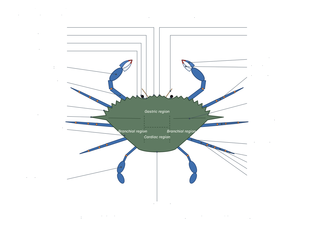
The top of the carapace is divided into regions named after the organs beneath them: the gastric region over the stomach, the cardiac region over the heart and the two branchial regions over the gills (Stachowitsch glossary). Along the front margin, the blue crab has two broad triangular frontal teeth between the eyes, a key feature that separates it from other Callinectes species. On each side are nine anterolateral teeth: the first is the outer orbital tooth beside the eye and the ninth is the long lateral spine (Williams, 1974; Chesapeake Bay Program).[18, 2, 21]
Under the front edge, the eyestalks lie in the orbits and the short antennules fold into pits called antennular fossae. In true crabs the antennae are short, and the outer maxillipeds are "operculiform": they close over the mouth like a pair of lids (Miers, 1886). The sides of the carapace curve down and in as the branchiostegites, the outer walls of the two gill chambers.[17, 18]
| Region | What you see | What it does |
|---|---|---|
| Carapace (top) | Gastric, cardiac and branchial regions; granules and low ridges | Shields the stomach, heart, gonads and gills[18, 2] |
| Front margin | Two frontal teeth, two orbits with the eyestalks | Protects the eyes and antennules[18, 2] |
| Side margins | Nine anterolateral teeth, the last a long lateral spine | Edge of the shell; teeth and spines identify species[2, 21] |
| Underside of the carapace | Pterygostomial and branchiostegal regions | Outer wall of the gill chambers[16, 18] |
| Underside of the body | Sternum, third maxillipeds, leg bases, folded apron | Floor of the body; holds the abdomen and genital openings[18, 17] |
Parts of a crab and their functions
A crab's appendages are, from front to back: antennules, antennae, mandibles, maxillules, maxillae, three pairs of maxillipeds, the chelipeds, three pairs of walking legs, the swimming legs and, under the abdomen, the pleopods. Each walking leg has six segments: coxa, a fused basis-ischium, merus, carpus, propodus and dactyl (FAO guide).[16, 18]
| Appendage | Pairs | Main job |
|---|---|---|
| Antennules | 1 | Smell: the tips carry olfactory sensilla (aesthetascs)[10] |
| Antennae | 1 | Touch and contact chemical sense; the green gland opens at the base[10, 18] |
| Mandibles | 1 | The jaws: tear and grind food[18, 15] |
| Maxillules and maxillae | 2 | Handle food; the maxilla's scaphognathite pumps water through the gill chamber[15, 18] |
| Maxillipeds | 3 | Hold and pass food, groom; the third pair closes over the mouth[15, 6, 17] |
| Chelipeds | 1 | Defence, grasping and crushing prey, holding a mate[24, 1] |
| Walking legs | 3 | Walking and support[21, 24] |
| Swimming legs | 1 | Paddle-shaped last legs for swimming (swimming crabs only)[24, 18] |
| Pleopods | 2 (male) or 4 (female) | Male gonopods transfer sperm; female pleopods carry the eggs[1, 18] |
Crab anatomy labeled: every part in the 3D model
This is the text version of the 3D model: every labeled structure of the blue crab, grouped by body system, with what it does.
Shell & body regions
- Carapace (Shell, dorsal shield)
- The broad, hard shield that covers the whole cephalothorax (head and thorax) of the crab. Protects the stomach, heart, digestive gland, gonads and gills, and forms the outer wall of the two gill chambers.
- Frontal teeth (Front, the crab's rostrum region)
- The two broad triangular teeth on the front edge of the carapace, between the eyes. Form the 'front' of the shell above the antennules; their shape is used to identify species.
- Orbits (Eye sockets)
- The two notches in the front margin of the carapace that hold the eyestalks. Enclose and protect the eyestalks.
- Anterolateral teeth (Marginal teeth)
- The row of sharp teeth along each front-side edge of the carapace. Edge the front of the shell; their number and shape are key features for identifying crab species.
- Lateral spines (Ninth anterolateral teeth)
- The long, sharp spine at each side of the carapace, the widest point of the crab. Mark the widest point of the shell; their length and direction help tell forms of the species apart.
- Gill chamber (Branchial chamber; its outer wall is the branchiostegite)
- The space under each side of the carapace that holds the gills, roofed by the branchiostegite. Protects the delicate gills and channels the breathing current over them.
- Sternum (Sternal plastron, thoracic sternites)
- The broad plate on the crab's underside between the leg bases, made of fused sternites. Floors the body, carries the leg joints and shelters the folded abdomen in a shallow cavity.
- Apron (male abdomen) (T-shaped abdomen of a 'jimmy') · males only
- The male's narrow, T-shaped abdomen, folded flat under the body. Covers and protects the male's gonopods; it hangs free during mating.
- Apron (female abdomen) (Broad rounded abdomen of a mature 'sook') · females only
- The mature female's broad, almost semicircular abdomen, folded under the body. Shelters the four pairs of pleopods that carry the eggs.
- Abdominal locking tubercles (Abdominal lock, 'snap fasteners') · males only
- A pair of small knobs on the sternum that hold the male's apron shut. Lock the folded abdomen against the body; the male releases it to mate.
Legs & mouthparts
- Mandibles (Jaws)
- The hard, toothed jaws on each side of the mouth, innermost of the six pairs of mouthparts. Tear and grind food and push it into the mouth.
- Maxillules (First maxillae)
- Small, thin mouthparts between the mandibles and the maxillae. Help hold and move food towards the mandibles.
- Maxillae (Second maxillae)
- Thin, lobed mouthparts between the maxillules and the first maxillipeds, each carrying the gill bailer. Handle food, and pump water through the gill chamber with the scaphognathite.
- First maxillipeds (Jaw-feet, pair 1)
- The first and innermost pair of maxillipeds, thin and leaf-like. Hold food at the mouth; their long epipods sweep the gills.
- Second maxillipeds (Jaw-feet, pair 2)
- The middle pair of maxillipeds, small and leg-like. Handle food and groom.
- Third maxillipeds (Outer mouthparts, 'mouth doors')
- Two broad, flat plates that close over the mouth field like a pair of doors. Cover and protect the other mouthparts, hold food, and groom.
- Chelipeds (Claw legs, first pereiopods (P1))
- The first and strongest pair of legs, which end in the claws. Defence, grasping and crushing prey, and holding the female during mating.
- Claws (chelae) (Pincers: palm, fixed finger (pollex) and dactyl)
- The pincers at the end of the chelipeds: a broad palm with a fixed finger and a movable finger. Grip and crush prey.
- First walking legs (Second pereiopods (P2))
- The first of three pairs of slender walking legs, just behind the claws. Walking, and holding the female during mating.
- Second walking legs (Third pereiopods (P3))
- The middle pair of walking legs, usually the longest. Walking and support.
- Third walking legs (Fourth pereiopods (P4))
- The last pair of true walking legs. Walking and support.
- Swimming legs (Fifth pereiopods (P5), paddles, 'backfin')
- The last pair of legs, whose final two segments are flattened into oval paddles. Swimming: the paddles rotate in a sculling action to drive the crab through the water.
Eyes & sense organs
- Compound eyes
- The two dark eyes at the tips of the eyestalks, each made of many units (ommatidia). Vision.
- Eyestalks (Ocular peduncles)
- The two short, movable stalks that carry the eyes. Hold the eyes up and move them; they also contain hormone glands.
- X-organ–sinus gland (Eyestalk neuroendocrine gland)
- A small hormone-releasing gland inside each eyestalk. Releases molt-inhibiting hormone, which holds back molting until conditions are right.
- Antennules (First antennae)
- The first, small pair of antennae, folded into pits under the front of the carapace. Smell: their tips carry the crab's olfactory sensilla (aesthetascs).
- Antennae (Second antennae)
- The second pair of antennae: short feelers between the antennules and the eyes. Touch and contact chemical sensing; the green gland opens on the base of each one.
Digestive
- Mouth
- The opening of the gut, hidden in the buccal frame behind all six pairs of mouthparts. Takes in food that the mouthparts have torn up.
- Oesophagus (Esophagus)
- A short tube running up from the mouth into the stomach. Carries food to the stomach.
- Cardiac stomach (Anterior chamber of the stomach)
- The large, thin-walled front chamber of the stomach, holding the gastric mill. Stores food and grinds it with the gastric mill.
- Gastric mill (Stomach teeth)
- Three hard teeth inside the back of the cardiac stomach that chew food. Grinds food: two lateral teeth work against a median tooth.
- Pyloric stomach (Posterior chamber of the stomach)
- The smaller back chamber of the stomach, which filters the ground food. Sorts food: only the finest particles pass through a filter into the digestive gland, coarser material goes to the midgut.
- Hepatopancreas (Digestive gland, 'mustard')
- A large, two-lobed, yellow to brown gland made of countless blind tubules, filling much of the body. Secretes digestive enzymes, absorbs food, and stores lipids, glycogen and minerals; it also makes the blood pigment hemocyanin.
- Midgut
- The short section of gut behind the stomach, where the digestive gland opens. Carries food on from the stomach to the intestine.
- Intestine (Hindgut)
- A straight tube from the midgut back and down into the folded abdomen. Forms and carries faeces to the anus.
- Anus
- The end of the gut, on the telson at the tip of the folded abdomen. Releases faeces.
Circulatory (open)
- Heart
- A flattened heart in the middle of the back, under the cardiac region of the shell. Pumps hemolymph (blood) out through seven arteries.
- Ostia
- Slit-like valved openings in the heart wall through which blood enters the heart. Let blood flow from the pericardial sinus into the heart, then close when it contracts.
- Anterior aorta (Ophthalmic artery, with the optic arteries)
- The artery that runs forward from the heart over the stomach to the brain and eyes. Supplies the brain (supraoesophageal ganglion) and the eyestalks through the optic arteries.
- Anterolateral arteries
- A pair of arteries running forward and outward from the front of the heart. Supply the gonads, hepatopancreas, stomach, antennal glands, mandibular muscles and the skin of the front of the body.
- Hepatic arteries
- A pair of arteries that leave the front underside of the heart and branch through the digestive gland. Supply the hepatopancreas; a side branch (the pyloric hepatic artery) supplies the pyloric stomach and midgut.
- Posterior aorta
- The smallest artery, running back from the heart into the abdomen. Supplies the hindgut and abdomen.
- Sternal artery (Descending artery)
- The largest artery: it drops straight down from the heart to the floor of the body. Feeds the ventral thoracic artery, which supplies all the legs and mouthparts.
- Ventral thoracic artery
- The artery along the floor of the body, with branches to every leg. Supplies the chelae, the mouthparts and each pereiopod.
Respiratory (gills)
- Gills (Phyllobranchs, 'dead man's fingers')
- Eight feathery, triangular gills on each side, made of stacks of thin plates. Take up oxygen from the water and release carbon dioxide; the back gills also help regulate salts.
- Scaphognathite (Gill bailer)
- A flat paddle on each maxilla that beats to pump water through the gill chamber. Drives the breathing current: water is pulled in near the claws, over the gills, and pushed out at the front.
- Maxilliped epipods (Gill cleaners)
- Long, flexible blades from the bases of the maxillipeds that reach into the gill chamber. Clean the gills of particles and fouling organisms.
- Inhalant openings (Afferent branchial openings)
- Openings near the base of each cheliped where water enters the gill chamber. Let fresh water into the gill chamber.
- Exhalant openings (Efferent branchial openings)
- Openings beside the mouth field where water leaves the gill chambers. Release the breathing current forward, away from the inflow.
Excretory (antennal glands)
- Antennal glands (Green glands)
- The crab's two 'kidneys', one at the front of the head on each side. Filter the blood, adjust its salts and water, and make urine.
- Bladders
- Thin sacs that store urine from each antennal gland. Store urine and may modify it before release.
- Excretory pores (Nephropores)
- Tiny openings on the base of each antenna where urine leaves the body. Release urine.
Nervous
- Brain (Supraoesophageal (cerebral) ganglion)
- A small nerve mass at the front of the head, above the oesophagus. Processes input from the eyes, antennules and antennae.
- Optic nerves
- Nerves running from the brain out to each eyestalk. Carry visual signals from the eyes to the brain.
- Circumoesophageal connectives
- Two long nerve cords that run from the brain around the oesophagus and back to the thoracic ganglion. Link the brain with the thoracic ganglion.
- Thoracic ganglion (Fused ventral ganglionic mass)
- One large, round nerve centre on the floor of the body, formed by fusion of the whole ventral nerve cord. Controls the legs, mouthparts and abdomen.
- Leg nerves (Pedal nerves and abdominal nerve)
- Nerves radiating from the thoracic ganglion to each leg, to the maxillipeds and back into the abdomen. Carry motor commands to the leg muscles and sensory signals back.
- Stomatogastric ganglion (STG)
- A tiny ganglion on top of the stomach that runs the gastric mill. Generates the rhythms that drive the gastric mill and the pyloric filter.
Reproductive
- Testes · males only
- Two slender, convoluted, white tubes in the front of the body, joined in the middle. Produce sperm.
- Vasa deferentia (Sperm ducts) · males only
- Paired, highly coiled ducts that carry sperm from the testes to the penes. Carry sperm and package it into spermatophores.
- Penes (Male genital openings) · males only
- A pair of short tubes at the bases of the swimming legs, where the sperm ducts open. Deliver sperm into the gonopods.
- Gonopods (First and second pleopods of the male (G1, G2)) · males only
- Two pairs of stiff, tube-like appendages under the male's apron used to transfer sperm. Transfer sperm to the female: the long first pair are the intromittent organs, the short second pair accessory organs.
- Ovaries · females only
- Paired, elongated orange organs on top of the digestive gland, joined in the middle. Produce eggs.
- Oviducts · females only
- Short tubes from each ovary down to the spermathecae. Carry eggs from the ovaries.
- Spermathecae (Seminal receptacles) · females only
- A pair of sacs that store sperm after mating; eggs are fertilised as they pass through them. Store sperm for later spawning.
- Vulvae (Female gonopores) · females only
- Two openings on the female's sternum, normally hidden under the apron. Receive the gonopods during mating and release eggs.
- Pleopods (female) (Swimmerets) · females only
- Four pairs of fringed appendages under the female's apron that hold the eggs. The eggs are glued to their hairs and carried as the 'sponge' until they hatch.
Crab internal anatomy
When the carapace is lifted off a crab, you see the stomach in the front middle, the heart just behind it, the yellow to brown hepatopancreas filling both sides, the gonads lying on top of it, and the feathery gills in a chamber on each side. The brain and antennal glands are at the very front, and one large thoracic ganglion sits on the floor of the body.[5, 4, 9]
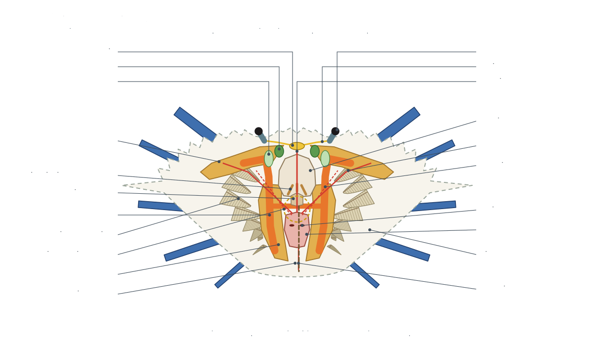
This layout comes from dissections of the related blue swimmer crab, where mature orange ovaries can completely hide the hepatopancreas and the intestine runs back into the folded abdomen to the anus on the telson (Tahon & Shaker, 2023).[5]
How do crabs breathe?
Crabs breathe with gills hidden in a gill chamber under each side of the carapace. A blue crab has eight gills per side. A paddle on each maxilla, the scaphognathite, beats to draw water in near the bases of the claws, across the gills and out of exhalant openings beside the mouth.[6, 18]
Wortham & Pascual (2017) counted eight gills and three gill-cleaning epipods in each blue crab gill chamber; the related blue swimmer crab also has eight (Tahon & Shaker, 2023). Crab gills are phyllobranchs: flat, leaf-like lamellae stacked along a central axis. The back gills also regulate salts (Felgenhauer, 1992). Cooks call them "dead man's fingers"; they are not poisonous (Maryland Sea Grant).[6, 5, 4, 22]
Where is a crab's heart? The circulatory system
A crab's heart lies in the middle of its back, just under the cardiac region of the shell and behind the stomach. It is a flattened sac inside the pericardial sinus. Blood enters it through valved openings called ostia and leaves through seven arteries.[5, 18, 4, 3]
Using resin casts, McGaw & Reiber (2001) mapped the seven arteries of the blue crab: the anterior aorta to the brain and eyes, two anterolateral arteries, two hepatic arteries, the small posterior aorta to the abdomen and the sternal artery. The sternal artery, the largest, drops straight down from the heart and feeds the ventral thoracic artery, which supplies every leg and the mouthparts. They found the arteries dividing into capillary-like vessels, and the return sinuses to be "discrete channels rather than random open spaces", so crab circulation is more organised than the textbook "open system" suggests. In crustaceans the heart is the first organ to receive oxygenated blood from the gills (Maus et al., 2020). Oxygen is carried by hemocyanin, a blue, copper-based respiratory protein that is made in the hepatopancreas (Todorovska et al., 2021).[3, 7, 8]
Crab digestive system: stomach, gastric mill and the mustard
Food torn up by the six pairs of mouthparts passes up a short oesophagus into the cardiac stomach, where the gastric mill grinds it. The pyloric stomach filters the result: the finest particles go to the hepatopancreas for digestion and absorption, and the rest travels along the midgut and intestine to the anus.[15, 4, 5]
The gastric mill is a set of three teeth: a median tooth on the urocardiac ossicle and two lateral teeth on the zygocardiac ossicles (Felgenhauer, 1992). It is driven by the stomatogastric ganglion, a cluster of about 26 neurons (Otopalik et al., 2017) that generates two stomach rhythms, the fast pyloric rhythm and the slower gastric mill rhythm (Haley et al., 2018). The yellow "mustard" in a cooked crab is the hepatopancreas, not fat; Maryland Sea Grant advises against eating it because chemical contaminants concentrate there. Because the stomach lining is part of the exoskeleton, crabs stop feeding shortly before they molt (Van Engel, 1958).[4, 11, 12, 22, 1]
Does a crab have a brain? The nervous system
Yes. A crab's brain, the supraoesophageal ganglion, sits at the front of the head above the oesophagus. Two connectives run around the gut to one large thoracic ganglion on the floor of the body, formed by fusion of the whole ventral nerve cord. Nerves fan out from it to every leg.[4, 9]
In brachyuran crabs the suboesophageal, thoracic and abdominal ganglia are fused into one thoracic ganglion (Kotsyuba & Dyachuk, 2026). A long-tailed decapod such as a crayfish keeps a ladder-like cord with a ganglion in each abdominal segment (Felgenhauer, 1992). The main sense organs are the stalked compound eyes and the antennules, whose olfactory sensilla give the crab its sense of smell; contact chemoreceptors on all the appendages let it taste what it touches (Krieger et al., 2015).[9, 4, 19, 10]
How crabs excrete: the antennal glands
Crabs excrete with two antennal glands, also called green glands, at the front of the head. Each filters the blood in an end sac, adjusts the salts in a labyrinth and canal, stores urine in a bladder, and releases it through an excretory pore on the base of the antenna (Felgenhauer, 1992; Stachowitsch glossary).[4, 18]
Male vs female crab anatomy
The quickest way to sex a blue crab is to turn it over. A male has a narrow apron shaped like an inverted T. A mature female has a broad, rounded apron, and an immature female a triangular one. Watermen call males "jimmies", immature females "sallies" and mature females "sooks" (Maryland Sea Grant; Chesapeake Bay Program).[21, 22, 1]
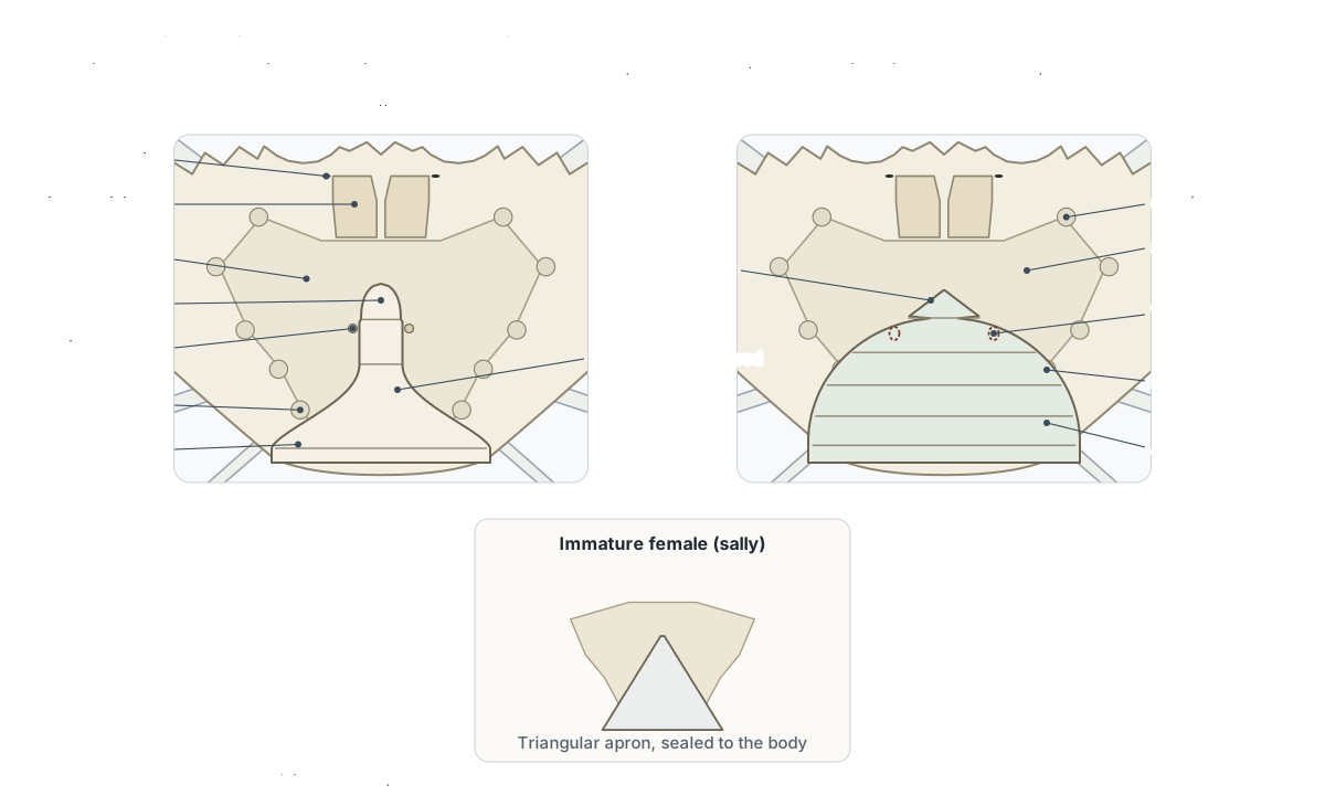
| Male (jimmy) | Mature female (sook) | |
|---|---|---|
| Apron | Narrow, T-shaped; segments 3–5 fused | Broad, almost semicircular; segments free[2, 1] |
| Pleopods under the apron | Two pairs of gonopods: long first pair, short second pair | Four pairs of fringed pleopods that carry the eggs[1] |
| Genital openings | Penes on the bases of the swimming legs | Two vulvae on the sternum[18, 4, 17, 5] |
| Gonads | Slender, convoluted white testes | Paired orange ovaries over the hepatopancreas[5, 4] |
| Sperm | Packed into spermatophores in the vasa deferentia | Stored in two spermathecae for at least a year[4, 1] |
| Claw fingers | Blue, tipped with red | Orange tipped with purple (Williams) or red-tipped (Chesapeake Bay Program)[2, 21] |
Sources: Van Engel (1958), Williams (1974), Felgenhauer (1992) and Tahon & Shaker (2023). A female mates once, right after her final molt while her shell is soft, and the male carries her beneath him before and after. Eggs are fertilised as they pass the stored sperm and are glued to the pleopods as a "sponge" of about 700,000 to over 2 million eggs (Van Engel, 1958; Chesapeake Bay Program: 750,000 to 2 million). Animal Diversity Web gives 2 to 8 million.[1, 23, 21]
How do crabs grow? Molting
A crab grows only by molting. It forms a new, soft shell under the old one, cracks the old shell open along fixed lines, backs out, swells with water and then hardens the new shell. Each molt adds about a quarter to a third to the crab's width (Van Engel, 1958).[24, 1]
| Stage | What happens | Timing |
|---|---|---|
| Peeler | New shell visible through the edge of the swimming paddles: white-rim, then pink-rim, then red-sign | Red-sign crabs molt within 1–3 days[1, 22] |
| Buster | The old shell cracks and the crab backs out | 2–3 hours for a 4–5 inch crab[1] |
| Soft crab | Takes up water and expands to full size | About 2 hours[1] |
| Paper shell | Papery or leathery shell | 9–12 hours after the molt[1, 22] |
| Buckram | Shell stiff but not yet hard | 12–24 hours after the molt[1, 22] |
| Hard crab | Shell rigid; calcium keeps being added | About three more days[1] |
Sources disagree on how many molts it takes to grow up: Van Engel (1958) counted 18 or more after the larval stages, Animal Diversity Web gives 18 to 20, and LSU says a crab may shed "as often as 25 times". Small crabs molt every 3 to 5 days, crabs over 4 inches every 20 to 50 days. About 5% of the old shell's calcium is stored, mainly in the hepatopancreas; the rest of the new shell's minerals come from water and food (Van Engel, 1958). Molting is held back by molt-inhibiting hormone from the sinus gland in the eyestalks, which suppresses the Y-organs that make the molting hormone ecdysteroid (Legrand et al., 2021; Kozma et al., 2023). Larvae hatch as zoeae, with four or five zoeal stages according to Van Engel (1958) and seven according to Animal Diversity Web, then become a megalops before the first crab stage.[1, 23, 24, 13, 14]
How other crabs differ from the blue crab
All true crabs share the blue crab's body plan, but details differ between groups, and several animals called "crabs" are not true crabs at all.[19, 20]
| Crab | Main difference from the blue crab | Source |
|---|---|---|
| Stone crab (Menippe) | Nine gills per side instead of eight | Wortham & Pascual, 2017[6] |
| Green crab (Carcinus) | A swimming crab without paddles: the last legs are pointed | FAO key[16] |
| Fiddler crabs | Males have one hugely enlarged claw used in courtship and fights | Queensland Museum[25] |
| Hermit and porcelain crabs | Not true crabs (Anomura): the fifth legs are reduced | Lowry, Australian Museum[20] |
| Horseshoe crabs | Not crabs at all: arthropods related to spiders and scorpions | Oxford University Museum of Natural History[26] |
Compared with a crayfish, a crab has the same organ systems (gills under the carapace, a gastric mill, green glands, an open circulation) but a reduced abdomen with no tail fan and a fused nervous system. See comparative anatomy for how crustaceans compare with other animals, and the spider anatomy page for the chelicerate body plan that horseshoe crabs share.[25, 16, 4]
Frequently asked questions
What are the main parts of a crab?
A crab has a cephalothorax covered by a hard carapace, and a small, flat abdomen (the apron) folded underneath. On the front are two stalked eyes and two pairs of antennae, and six pairs of mouthparts cover the mouth. It has ten legs: two chelipeds with claws and eight walking legs; in swimming crabs such as the blue crab the last pair are paddles.
What is the apron on a crab?
The apron is the crab's abdomen, folded flat under the body. Its shape shows the sex. In blue crabs, males have a narrow T-shaped apron, immature females a triangular one, and mature females a broad, rounded one. Under the apron, males carry two pairs of gonopods and females four pairs of pleopods that hold the eggs.
Where is a crab's heart?
In the middle of the back, just under the shell, behind the stomach, in the cardiac region of the carapace. The heart is a flattened sac. In the blue crab it pumps blood into seven arteries, the largest of which drops straight down to the floor of the body. Blood returns through the gills to a sinus around the heart and enters it through valved openings, the ostia.
How do crabs breathe?
With gills hidden in a chamber under each side of the carapace. A blue crab has eight gills per side, each a stack of thin plates. A paddle on each maxilla, the scaphognathite, beats to pull water in near the bases of the claws, across the gills and out beside the mouth. Long epipods from the maxillipeds sweep the gills clean.
What is the yellow stuff inside a crab?
The yellow 'mustard' is the hepatopancreas, the crab's digestive gland, not fat. It makes digestive enzymes, absorbs food and stores reserves. Maryland Sea Grant advises against eating it because chemical contaminants concentrate in this organ. The orange material in a female is the ovary.
Do crabs have a brain?
Yes. A crab has a small brain, the supraoesophageal ganglion, above the oesophagus at the front of the head. Two connectives loop around the gut to a large thoracic ganglion on the floor of the body. In crabs the whole ventral nerve cord is fused into this one mass, from which nerves run to every leg.
How can you tell a male crab from a female?
Turn it over and look at the apron. A male blue crab ('jimmy') has a narrow apron shaped like an inverted T. A mature female ('sook') has a broad, rounded apron, and an immature female ('sally') a triangular one. Claw colour helps: males have blue claws with red tips, while mature females have orange or red-tipped fingers.
How many legs does a crab have?
Ten, which is why crabs are called decapods. The first pair are the chelipeds, which carry the claws. Behind them are four more pairs. In the blue crab three pairs are walking legs and the last pair are flat swimming paddles. Hermit and porcelain crabs are not true crabs; their fifth legs are reduced, so they seem to have fewer.
References
Every source below was opened and checked on 25 October 2026.
- Van Engel, W. A. (1958). The blue crab and its fishery in Chesapeake Bay. Part 1: Reproduction, early development, growth, and migration. Commercial Fisheries Review 20(6): 6–17.
- Williams, A. B. (1974). The swimming crabs of the genus Callinectes (Decapoda: Portunidae). Fishery Bulletin 72(3): 685–798.
- McGaw, I. J. & Reiber, C. L. (2001). Cardiovascular system of the blue crab Callinectes sapidus. Journal of Morphology 251(1): 1–21.
- Felgenhauer, B. E. (1992). Internal anatomy of the Decapoda: an overview. In Microscopic Anatomy of Invertebrates, vol. 10: Decapod Crustacea, pp. 45–75. Wiley-Liss.
- Tahon, R. R. & Shaker, N. A. (2023). Topographical anatomical studies of the internal organs of the blue swimmer crab (Portunus pelagicus) in conjunction with its external features. Advances in Animal and Veterinary Sciences 11(1): 166–175.
- Wortham, J. L. & Pascual, S. (2017). Grooming behaviors and gill fouling in the commercially important blue crab (Callinectes sapidus) and stone crab (Menippe mercenaria). Nauplius 25: e2017028.
- Maus, B., Gutsfeld, S., Bock, C. & Pörtner, H.-O. (2020). Non-invasive MRI studies of ventilatory and cardiovascular performance in edible crabs Cancer pagurus. Frontiers in Physiology 11: 596529.
- Todorovska, E. et al. (2021). Molecular cloning, structure and phylogenetic analysis of a hemocyanin subunit from the Black Sea crustacean Eriphia verrucosa. Genes 12(1): 93.
- Kotsyuba, E. & Dyachuk, V. (2026). Parasite-induced changes in the nervous system of the shore crab Hemigrapsus sanguineus. International Journal of Molecular Sciences 27(13): 5993.
- Krieger, J. et al. (2015). Comparative analyses of olfactory systems in terrestrial crabs (Brachyura): evidence for aerial olfaction? PeerJ 3: e1433.
- Otopalik, A. G. et al. (2017). Sloppy morphological tuning in identified neurons of the crustacean stomatogastric ganglion. eLife 6: e22352.
- Haley, J. A., Hampton, D. & Marder, E. (2018). Two central pattern generators from the crab, Cancer borealis, respond robustly and differentially to extreme extracellular pH. eLife 7: e41877.
- Legrand, E. et al. (2021). Understanding molt control switches: genes involved in ecdysteroidogenesis in the Y-organ of the blue crab, Callinectes sapidus. PLOS ONE 16(9): e0256735.
- Kozma, M. T. et al. (2023). In silico analysis of crustacean hyperglycemic hormone family G protein-coupled receptor candidates. Frontiers in Endocrinology 14: 1322800.
- Crain, J. A. (1999). Functional morphology of prey ingestion by Placetron wosnessenskii zoeae (Crustacea: Anomura: Lithodidae). Biological Bulletin 197: 207–218.
- Tavares, M. (2002). True crabs. In K. E. Carpenter (ed.), The Living Marine Resources of the Western Central Atlantic, vol. 1. FAO.
- Miers, E. J. (1886). Report on the Brachyura. Report on the Scientific Results of the Voyage of H.M.S. Challenger, Zoology 17.
- Crabs of Japan glossary (Naturalis Biodiversity Center), definitions after Stachowitsch, M. (1992), The Invertebrates: an Illustrated Glossary.
- Lowry, J. K. Crustacea, the higher taxa: Brachyura. Australian Museum.
- Lowry, J. K. Crustacea, the higher taxa: Anomura. Australian Museum.
- Chesapeake Bay Program. Blue crab (field guide).
- Maryland Sea Grant (2012). Glossary of blue crab biology. Chesapeake Quarterly 11(2).
- Bodden, S. (2011). Callinectes sapidus, blue crab. Animal Diversity Web, University of Michigan.
- Louisiana Fisheries, LSU AgCenter and Louisiana Sea Grant. Blue crab.
- Queensland Museum. Crabs.
- Oxford University Museum of Natural History. Horseshoe crabs are not crabs (Learning Zone).
More interactive anatomy models
- Crayfish anatomyExternal appendages and internal organs of the crayfish.
- Crayfish dissectionGuided crayfish dissection: gills, green gland, stomach and heart.
- Human anatomy1,000+ bones, muscles, organs, vessels and nerves from real anatomical data.
- Octopus anatomyThree hearts, a ring-shaped brain, eight arms and a beak, in 3D.
- Squid anatomyPen, eight arms, two tentacles, giant axons and three hearts of the longfin squid.
- Squid dissectionStep-by-step guided squid dissection with every structure labeled.