

Rat anatomy at a glance
The reference animal here is the laboratory rat, the brown or Norway rat (Rattus norvegicus) bred for research, modelled as an albino strain. It was the first animal domesticated mainly for science and is the most used laboratory animal after the mouse.[8] Rats have every organ system that people have, but several organs work differently. Biologists use four direction words for a four-legged animal: cranial (towards the head), caudal (towards the tail), dorsal (the back) and ventral (the belly).
Rat body by the numbers
| Feature | Laboratory rat | Source |
|---|---|---|
| Head and body length | 23–28 cm (9–11 in), plus a 18–23 cm tail | [2] |
| Adult weight | Males 450–650 g, females 350–450 g (MSD); males 450–520 g, females 250–320 g (UTSA) | [2, 8] |
| Teeth | 16: one incisor and three molars in each half of each jaw | [11, 12] |
| Vertebrae | 7 neck, 13 chest, 6 lumbar, 4 sacral, about 28 tail | [1] |
| Resting heart rate | 250–400 a minute (MSD); 250–450 (UTSA) | [5, 8] |
| Breathing rate | 70–115 breaths a minute | [8] |
| Body temperature | 35.9–37.5 °C | [8] |
| Life span | About 1.5–3 years (MSD); 2–3.5 years (UTSA) | [2, 8] |
External anatomy of a rat
The outside of a rat shows a pointed snout with long whiskers, large ever-growing front teeth, eyes and ears on the sides of the head, a furred body, four paws and a long tail with hairless skin covered in scales.[1, 19]
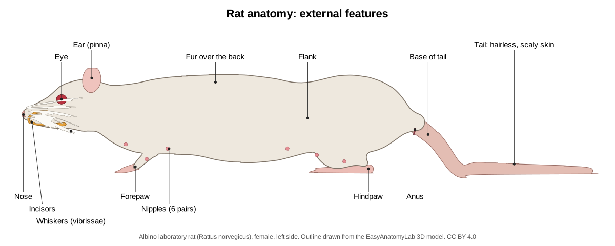
Whiskers (vibrissae) are long touch hairs in rows on each side of the snout. A rat sweeps them back and forth, called whisking, to feel its surroundings; tracking in one study measured an average of about 8.5 sweeps a second, slower than a mouse's 11.3.[16]
Eyes and ears. Rat hearing is acute and reaches into ultrasound, but vision is poor.[8] Sources disagree on colour: a university handout says rats cannot see colour and are blind to red light,[8] while tests of rat cones found two types, one sensitive to ultraviolet and one to green, and evidence of two-colour (dichromatic) vision.[17]
The tail: a radiator for body heat
The rat's tail is its main organ for losing heat.[8, 19] Heat lost without evaporating water leaves mainly through the tail, whose blood vessels widen when the brain senses warmth.[18] Nerves that narrow these vessels control the flow, so a cold rat keeps warm blood away from the tail.[18] The tail makes up about 9% of a rat's body surface but can dissipate an estimated 17% of its body heat, far more than a mouse's tail.[20] Hairless skin elsewhere, such as the soles of the paws, helps too.[19]
Rat teeth: why the incisors never stop growing
A rat has 16 teeth: 2 × (I 1/1, C 0/0, P 0/0, M 3/3). It has no canines or premolars, and a long toothless gap, the diastema, separates the incisors from the molars.[11, 12] The incisors are open-rooted and keep growing and erupting throughout life, while the molars have true roots and stop growing.[11, 12] Only the front of each incisor is covered with hard enamel over softer dentine, which makes the incisors self-sharpening chisels.[11, 14, 1]
In young rats a lower incisor that does not meet its partner erupts about 1.0 mm a day, and a normally worn one about 0.6 mm a day; the shorter the tooth is cut, the faster it grows.[13] Rats need to gnaw to keep them short: overgrown incisors are common in pet rats and can grow into the nasal cavity.[4, 9]
Why are rat teeth orange? The enamel of rodent incisors contains iron, which was long thought to give the colour. A 2024 study of seven rodents, including laboratory rats, found that the iron-rich layer looks like ordinary enamel; the orange-brown colour comes mainly from a thin surface layer and the zone under it, and the thicker that layer, the darker the tooth.[14, 15]
Rat skeleton
The rat's backbone has 7 cervical, 13 thoracic, 6 lumbar, 4 sacral and about 28 caudal (tail) vertebrae. There are 13 pairs of ribs: the first 7 pairs reach the sternum, and of the 6 pairs of false ribs the last 3 float free.[1] The rat has a collarbone, and the clavicle is the only bony link between the shoulder blade and the trunk.[1] Turn on Skeletal and switch off the other systems in the viewer to see every bone.
Rat internal anatomy: where the organs are
A muscular diaphragm divides the chest, which holds the heart, lungs and thymus, from the abdomen, which holds the liver, stomach, spleen, pancreas, intestines, kidneys, bladder and reproductive organs.[1]
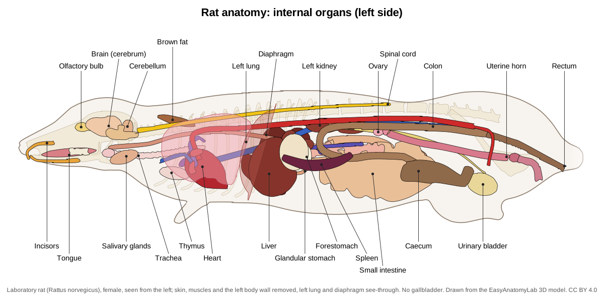
| Organ | Where it is | Rat-specific facts |
|---|---|---|
| Heart | Middle of the chest between the lungs | Four chambers, as in humans[1] |
| Lungs | Chest, either side of the heart | Left lung 1 lobe; right lung 4 (cranial, middle, caudal, accessory)[1, 25] |
| Liver | Against the diaphragm | Deeply cleft into lobes; no gallbladder[1, 23] |
| Stomach | Left front of the abdomen | Non-glandular forestomach plus glandular stomach[22] |
| Caecum | Where small and large intestine meet | A large blind sac about the size of the stomach[22] |
| Kidneys | Under the lumbar spine | The right one sits further forward, in a hollow of the liver[1] |
Rat digestive system: no gallbladder, no vomiting and a big caecum
The rat stomach is a half-moon-shaped sac with two regions: a non-glandular forestomach, about three fifths of its area, and a glandular stomach, divided by a raised limiting ridge. The human stomach is glandular only.[22, 1]
Why can't rats vomit? Vomiting is widespread among mammals, from cats and dogs to humans, but rats and other rodents do not do it. A comparative study found that rodents have a less muscular diaphragm and a less funnel-shaped stomach than vomiting species, yet the main reason is in the brain: rodents lack the brainstem circuit that drives the coordinated mouth, throat and breathing movements of a vomit.[21] Because rodents do not vomit, lab rats are not usually fasted before an anaesthetic.[8]
Liver without a gallbladder. The rat liver is cleft into distinct lobes. Hunt's manual names four main lobes (median, left lateral, right lateral and caudate), while a 2016 surgical study counts six by splitting the median and adding a quadrate lobe; authors simply name the lobes differently.[1, 23] It weighs about 5–6% of the body.[23] There is no gallbladder: the hepatic ducts join into one common hepatic duct that carries bile straight to the duodenum.[1, 23]
Intestines and caecum. In one surgical study the small intestine was about 1.49 m long and the large intestine 0.26 m; the caecum measured 45–70 mm and was as large as the stomach, and the colon was simple, without the pouches of the human colon.[22] Hunt gave smaller figures, about 2.5 cm for the caecum and 20 cm for the whole large intestine.[1] Rats are hindgut fermenters: bacteria in the large intestine ferment plant material, and rats eat some of their droppings to recover the amino acids and vitamins made there.[10]
Heart, lungs and brown fat
The heart has four chambers, two atria and two ventricles,[1] and beats 250–400 times a minute at rest, against 450–750 in a mouse.[5] The left lung is a single lobe and the right lung has four: cranial, middle, caudal and accessory.[1, 25]
Rats keep a pad of brown fat between the shoulder blades (the interscapular depot). Brown fat makes heat directly, a process called non-shivering thermogenesis, using a protein unique to it, uncoupling protein 1 (UCP1).[26]
Brain, senses and the Harderian gland
The rat's cerebrum is smooth: it lacks the grooves (sulci) and ridges (gyri) seen on the brains of many larger mammals. Large olfactory bulbs at its front end serve the sense of smell.[1]
Behind each eyeball lies the Harderian gland.[4, 9] It occurs in most land vertebrates, and in rodents it makes lipids, porphyrins and indoles.[27] Its reddish-brown secretion lubricates the eye; around the eyes and nose it looks like blood but contains none, which is why it is called red tears.[4] The pigments are porphyrins that fluoresce pink under ultraviolet light, and heavy staining (chromodacryorrhea) usually signals stress or illness.[6, 9]
Male vs female rat anatomy
Male and female rats differ in size, in the distance between the anus and the genital opening, in nipples and in their reproductive organs. Use the Female/Male switch in the viewer to compare them, and see the urogenital diagram in the dissection guide for every gland.
| Male rat (buck) | Female rat (doe) | |
|---|---|---|
| Adult weight | 450–650 g | 350–450 g[2] |
| Anogenital distance | About twice as long | Short[29] |
| Nipples | None | 12, in six pairs[29, 28] |
| Under the tail | Anus, then the scrotum (about 4 cm long in a large male), then the penis | Anus, vaginal opening and a separate urinary opening[1] |
| Gonads | Testes, which can be pulled back into the abdomen | Ovaries, each in a thin capsule (bursa)[1] |
| Accessory organs | Vesicular glands, prostate, preputial glands | Paired clitoral glands; uterus with two horns and two cervical canals[1, 30] |
| Sexual maturity | 6–10 weeks (MSD); breeding from 65–110 days (UTSA) | 8–12 weeks (MSD); breeding from 65–110 days (UTSA)[3, 8] |
Male. The inguinal canal stays open in rats, so the cremaster muscle can withdraw the testes into the abdomen; preserved lab rats often show them partly retracted. The large, warty vesicular glands were once called seminal vesicles, although they hold no sperm.[1]
Female. The uterus has two long horns with two separate cervical canals opening into a single vagina (uterus duplex bicollis).[30, 1] Pregnancy lasts 21–23 days[3, 8] and a litter has 8–18 pups.[3] Mammary tissue is spread over a wide area of the belly, and mammary tumours are the most common cancer in pet rats.[9]
Rat vs mouse anatomy
Rats and mice share the same body plan, but a rat is more than ten times heavier and differs in a few organs. For the mouse in 3D, see mouse anatomy.
| Feature | Laboratory rat | House mouse |
|---|---|---|
| Adult weight | 350–650 g[2] | 20–40 g[7] |
| Gallbladder | Absent[23] | Present[24] |
| Mammary glands (female) | 6 pairs | 5 pairs[28] |
| Resting heart rate | 250–400 a minute | 450–750 a minute[5] |
| Whisking (mean sweeps a second) | 8.5 | 11.3[16] |
| Tail: share of body surface, of heat loss | About 9%, up to 17% | About 6%, 5–8%[20] |
| Life span | About 1.5–3 years | 1.5–2.5 years[2, 7] |
Rat vs human anatomy
| Feature | Rat | Human |
|---|---|---|
| Teeth | 16; incisors grow for life[11] | 32 permanent teeth[33] |
| Vertebrae | C7, T13, L6, S4, about 28 caudal[1] | C7, T12, L5, sacrum (5 fused), coccyx (4 fused)[31] |
| Lung lobes (left, right) | 1, 4[25] | 2, 3[32] |
| Gallbladder | None[23] | Stores and concentrates bile[35] |
| Stomach lining | Non-glandular forestomach plus glandular part[22] | Glandular only[22] |
| Can vomit? | No[21] | Yes[21] |
| Small intestine | About 1.5 m[22] | About 3 m in a living person (OpenStax); 5.5–6.4 m (Vdoviaková)[34, 22] |
| Colon | Smooth, not pouched[22] | Pouched (haustra)[34] |
The two human small-intestine figures disagree: OpenStax gives the length in a living person, and the larger figure is the one quoted in the rat surgical study.
Rat anatomy labeled: every structure in the 3D model
This is the text version of the 3D rat: all 114 labeled structures, grouped by body system, with what each does. Structures marked "males only" or "females only" appear when you switch sex in the viewer. For a step-by-step bench dissection with a worksheet and answer key, use the rat dissection guide.
External
- Skin and fur (Integument, pelage)
- Loose skin covered by fur, white in albino laboratory rats and grey-brown above, paler below, in wild Norway rats. Protects the body, holds heat and carries touch receptors. The skin is loosely attached, so it lifts easily from the muscles in a dissection.
- Nose and nostrils (Rhinarium, external nares)
- The hairless tip of the snout, with two nostrils (external nares). Takes in air and odours. Rats rely heavily on smell; the nasal cavity leads back to the large olfactory bulbs of the brain.
- Vibrissae (Whiskers, mystacial vibrissae)
- Long, stiff sensory hairs arranged in rows on each side of the snout. Touch organs. Rats sweep (whisk) them back and forth to feel surfaces and gaps. Hunt (1924) noted that rats run faster along a wall, guided by touch through the vibrissae.
- Pinna (External ear, auricle)
- The thin, rounded, nearly hairless external ear flap. Collects sound and funnels it into the ear canal. Rats and mice hear well into ultrasound.
- Forepaw (Manus, front foot)
- The front foot: four clawed fingers and a tiny thumb with a flat, nail-like claw. Used for walking, climbing, digging and holding food while the rat sits up and gnaws.
- Hindpaw (Pes, hind foot)
- The long hind foot with five clawed toes; the rat walks on the whole sole (plantigrade). Pushes the body forward and supports the rat when it sits or stands up.
- Tail (Cauda)
- A long, nearly hairless tail covered in rings of overlapping scales. Balance when climbing, and heat loss: blood flow through the tail changes to keep the body temperature steady.
- Nipples (teats) (Papillae mammae) · females only
- Six pairs of nipples on the underside of a female rat: three pairs on the chest and three on the belly and groin. Each nipple drains one mammary gland; the pups suckle here.
- Scrotum (Scrotal sacs) · males only
- The large swelling below the base of the tail that holds the two testes. Keeps the testes slightly cooler than the body, which sperm production needs.
- Prepuce and urogenital opening (Foreskin, external urethral opening) · males only
- The furry fold that surrounds the tip (glans) of the penis; the single urogenital opening of the male lies here. Protects the glans. Urine and semen both leave the body through this opening.
- Urethral papilla (Urinary papilla, clitoral prepuce) · females only
- A small raised cone in front of the vaginal opening, with the urethral opening at its tip. Urine leaves through it. It is the female counterpart (homologue) of the male prepuce and contains the clitoris.
- Vaginal opening (Vaginal orifice) · females only
- The slit-like opening between the urethral papilla and the anus. Entrance to the reproductive tract: receives the penis at mating, and the pups are born through it.
- Anus
- The opening at the end of the digestive tract, just below the base of the tail. Faeces leave the body here as firm pellets formed in the colon.
Skeletal
- Skull (cranium) (Braincase, rostrum, zygomatic arches)
- The long, narrow skull: a braincase behind, a long snout (rostrum) in front, and a zygomatic arch on each side. Protects the brain and sense organs and anchors the jaw muscles.
- Mandible (Lower jaw (paired dentaries))
- The lower jaw, made of two halves joined loosely at the chin. Holds the lower incisors and molars. It slides forward to gnaw (incisors meet) and backward to chew (molars meet).
- Cervical vertebrae (7) (Neck vertebrae, atlas and axis)
- Seven neck vertebrae, as in almost all mammals. Support and move the head. The first (atlas) rocks on the skull; the second (axis) lets the head turn.
- Thoracic vertebrae (13)
- Thirteen vertebrae, each carrying a pair of ribs. Support the ribs and the trunk muscles.
- Lumbar vertebrae (6)
- Six large vertebrae of the lower back, with long transverse processes and no ribs. Carry the powerful back and hip muscles used in running and jumping.
- Sacrum (4 sacral vertebrae)
- Four sacral vertebrae, more or less fused into one bone. The first two sacral vertebrae join the ilium of each hip bone, so the weight of the hind body passes through them (Hunt 1924).
- Caudal vertebrae (Tail vertebrae)
- About 28 small tail vertebrae (rat) that become simpler and longer towards the tip. Support the tail and its tendons.
- Ribs (13 pairs)
- Thirteen pairs of ribs: 7 true ribs reach the sternum; 6 false ribs do not, and the last 3 of these float free. Protect the heart and lungs and move during breathing.
- Sternum (Breastbone, sternebrae, xiphoid process)
- The breastbone, made of six segments (sternebrae): the manubrium in front, four segments of the body and the xiphoid process behind. Anchors the true ribs and the chest muscles. In a dissection you cut beside it to open the thorax.
- Scapula (Shoulder blade)
- The flat, triangular shoulder blade with a ridge (spine) on its outer face. Anchors the shoulder muscles and forms the socket of the shoulder joint.
- Clavicle (Collarbone)
- A slender collarbone joining the shoulder to the sternum. Braces the shoulder, which helps rats and mice climb and handle food. (Many running mammals, such as dogs and horses, lack a functional clavicle.)
- Humerus
- The bone of the upper arm. Lever for the shoulder and elbow muscles.
- Radius and ulna
- The two forearm bones. The ulna forms the elbow hinge; the radius carries the wrist.
- Carpals, metacarpals and phalanges (Bones of the forepaw)
- The small wrist bones (carpals) in two rows, five metacarpals and the finger bones (phalanges). Support the hand. Each finger has three phalanges, except the thumb, which has two (Hunt 1924).
- Pelvis (os coxae) (Hip bones: ilium, ischium, pubis)
- Two hip bones, each made of the fused ilium, ischium and pubis, joined at the pubic symphysis. Carries the hindlimbs and protects the bladder, rectum and reproductive tract. The femur sits in its socket, the acetabulum.
- Femur (Thigh bone)
- The thigh bone. Lever for the large thigh muscles.
- Patella (Kneecap)
- A small bone in the tendon of the quadriceps, in front of the knee. Improves the leverage of the knee extensors.
- Tibia and fibula (Shin bones)
- The two bones of the lower leg; in rats and mice the slender fibula fuses to the tibia in its lower half. Support the body and anchor the calf and shin muscles.
- Tarsals, metatarsals and phalanges (Bones of the hind foot)
- Ankle bones (tarsals, including the heel bone, the calcaneus), five long metatarsals and the toe bones. Form the long, flat-footed lever used in running and jumping.
Muscular
- Masseter
- The big cheek muscle that closes the jaw and pulls it forward for gnawing. Rodents (the name means 'gnawers') have a very large, complex masseter that moves the jaw forward so the incisors meet.
- Diaphragm
- A thin dome of muscle and tendon that separates the thorax from the abdomen. The main breathing muscle: it flattens to draw air into the lungs.
- Quadriceps femoris (Rectus femoris, vastus muscles)
- Four muscles on the front of the thigh that straighten the knee. Knee extension when running and jumping.
- Gastrocnemius (Calf muscle)
- The two-headed calf muscle that inserts on the heel through the calcaneal (Achilles) tendon. Extends the ankle (plantarflexion) and helps bend the knee.
- Tibialis anterior
- The muscle on the front of the shin. Flexes the ankle (dorsiflexion), lifting the foot as the leg swings forward.
Nervous & senses
- Eye (Eyeball)
- A large, bulging eye on each side of the head; pink-red in albino laboratory rats, which lack pigment. Vision in dim light. The retina is dominated by rods; rats see poorly in bright daylight (Hunt 1924).
- Lens
- The clear, nearly spherical lens, which fills a large part of the small rodent eye. Focuses light on the retina.
- Olfactory bulbs
- Two large knobs at the front of the brain that receive the nerves of smell. First processing centre for odours. Their size reflects how much rats and mice depend on smell.
- Cerebrum (Cerebral hemispheres, cerebral cortex)
- The two smooth cerebral hemispheres, without the folds (gyri) of the human brain. Senses, movement, learning and memory.
- Hippocampus
- A curved, C-shaped structure inside each hemisphere, beneath the cortex. Memory and spatial navigation. Much of what is known about 'place cells' comes from rats and mice.
- Cerebellum
- The folded 'little brain' behind the cerebrum, with a central vermis and two side lobes. Balance and the timing and coordination of movement.
- Brainstem (medulla oblongata)
- The stalk of the brain that continues into the spinal cord. Controls breathing, heart rate and reflexes such as swallowing. Horn et al. (2013) found rat and mouse brainstems lack the coordinated motor pattern that produces vomiting.
- Spinal cord
- The cord of nerve tissue inside the vertebral canal. Carries signals between the brain and body and runs spinal reflexes.
- Sciatic nerve
- The largest nerve of the body, running down the back of the thigh. Supplies the muscles and skin of the hind leg.
Digestive & oral
- Upper incisors
- Two long, curved, chisel-like front teeth that grow throughout life. Gnawing. Hard enamel covers only the front face, so the softer dentine behind wears faster and the edge stays sharp (Hunt 1924).
- Lower incisors
- Two long lower front teeth, rooted far back in the mandible. Work against the upper incisors like scissors when the jaw is pulled forward.
- Molars (Cheek teeth)
- Three small grinding teeth in each half of each jaw (12 in total). Grind food. Unlike the incisors, the molars of rats and mice do not keep growing.
- Tongue
- A thick, muscular tongue with a raised hump at the back. Moves food to the molars and starts swallowing.
- Salivary glands (Submandibular (submaxillary) and parotid glands)
- Paired glands under the skin of the throat and below the ears: the large submandibular, the smaller sublingual beside it, and the flat, lobulated parotid. Produce saliva that moistens food and starts the digestion of starch.
- Oesophagus (Esophagus, gullet)
- The muscular tube that carries food from the throat to the stomach. Moves swallowed food by waves of contraction (peristalsis).
- Forestomach (non-glandular stomach) (Cardiac sac, nonglandular stomach)
- The thin-walled left part of the stomach, lined by tough, keratinised squamous epithelium like the oesophagus. Stores food before digestion. It makes no gastric juice.
- Glandular stomach (Corpus and pyloric antrum)
- The thick, opaque right part of the stomach, lined with gastric glands. Secretes acid and pepsin. Parietal cells make acid; chief cells make pepsinogen (NTP atlas).
- Limiting ridge (Margo plicatus, boundary fold)
- A raised fold on the inside of the stomach that marks the border between the forestomach and the glandular stomach. Separates the two linings. The oesophagus opens into the forestomach just beside it.
- Duodenum
- The first, U-shaped part of the small intestine, starting at the pylorus. Receives bile and pancreatic juice through the bile duct, which enters it about 3 cm from the pylorus (Hunt 1924).
- Jejunum
- The long, coiled middle part of the small intestine. Main site of digestion and absorption of nutrients.
- Ileum
- The last part of the small intestine, darker than the jejunum in a dissected rat. Absorbs remaining nutrients, vitamin B12 and bile salts.
- Caecum (Cecum)
- A large, comma-shaped blind sac where the small intestine meets the colon; often the biggest single loop in the abdomen. Fermentation chamber: bacteria break down plant fibre. Rats and mice also eat some of their own faeces (coprophagy) to recover vitamins made there.
- Colon (Ascending, transverse and descending colon)
- The large intestine: it runs forward from the caecum, crosses to the left, and runs back along the midline as a chain of faecal pellets. Absorbs water and forms the faecal pellets, which are shaped like cotton seeds (Hunt 1924).
- Rectum
- The final straight part of the large intestine, running through the pelvis to the anus. Stores pellets until they are passed.
- Liver: median lobe (Middle lobe)
- The largest liver lobe, at the front middle of the abdomen, partly split by a deep notch. Part of the liver: makes bile, stores glycogen, processes nutrients from the portal vein.
- Liver: left lateral lobe
- A large, thick lobe on the left, behind the median lobe. Part of the liver. With the median lobe it is removed in the classic two-thirds partial hepatectomy used to study liver regeneration.
- Liver: right lateral lobe (Right lobe (anterior and posterior lobules))
- The right lobe, nearly divided into a front and a back lobule. Part of the liver.
- Liver: caudate lobe (Spigelian lobe)
- Two small lobules that fit around the lesser curvature of the stomach. Part of the liver.
- Bile duct (Common hepatic duct, ductus choledochus)
- The thin duct that carries bile from the liver to the duodenum. Carries bile continuously, because the rat has no gallbladder to store it. Small pancreatic ducts join it as it crosses the pancreas (Hunt 1924).
- Pancreas
- A soft, pinkish, branching gland spread thinly through the mesentery, not a compact organ. Makes digestive enzymes (exocrine) and the hormones insulin and glucagon (islets, endocrine).
- Mesentery (Mesentery proper, greater omentum)
- The thin, transparent double sheet of peritoneum that holds the intestines and carries their blood vessels. Suspends the gut, carries vessels, nerves and lymphatics, and stores fat.
Respiratory
- Larynx (Voice box)
- The cartilage box at the top of the trachea. Guards the airway when swallowing and produces sound, including ultrasonic calls.
- Trachea (Windpipe)
- The windpipe, kept open by C-shaped rings of cartilage. Carries air to and from the lungs.
- Left lung (one lobe)
- A single, undivided lobe: the largest single lung lobe. Gas exchange.
- Right cranial lobe (Right anterior / apical lobe)
- The front lobe of the right lung. Gas exchange.
- Right middle lobe (Cardiac lobe)
- The middle lobe of the right lung, beside the heart. Gas exchange.
- Right caudal lobe (Right posterior lobe)
- The largest lobe of the right lung. Gas exchange.
- Accessory lobe (Postcaval / azygous / median lobe)
- A small, pyramid-shaped lobe of the right lung that sits behind the heart, mostly left of the midline. Gas exchange.
Heart & vessels
- Heart (ventricles)
- The four-chambered heart in its pericardial sac; this part shows the right and left ventricles, with the apex pointing back and to the left. The right ventricle pumps blood to the lungs; the thick-walled left ventricle pumps it to the body.
- Right atrium
- The thin-walled chamber that receives blood from the body. Receives oxygen-poor blood from the two cranial venae cavae and the caudal vena cava.
- Left atrium
- The chamber that receives oxygen-rich blood from the lungs. Passes blood from the pulmonary veins to the left ventricle.
- Aorta (Aortic arch, dorsal (abdominal) aorta, iliac arteries)
- The main artery: it arches over the heart, runs back under the spine and splits into the iliac arteries for the legs. Carries oxygen-rich blood to the body.
- Pulmonary trunk (Pulmonary arteries)
- The vessel that leaves the right ventricle and splits into the left and right pulmonary arteries. Carries oxygen-poor blood to the lungs.
- Cranial venae cavae (left and right) (Superior venae cavae, precaval veins)
- Two large veins, one on each side, that return blood from the head and forelimbs to the right atrium. Venous return from the front of the body.
- Caudal vena cava (Inferior vena cava, postcava)
- The large vein that returns blood from the hind body; it passes through the liver and diaphragm to the heart. Venous return from the legs, kidneys and gut (via the liver).
- Common carotid arteries
- Paired arteries running up each side of the trachea to the head. Supply the brain, face and head.
- Jugular veins (External jugular veins)
- Large, superficial veins on each side of the neck. Return blood from the head.
- Hepatic portal vein (Portal vein)
- The large vein that carries blood from the stomach, intestines, pancreas and spleen to the liver. Takes nutrients and other substances absorbed from the gut straight to the liver, which processes them before the blood reaches the rest of the body.
- Renal arteries and veins
- Short vessels linking each kidney with the aorta and caudal vena cava. About a fifth of the blood pumped by the heart passes through the kidneys to be filtered.
Lymphoid & brown fat
- Thymus
- A pale, two-lobed gland lying over the front of the heart; large in young rats, smaller in adults. Trains T lymphocytes (a type of white blood cell).
- Spleen
- A long, flat, dark red organ along the greater curvature of the stomach. Filters the blood, removes old red cells and makes immune cells.
- Lymph nodes (Cervical, axillary, inguinal, iliac and mesenteric nodes)
- Small, firm, bean-shaped nodes in the neck, armpits, groin, along the aorta and in the mesentery. Filter lymph and start immune responses.
- Brown fat (interscapular) (Brown adipose tissue, BAT)
- A butterfly-shaped pad of brownish fat between the shoulder blades. Produces heat (non-shivering thermogenesis) using uncoupling protein 1 (UCP1) in its mitochondria (Virtue & Vidal-Puig 2013).
Urinary & endocrine
- Thyroid gland (Thyroid lobes and isthmus; parathyroid glands)
- Two small, dark red lobes pressed against the sides of the trachea just behind the larynx, joined by a thin isthmus across the ventral trachea. Endocrine gland (it has no duct): makes the thyroid hormones (T4 and T3) that set the metabolic rate, and calcitonin from its C cells.
- Kidneys
- Two bean-shaped, dark red kidneys against the dorsal body wall; the right kidney is more cranial than the left. Filter blood and make urine.
- Adrenal glands (Suprarenal glands)
- Small, round, pinkish glands at the front end of each kidney. Endocrine glands with no duct: they release adrenaline and steroid hormones (corticosterone in rats) into the blood.
- Ureters
- Thin tubes running from each kidney back to the bladder. Carry urine to the bladder.
- Urinary bladder
- A small, thin-walled, oval sac in the floor of the pelvis. Stores urine.
- Urethra (female) · females only
- The tube from the bladder to the urethral papilla; separate from the vagina. Carries urine only.
- Urethra (male) · males only
- The tube from the bladder through the pelvis and penis; it carries both urine and semen. Receives the vasa deferentia and accessory gland ducts near the bladder neck.
Reproductive
- Testes · males only
- Two large, oval testes, about 2 cm long in an adult rat (Hunt 1924). Make sperm and testosterone.
- Epididymis (Caput, corpus and cauda epididymidis) · males only
- A coiled duct along the edge of each testis, with a head, body and tail. Stores and matures sperm.
- Vas deferens (Ductus deferens) · males only
- The sperm duct from the tail of the epididymis to the urethra. Carries sperm at ejaculation.
- Seminal vesicles (Vesicular glands) · males only
- A pair of large, knobbly glands shaped like question marks: the most conspicuous male organ in the lower abdomen. Produce a thick secretion that forms part of the semen and the copulatory plug. Despite the old name, they store no sperm (Hunt 1924).
- Coagulating glands (Anterior prostate) · males only
- Thin, pale glands lying along the inner (concave) curve of each seminal vesicle. Their secretion clots the seminal-vesicle fluid to form a copulatory plug in the female's vagina.
- Prostate (Ventral and dorsolateral prostate) · males only
- Lobed glands around the neck of the bladder: two ventral lobes under the bladder and a dorsal part on the urethra. Adds fluid to the semen.
- Bulbourethral glands (Cowper's glands) · males only
- Small, oval glands at the root of the penis. Secrete fluid into the urethra before ejaculation.
- Preputial glands · males only
- A pair of flat, yellowish glands under the skin on each side of the penis. Produce a waxy, scented secretion used in social and sexual communication (Hunt 1924).
- Penis · males only
- The penis, bent in a figure-7 shape, lying under the skin with its tip (glans) inside the prepuce. Carries urine and semen out of the body.
- Os penis (Baculum, penile bone) · males only
- A small bone inside the tip of the penis. Stiffens the glans. Rats and mice have one; humans do not.
- Ovaries · females only
- Two small, knobbly ovaries, each wrapped in a thin capsule (ovarian bursa) and set in fat. Make eggs and the hormones oestrogen and progesterone.
- Oviducts (Uterine (Fallopian) tubes) · females only
- Tiny, tightly coiled tubes between each ovary and its uterine horn. Carry eggs to the uterus; fertilisation happens here.
- Uterine horns (Cornua uteri (duplex uterus)) · females only
- Two long, separate horns of the uterus, forming a Y with the vagina; the embryos develop in a row in each horn. Hold the embryos during the roughly three-week pregnancy.
- Cervix · females only
- The firm neck where the uterine horns open into the vagina. Closes the uterus. It contains two separate canals, one for each horn, in rats (Hunt 1924) and in mice (Bertram et al. 2019).
- Vagina · females only
- The muscular tube from the cervix to the vaginal opening. Receives the penis and is the birth canal.
- Clitoral glands (Female preputial glands) · females only
- Paired glands under the skin beside the urethral papilla, the female counterpart of the preputial glands. Produce a waxy, scented secretion (Hunt 1924).
- Mammary glands · females only
- Thin sheets of glandular tissue under the skin of the chest and belly, one gland per nipple. Make milk after the pups are born.
Frequently asked questions
Do rats have a gallbladder?
No. Laboratory rats have no gallbladder, so bile is not stored. The liver's ducts join into a common hepatic duct that carries bile straight to the duodenum. Mice and humans do have a gallbladder; in humans it stores and concentrates bile and releases it into the duodenum when needed.
Why can't rats vomit?
A 2013 comparative study found that rats and other rodents lack the brainstem circuit that coordinates the mouth, esophagus and breathing muscles during vomiting. Rodents also have a less muscular diaphragm and a less funnel-shaped stomach than animals that vomit, but the missing brainstem component is the most likely cause.
How many teeth does a rat have?
Sixteen: in each half of each jaw, one incisor and three molars, giving the dental formula 2 × (I 1/1, C 0/0, P 0/0, M 3/3). Rats have no canines or premolars; the long toothless gap between the incisors and molars is the diastema. Only the incisors grow throughout life.
Why are rat teeth orange?
The orange-brown colour sits on the front of the incisors, the only face covered by enamel. Rodent incisor enamel contains iron, but a 2024 study of seven rodents including rats found that the colour comes mainly from a thin surface layer and the zone beneath it; the thicker that layer, the darker the tooth.
How does a rat use its tail?
The hairless, scaly tail is a radiator. When a rat is warm, the tail's blood vessels widen and blood carries heat to its surface; when cold, they narrow and heat is saved. The tail makes up about 9% of a rat's surface but can shed an estimated 17% of its body heat.
What are the red tears around a rat's eyes?
They are porphyrins, reddish-brown pigments made by the Harderian gland, which lies behind each eyeball. The secretion looks like blood but contains none, and it fluoresces pink under ultraviolet light. Large amounts of this staining, called chromodacryorrhea, usually mean the rat is stressed or ill.
How can you tell a male rat from a female?
Lift the tail. The distance from the anus to the genital opening is about twice as long in males, and adult males have a large scrotum. Females have separate vaginal and urinary openings and twelve nipples in six pairs; male rats have no nipples. Males are also heavier.
Is a rat's stomach like a human stomach?
Only in part. A rat's stomach is a half-moon-shaped sac with two regions: a non-glandular forestomach, which covers about three fifths of the stomach, and a glandular stomach, separated by a raised limiting ridge. The human stomach is a pear-shaped sac without a non-glandular forestomach.
References
Every source below was opened and checked on 25 October 2026.
- Hunt, H. R. (1924). A Laboratory Manual of the Anatomy of the Rat. New York: Macmillan (Internet Archive)
- MSD Veterinary Manual (pet owner version): Description and physical characteristics of rats
- MSD Veterinary Manual (pet owner version): Breeding and reproduction of rats
- MSD Veterinary Manual (pet owner version): Routine health care of rats
- MSD Veterinary Manual: Table, resting heart rates
- Merck Veterinary Manual: Image, chromodacryorrhea (red tears), rat
- MSD Veterinary Manual (pet owner version): Description and physical characteristics of mice
- University of Texas at San Antonio, Laboratory Animal Resources Center: Rat biomethodology handout (PDF)
- RSPCA Knowledgebase: What are common health problems in rats?
- University of Illinois, ANSC 207 lecture notes: Rodents (week 13)
- Mancinelli, E. & Capello, V. (2016). Anatomy and disorders of the oral cavity of rat-like and squirrel-like rodents. Veterinary Clinics of North America: Exotic Animal Practice 19, 871–900 (PMC7110795)
- Salter, R. (2007). Rabbit and rodent dentistry. World Small Animal Veterinary Association World Congress Proceedings (VIN)
- Law, K.-T. et al. (2003). The relationship between eruption and length of mandibular incisors in young rats. Medical Science Monitor 9, BR47–53
- Srot, V. et al. (2024). Ingenious architecture and coloration generation in enamel of rodent teeth. ACS Nano 18, 11270–11283 (PMC11064225)
- Max Planck Society (2024): Rodents inspire dentistry
- Mitchinson, B. et al. (2011). Active vibrissal sensing in rodents and marsupials. Philosophical Transactions of the Royal Society B 366, 3037–3048 (PMC3172598)
- Jacobs, G. H., Fenwick, J. A. & Williams, G. A. (2001). Cone-based vision of rats for ultraviolet and visible lights. Journal of Experimental Biology 204, 2439–2446 (Europe PMC)
- Tanaka, M. et al. (2002). Role of the medullary raphé in thermoregulatory vasomotor control in rats. Journal of Physiology 540, 657–664 (PMC2290245)
- Tanaka, M. et al. (2007). Independent vasomotor control of rat tail and proximal hairy skin. Journal of Physiology 582, 421–433 (PMC2075273)
- Škop, V. et al. (2020). The contribution of the mouse tail to thermoregulation is modest. American Journal of Physiology: Endocrinology and Metabolism 319, E438–E446 (PMC7473913)
- Horn, C. C. et al. (2013). Why can't rodents vomit? A comparative behavioral, anatomical, and physiological study. PLoS ONE 8, e60537 (PMC3622671)
- Vdoviaková, K. et al. (2016). Surgical anatomy of the gastrointestinal tract and its vasculature in the laboratory rat. Gastroenterology Research and Practice 2016, 2632368 (PMC4706906)
- Vdoviaková, K. et al. (2016). Importance of rat liver morphology and vasculature in surgical research. Medical Science Monitor 22, 4716–4728 (PMC5153322)
- Ruehl-Fehlert, C. et al. (2003). Revised guides for organ sampling and trimming in rats and mice, part 1: liver and gall bladder (RENI)
- Olivares Pérez-Montt, R. & Gil, J. (2016). Lobulación pulmonar de la rata blanca (Rattus norvegicus albinus). International Journal of Morphology 34, 901–903
- Virtue, S. & Vidal-Puig, A. (2013). Assessment of brown adipose tissue function. Frontiers in Physiology 4, 128
- Payne, A. P. (1994). The harderian gland: a tercentennial review. Journal of Anatomy 185, 1–49 (PMC1166813)
- National Toxicology Program, Nonneoplastic Lesion Atlas: Mammary gland
- Schwartz, C. L. et al. (2021). On the use and interpretation of areola/nipple retention as a biomarker for anti-androgenic effects in rat toxicity studies. Frontiers in Toxicology 3, 730752
- Bertram, C. A. et al. (2019). Uterus duplex bicollis, vagina simplex in laboratory guinea pigs, rats and mice. Anatomia, Histologia, Embryologia 48, 388–393 (Europe PMC)
- OpenStax Anatomy and Physiology 2e, 7.3: The vertebral column
- OpenStax Anatomy and Physiology 2e, 22.2: The lungs
- OpenStax Anatomy and Physiology 2e, 23.3: The mouth, pharynx, and esophagus
- OpenStax Anatomy and Physiology 2e, 23.5: The small and large intestines
- OpenStax Anatomy and Physiology 2e, 23.6: Accessory organs in digestion: the liver, pancreas, and gallbladder
More interactive anatomy models
- Rat dissectionGuided rat dissection: organs, caecum, vessels and reproductive system.
- Mouse anatomyLaboratory mouse anatomy: brain, heart, organs and skeleton.
- Human anatomy1,000+ bones, muscles, organs, vessels and nerves from real anatomical data.
- Octopus anatomyThree hearts, a ring-shaped brain, eight arms and a beak, in 3D.
- Squid anatomyPen, eight arms, two tentacles, giant axons and three hearts of the longfin squid.
- Squid dissectionStep-by-step guided squid dissection with every structure labeled.