

Turtle anatomy at a glance
The reference animal is the red-eared slider, Trachemys scripta elegans, a pond turtle named for the bright red streak behind each eye.[3] It has webbed feet with five digits on each limb for swimming.[3] Where tortoises and sea turtles differ, this page says so. Four words describe the shell and body: the carapace (top shell), the plastron (bottom shell), the bridge joining them at the sides, and the cloaca, the single opening for waste and reproduction under the tail.[1]
Red-eared slider by the numbers
| Feature | Red-eared slider | Source |
|---|---|---|
| Average adult shell length | Females 25.4 cm, males 17.8 cm | [3] |
| Size at sexual maturity | Males about 12.0 cm and females 12.5 cm carapace length (Brazil); males 98 mm plastron length, at 3–5 years (Illinois) | [4, 5] |
| Sex of hatchlings | Set by nest temperature: all males below 28.6 °C, all females above 29.6 °C | [3, 6] |
| Hearing range tested | 0.2–1.1 kHz | [17] |
| Life span | Up to 30 years in the wild, 41.3 in captivity | [3] |
The shell: carapace, plastron and scutes
A turtle's shell is bone covered by thin plates of keratin called scutes; in the leatherback sea turtle, blubber and waxy skin with small embedded bony plates take the place of scutes.[1] The seams between the scutes do not line up with the sutures between the bones underneath.[1] The ribs are built into the shell,[7] so a turtle cannot leave it: the bony carapace is part of its skeleton. For every shell bone and scute by name, see the turtle skeleton guide.
Skeleton inside the shell. Because the shell is built from broadened ribs, the shoulder blades end up underneath it, inside the rib cage; in the embryo a pair of ribs grows outwards and forwards over the shoulder blade.[8] The skeleton guide shows the shoulder and hip girdles bone by bone.
How do turtles breathe?
Turtles breathe by moving their organs, not their ribs. Their intercostal muscles are lost before hatching and their ribs are locked into the shell, so two pairs of abdominal muscles do the work instead.[7] The transversus wraps around the organs like a sling: when it contracts it presses them up against the lungs, which lie on top, and the turtle breathes out. The obliquus abdominis, a dome of muscle in the flank between the hind leg and the bridge, flattens to pull the organs back down, and air flows in.[7] Fossils suggest this muscle-driven breathing evolved about 50 million years before the fully bony shell.[7]
The two lungs are spongy and lie above all the other organs,[2] attached to the carapace and the vertebral column, with each bronchus running almost to the back of its lung.[1] The windpipe varies: a study of 19 species found a very short trachea in Testudo tortoises, and a shorter, narrower glottis (the opening to the windpipe) in aquatic turtles than in tortoises.[9]
Can turtles breathe through their butts?
A few can. Some Australian side-necked turtles, such as the Fitzroy River turtle, have cloacal bursae, sacs off the cloaca through which they take up oxygen from water, and they rely on them heavily under water.[10] In a related species the bursae let the turtle stay on long, aerobic dives in winter.[11] Sea turtles instead have two small accessory urinary bladders beside the bladder neck, which are seldom filled.[1]
Turtle internal anatomy: where the organs are
Inside the shell the organs are stacked in layers. The lungs lie on top, against the carapace; below them are the heart on the midline, the large liver on either side of it, the stomach on the left and the intestines, mostly on the right, with the kidneys, gonads and bladder at the back.[2, 1]
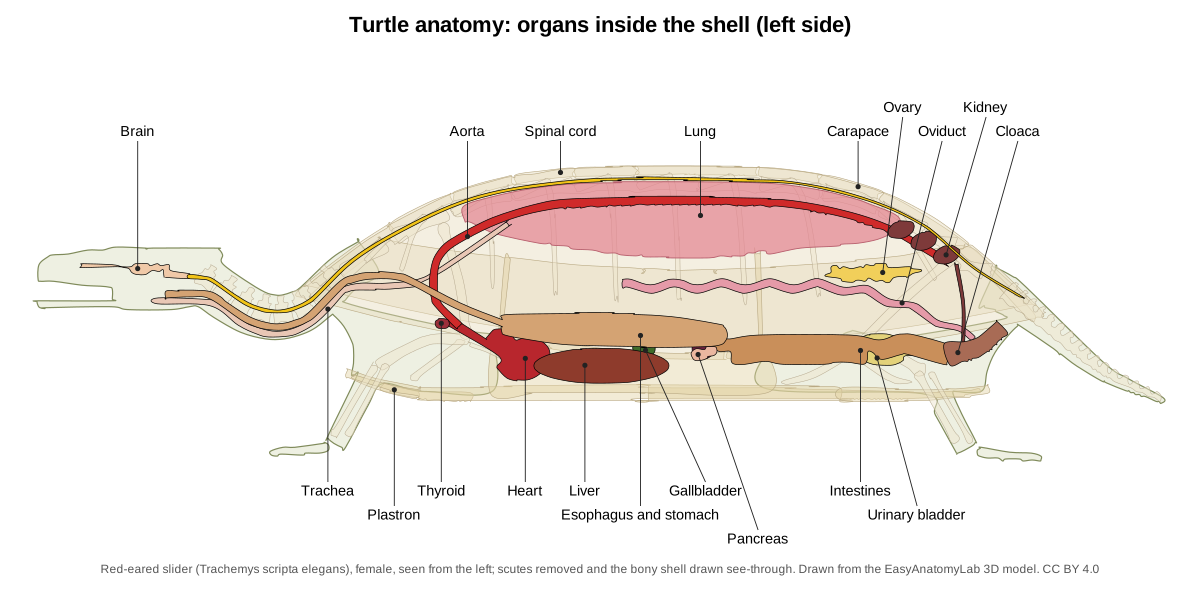
| Organ | Where it is in a red-eared slider | Notes |
|---|---|---|
| Lungs | Top of the body cavity, above all other organs | Spongy and mostly pink[2] |
| Heart | Midline, front half of the body cavity, in its own membrane sac | Two atria, one ventricle[2, 1] |
| Liver | Lowest organ, just above the plastron | Right, left and intermediate lobes; dark red-brown[2] |
| Gallbladder | On the right liver lobe | Stores bile[2, 1] |
| Stomach | On the upper surface of the left liver lobe | Ends in the pylorus[2, 1] |
| Intestines | Right side, behind the stomach | The colon starts with a small caecum[2, 1] |
| Pancreas and spleen | Behind the liver along the duodenum; spleen at the pancreas's far end | The spleen is lymphoid, not digestive[2, 1] |
| Kidneys | Back of the body cavity, behind the lungs | Outside the body-cavity lining, against the shell[2, 1] |
| Ovaries or testes | Behind the lungs, below the kidneys | Ovaries hold yellow follicles[2, 1] |
| Urinary bladder | Bottom of the body cavity, near the pelvis | Fills from the cloaca[2, 1] |
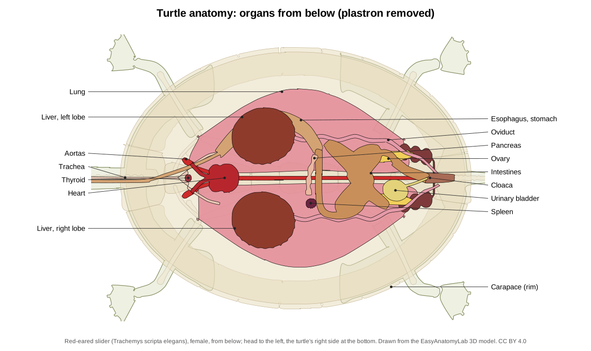
Turtle heart: three chambers and a partly divided ventricle
A turtle heart has three chambers: two atria and a single ventricle.[12] Inside, the ventricle has three spaces, the cavum venosum, cavum arteriosum and cavum pulmonale, separated only partly by a muscular ridge and septa.[13, 1] Wyneken's sea turtle manual counts a fourth chamber, the thin-walled sinus venosus, which collects blood from the body before it enters the right atrium.[1]
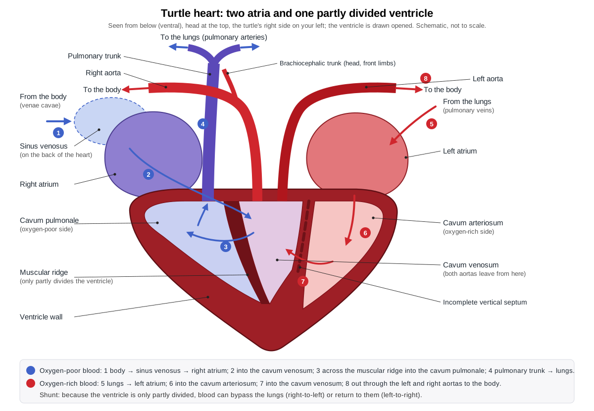
Blood from the body passes through the right atrium and the cavum venosum into the cavum pulmonale, which sends it to the lungs. Oxygen-rich blood returns to the left atrium and the cavum arteriosum, then crosses to the cavum venosum, where both aortas begin.[12] The right aorta gives off the brachiocephalic trunk to the head and front limbs and runs back to join the left aorta; in sea turtles the left aorta supplies the gut.[1]
Shunts. Because the ventricle is not fully divided, blood can skip the lungs (a right-to-left shunt) or go back to them (left-to-right).[12] In free-swimming red-eared sliders, shunting happens during routine activity as well as dives,[15] and sliders acclimated to cold had a 2.3-fold higher right-to-left shunt flow when measured under anaesthesia.[16] Even the leatherback, the largest turtle, has this typical reptile heart, only larger: a 950 ml ventricle in a 300 kg turtle, about twice the relative size of other reptiles'.[14] Pythons and monitor lizards, by contrast, have a well-developed septum.[14]
Turtle digestive system
Turtles have no teeth; a keratin beak, the rhamphotheca, covers the jaws, and in sea turtles the tongue is fixed to the floor of the mouth and cannot be pushed out.[1] The esophagus runs down the neck and bends sharply to the left to join the stomach, which is smooth-walled and ends in a muscular pylorus. Sea turtles have sharp, keratinized spines lining the esophagus that point towards the stomach and are thought to hold food while water is pushed out.[1]
The liver is the largest visceral organ.[1] In sliders it has right, left and intermediate lobes,[2] while the sea turtle manual describes two lobes joined by strips of liver tissue.[1] The gallbladder sits on the right lobe: on its upper surface in sliders,[2] and on its rear or lower surface in sea turtle descriptions.[1] The pancreas makes digestive enzymes as well as insulin and glucagon.[1] Young sliders are mostly carnivorous; adults develop gut microbes that let them digest plants.[3]
Kidneys, bladder and cloaca
The kidneys lie behind the lungs, outside the lining of the body cavity, against the shell. Their nephrons have no loop of Henle, and the ureters empty into the cloaca, not into the bladder; urine reaches the bladder from the cloaca.[1] The cloaca has three regions: the coprodeum receives faeces, the urodeum the ureters, genital ducts and bladder, and the proctodeum lies at the vent.[1]
Tortoises put the bladder to work: desert tortoises use it as a water reserve, reabsorbing water from it in dry weather, and those that empty it when handled by researchers are less likely to survive the year.[19]
Turtle senses: eyes, ears and smell
Red-eared sliders see a wide range of colours: their eyes have receptors for ultraviolet, violet, blue, green and red light.[3] Each ear has a tympanum (eardrum) at the edge of an air-filled middle ear cavity; its shape is similar across turtle families and suits hearing under water.[18] Sliders hear low sounds: tests ran from 0.2 to 1.1 kHz, and females were more sensitive than males at most frequencies.[17] The brain has olfactory bulbs at the front for smell, and sea turtles have large salt glands above and behind each eye that remove excess salt from the body.[1]
Male vs female turtle anatomy
In red-eared sliders, males have long front claws, a tail that is wider at the base and a vent that opens beyond the edge of the shell; females are larger, with a narrower tail and the vent at the shell's edge.[4] Use the Female/Male switch in the viewer to compare the gonads.
| Male slider | Female slider | |
|---|---|---|
| Body size | Smaller: average shell 17.8 cm | Larger: average shell 25.4 cm[3] |
| Front claws | Long; used to vibrate water near the female's eyes in courtship | Short[4, 3] |
| Tail and vent | Tail wide at the base; vent beyond the shell edge | Tail narrower; vent at the shell edge[4] |
| Colour with age | Older males often turn almost black (melanism)[3] | — |
| Gonads | Testes and coiled vas deferens; one penis in the floor of the cloaca | Ovaries with yellow follicles; oviducts[1] |
Claw length is the most reliable single sign in sliders: in a Brazilian study it was the only external trait used to identify males, confirmed by dissection.[4] In box turtles and gopher tortoises the male's plastron is concave, and male box turtles have red rather than brown eyes.[21, 20] A slider's sex is set by the temperature of its nest: warm nests produce females and cool nests males.[3, 6]
Turtle vs tortoise vs sea turtle anatomy
| Feature | Pond turtle (slider) | Tortoise | Sea turtle |
|---|---|---|---|
| Limbs | Webbed feet, five digits[3] | Shovel-like front legs; flat, stumpy, unwebbed hind feet (gopher tortoise)[20] | Flippers[1] |
| Windpipe | Shorter, narrower glottis than tortoises[9] | Very short trachea in Testudo[9] | — |
| Water and salt | — | Bladder used as a water store (desert tortoise)[19] | Large salt glands by the eyes[1] |
| Esophagus | — | — | Lined with keratin spines[1] |
| Male plastron | — | Concave (gopher tortoise)[20] | — |
| Shell covering | — | — | Scutes, or blubber and waxy skin with small bony plates in the leatherback[1] |
A dash means the feature was not compared in the sources used for this page.
Turtles are reptiles, like lizards and snakes, which also have a single, partly divided ventricle; the bony shell, the ribs fused into it and the muscle-driven breathing are what set turtles apart.[14, 7]
Turtle anatomy labeled: every structure in the 3D model
This is the text version of the 3D turtle: all 86 labeled structures, grouped by body system. Structures marked "males only" or "females only" appear when you switch sex in the viewer.
Lungs & airway
- Lungs
- Two large, spongy lungs pressed against the underside of the carapace. Breathe air. They fill as the flank muscles pull the organs away and empty as other muscles squeeze the organs up against them.
- Trachea & bronchi (Windpipe)
- The windpipe running down the neck, splitting into two unbranched bronchi that enter the lungs. Carries air between the glottis and the lungs, even when the neck is folded back into the shell.
Heart & blood
- Heart
- A heart with two atria and a single ventricle that is only partly divided inside; usually called three-chambered. Pumps blood to the lungs and the body. Because the ventricle is not fully divided, blood can be sent past the lungs (a shunt) when the turtle is not breathing.
- Spleen
- A small, round to oval, dark red organ of the immune system. A lymphoid organ involved in immune defence. Despite its position next to the gut, it is not a digestive gland (Wyneken, 2001).
- Aortas (left and right) (Aortic arches, dorsal aorta)
- Two aortas leave the front of the heart and join behind it as the dorsal aorta. Carry blood from the heart to the body. In sea turtles the right aorta supplies the head, front limbs and lower body and the left aorta the gut, before the two join (Wyneken, 2001).
Digestive
- Liver
- The largest organ in the body cavity: dark red-brown, with a right and a left lobe on either side of the heart. Makes bile, processes the nutrients absorbed from the gut, stores energy and makes blood proteins such as albumin and clotting factors.
- Esophagus and stomach (Oesophagus, gullet)
- The esophagus runs down the neck beside the trachea and turns sharply left into a J-shaped, muscular stomach. Swallows food whole or in chunks (turtles have no teeth) and begins digesting it; the pylorus, a muscular ring at the end of the stomach, controls passage into the duodenum.
- Intestines
- The coiled small intestine (duodenum, jejunum, ileum) and the shorter, wider large intestine (colon), which ends at the rectum. Digest and absorb food and reabsorb water.
- Cloaca
- The shared chamber at the end of the gut, urinary and reproductive tracts, opening at the vent under the tail. Receives faeces from the rectum, urine from the ureters and bladder, and eggs or sperm, and releases them through the vent.
- Gallbladder
- A small, round, dark green sac that stores bile. Stores bile made by the liver and sends it down the common bile duct to the duodenum when food arrives.
- Pancreas
- A long, narrow, pink to yellow-orange gland lying along the duodenum. Makes digestive enzymes that flow into the duodenum, and hormones: insulin and glucagon, which control blood sugar (Wyneken, 2001).
Urinary & reproductive
- Kidneys and ureters (Metanephric kidneys)
- Two flattened, lobed kidneys pressed against the inside of the carapace at the back of the body cavity, each drained by a ureter. Filter the blood and make urine, which runs down the ureters to the cloaca.
- Urinary bladder
- A thin-walled sac that stores urine; turtles have one, unlike snakes. Stores urine and lets water be reabsorbed from it.
- Testes and vas deferens · males only
- Two smooth, yellow to tan, spindle-shaped testes, each drained by a coiled vas deferens. Make sperm, which pass through the epididymis and vas deferens to the cloaca at the base of the penis.
- Ovaries · females only
- Two long, narrow ovaries studded with round yellow follicles (developing yolks). Make the eggs: each follicle is a yolk that is released into the oviduct.
- Oviducts (Uterine tubes) · females only
- Two long tubes that carry eggs from the ovaries to the cloaca. Carry the follicles, then the finished eggs, to the cloaca for laying.
Brain & glands
- Thyroid gland
- A single, round, red to brown gland at the base of the neck. A hormone (endocrine) gland in the neck.
- Brain
- A small, elongated, white brain inside the braincase. Controls the body and processes the senses. From front to back: olfactory bulbs (smell), cerebral hemispheres, optic lobes (vision), cerebellum (movement) and medulla (Wyneken, 2001).
- Spinal cord
- The cord of nerve tissue running from the brain down the neck and along the backbone to the tail. Carries signals between the brain and the body and limbs.
Scutes & skin
- Cervical scute (Nuchal scute, precentral scute)
- The small single scute at the front midline of the carapace, above the neck. Covers and protects the front rim of the shell over the neck opening.
- Vertebral scutes (Central scutes)
- Five broad keratin plates running in a row down the middle of the carapace. Form the protective, waterproof surface over the backbone and neural bones.
- Pleural scutes (Costal or lateral scutes)
- Four pairs of large keratin plates on the sides of the carapace, between the vertebrals and marginals. Cover the flanks of the carapace.
- Marginal scutes
- The ring of small scutes around the rim of the carapace: 12 on each side, counting the supracaudals. Protect the shell edge; the rear marginals of sliders are slightly serrated.
- Supracaudal scutes (12th marginals, postcentral scutes)
- The last pair of marginal scutes, meeting at the midline above the tail. Cover the rear rim of the shell above the tail opening.
- Gular scutes
- The front pair of plastron scutes, under the throat. Protect the front lobe of the plastron under the neck.
- Humeral scutes
- The second pair of plastron scutes, under the front legs (humerus). Cover the front lobe, which shields the head and forelimbs when they are pulled in.
- Pectoral scutes
- The third pair of plastron scutes, at chest level. Cover the chest region of the plastron.
- Abdominal scutes
- The fourth and usually largest pair of plastron scutes, at the bridge. Cover the belly and the central plastron.
- Femoral scutes
- The fifth pair of plastron scutes, in front of the hind legs (femur). Cover the rear lobe of the plastron.
- Anal scutes
- The last pair of plastron scutes, at the rear notch near the tail. Cover the tail end of the plastron; the anal notch between them leaves room for the tail.
- Claws
- Keratin claws on the tips of the digits. Grip the bottom and banks, tear food and dig nests.
- Skin (Scaly skin of the head, neck, limbs and tail)
- Soft, scaly, olive-green skin with yellow stripes and the red patch behind each eye that names the red-eared slider. Protects the soft parts; the webbed skin between the toes makes the feet into paddles.
Carapace bones
- Nuchal bone
- The single, broad bone at the front midline of the carapace, above the neck. Forms the front of the shell and anchors the neck muscles; the last neck vertebra connects to the shell beneath it.
- Neural bones (Neurals, vertebral plates)
- Usually eight small, hexagonal plates down the midline of the carapace, fused to the vertebrae below. Form the roof of the trunk vertebrae inside the shell; the spinal cord runs in the space between the neural bones and the vertebral bodies.
- Pleural bones (Costal plates, costals)
- Eight pairs of broad plates on the sides of the carapace, each a rib fused with a sheet of dermal bone. Make up most of the domed carapace and enclose the lungs and other organs.
- Peripheral bones (Peripherals)
- A ring of narrow bones (usually 11 on each side) that forms the rim of the carapace. Form the shell edge; the middle peripherals join the bridge to the plastron.
- Suprapygal bone (Suprapygals (one or two, by species))
- A midline bone (or pair of bones) between the last neural and the pygal. Closes the back of the carapace between the last pleurals.
- Pygal bone
- The single bone at the rear midline of the carapace rim, above the tail. Forms the rear edge of the shell over the tail.
Plastron & bridge
- Epiplastra (Epiplastron (singular))
- The front pair of plastron bones. Form the front edge of the plastron under the neck.
- Entoplastron
- The single, unpaired, diamond- or T-shaped bone in the front of the plastron. Ties the front plastron bones together; the acromion processes of the shoulder girdles attach to it by ligaments.
- Hyoplastra (Hyoplastron (singular))
- The large second pair of plastron bones, from behind the entoplastron to the middle of the bridge. Form much of the plastron and the front strut of the bridge (axillary buttress) that joins the carapace.
- Hypoplastra (Hypoplastron (singular))
- The large third pair of plastron bones, from the middle of the bridge back to the hind legs. Form the rear strut of the bridge (inguinal buttress) and the central plastron.
- Xiphiplastra (Xiphiplastron (singular))
- The rear pair of plastron bones, with the anal notch between them. Form the rear tip of the plastron in front of the tail.
- Bridge (Axillary and inguinal buttresses)
- The upturned sides of the plastron that join it to the carapace on each side, between the front and hind legs. Locks top and bottom shell into one rigid box. Inside, the axillary and inguinal buttresses brace it against the carapace.
Skeleton
- Premaxillae
- The small paired bones at the tip of the upper jaw, below the nostril opening. Form the front of the upper jaw that carries the horny beak.
- Maxillae
- The main bones of the upper jaw on each side, below the orbit. Carry the cutting edge of the upper beak and form part of the palate.
- Prefrontals
- Paired bones roofing the snout in front of the eyes. Form the front of the skull roof and the front rim of each orbit.
- Frontals
- Paired roofing bones between the eyes. Roof the front of the braincase and the upper rims of the orbits.
- Parietals
- The paired bones forming the roof of the braincase behind the eyes. Cover the brain and anchor the large jaw-closing muscles.
- Postorbitals
- Bones forming the back rim of each orbit. Close the rear of the orbit and brace the skull roof.
- Jugals (Cheek bones)
- Small cheek bones below and behind each orbit. Form part of the cheek and the lower edge of the orbit.
- Quadratojugals
- Cheek bones in front of the ear region. Link the cheek to the quadrate, which carries the jaw joint.
- Squamosals
- Bones at the upper back corners of the skull. Form the rear corner of the skull and anchor neck and jaw muscles.
- Quadrates (With the middle ear (tympanic) cavity)
- The bone on each side that carries the jaw joint and surrounds the middle ear. The lower jaw hinges on it; its wide, rounded rim supports the eardrum.
- Supraoccipital crest
- A blade-like crest projecting backwards from the top of the braincase. Anchors the large jaw-closing (adductor) muscles.
- Braincase & occipital condyle (Basioccipital, exoccipitals, prootic, opisthotic)
- The bones enclosing the brain and inner ear, ending in the single knob that joins the neck. Protects the brain and inner ear; the occipital condyle articulates with the atlas (first neck vertebra).
- Palate (Vomer, palatines, pterygoids)
- The bones forming the roof of the mouth. Separate the mouth from the nasal passage and anchor jaw muscles.
- Orbit & scleral ring (Scleral ossicles)
- The large eye socket and the ring of small bony plates that supports the eyeball. Houses and protects the eye; the scleral ossicles keep its shape.
- Hyoid apparatus (Hyoid body and horns)
- A bony body and paired horns that hang between and behind the lower jaws. Supports the tongue and glottis and anchors throat muscles used in feeding and breathing.
- Dentaries
- The front bones of the lower jaw, joined at the chin. Carry the lower beak and the bite force.
- Post-dentary bones (Surangular, angular, articular, prearticular, coronoid, splenial)
- The smaller bones at the back of each lower jaw. Form the jaw joint and the attachment for the jaw muscles.
- Upper rhamphotheca (Upper beak)
- The horny keratin sheath covering the upper jaw bones. Works as the cutting and crushing surface instead of teeth.
- Lower rhamphotheca (Lower beak)
- The keratin sheath covering the front of the dentaries. Bites against the upper beak to slice plants and prey.
- Cervical vertebrae (Neck vertebrae C1–C8)
- Eight neck vertebrae, from the atlas behind the skull to the eighth, which meets the shell. Let the head reach, strike and fold back into the shell.
- Trunk vertebrae (Dorsal or thoracic vertebrae (10))
- Ten trunk vertebrae fused to the underside of the carapace midline. Carry the ribs and spinal cord; they cannot bend, because they are part of the shell.
- Ribs (Dorsal ribs fused to the pleurals)
- Broad ribs that run outward under the carapace and are fused into the pleural bones. Form the frame of the carapace; because they cannot move, turtles breathe with abdominal muscles instead.
- Sacral vertebrae
- Two (sometimes three) vertebrae whose rib-like side processes support the hips. Connect the backbone to the pelvis through the ilia.
- Caudal vertebrae (Tail vertebrae)
- A tapering series of small tail vertebrae. Support the tail, which steers in water and, in males, positions the cloaca during mating.
- Scapula (Shoulder blade (dorsal process))
- A rod-like shoulder blade that rises from the shoulder joint to the underside of the carapace. Braces the shoulder joint against the shell.
- Acromion process
- A long process that runs from the scapula inward along the front of the shell. Ties the shoulder girdle to the plastron (entoplastron) by ligaments and anchors limb muscles.
- Coracoid (Procoracoid in some texts)
- A flat, backward-pointing bone that completes the shoulder girdle. Forms part of the shoulder socket (glenoid) and anchors the muscles that pull the forelimb back.
- Ilium
- The hip bone that rises from the hip socket to the sacral vertebrae. Connects the pelvis to the backbone and, by ligaments, to the carapace.
- Pubis
- The front, lower hip bone, with a forward-pointing process. Forms the front of the pelvis and anchors belly and thigh muscles.
- Ischium
- The rear, lower hip bone. Forms the back of the pelvis; the large opening between pubis and ischium is the thyroid fenestra.
- Humerus
- The curved upper-arm bone, whose head sits in the shoulder socket inside the shell. Moves the front leg forward and back for walking and swimming.
- Radius
- One of the two forearm bones, on the thumb side. Supports the forearm and wrist.
- Ulna
- The second forearm bone, on the little-finger side. Supports the forearm and forms the elbow joint with the humerus.
- Carpals (Wrist bones: radiale, intermedium, ulnare, centrale, pisiform, distal carpals)
- A cluster of small wrist bones. Make the wrist flexible.
- Metacarpals
- Five slender bones of the palm. Support the webbed front foot.
- Phalanges of the front foot (Finger bones)
- The bones of the five fingers. Spread the web for swimming and carry the claws.
- Femur
- The thigh bone, from the hip socket inside the shell to the knee. Moves the hind leg for walking, swimming and digging nests.
- Tibia
- The larger shin bone. Carries the weight of the hind leg.
- Fibula
- The slender outer bone of the lower leg. Supports the outer side of the ankle.
- Tarsals (Astragalus, calcaneum, distal tarsals)
- The short ankle, made of a few large and several small bones. Form the ankle joint.
- Metatarsals
- Five bones of the sole of the hind foot. Support the broad, webbed hind foot, the slider's main paddle.
- Phalanges of the hind foot (Toe bones)
- The bones of the toes. Spread the web for swimming and carry the claws used for digging.
Frequently asked questions
How many chambers does a turtle heart have?
Three, in the usual count: two atria and one ventricle. The ventricle is partly divided by a muscular ridge into three linked spaces, the cavum venosum, cavum arteriosum and cavum pulmonale, which keeps oxygen-rich and oxygen-poor blood partly apart. Wyneken's sea turtle manual counts four chambers by adding the sinus venosus, which collects blood before the right atrium.
Where are a turtle's lungs?
On top of everything else. The two large, spongy lungs lie in the upper part of the body cavity, pressed against the underside of the carapace and attached along their inner edges to the backbone. The heart, liver, stomach and intestines lie below them, just above the plastron.
Do turtles have a diaphragm?
Not a mammal-like one. Turtles breathe with two pairs of abdominal muscles instead. The transversus wraps around the organs like a sling and squeezes them up against the lungs to breathe out; the obliquus abdominis, in the flank between each hind leg and the shell, flattens to pull the organs down and breathe in. Some anatomists call part of the transversus the diaphragmaticus.
Can turtles breathe through their butts?
A few can take up some oxygen that way. Australian side-necked turtles such as the Fitzroy River turtle have cloacal bursae, paired sacs off the cloaca that extract oxygen from water pumped in and out, which helps them stay under for long dives. Turtles without bursae, such as red-eared sliders, breathe air with their lungs.
How can you tell if a turtle is male or female?
In red-eared sliders, adult males have much longer front claws, a tail that is wider at the base and a vent (cloacal opening) that lies beyond the edge of the shell; females have shorter claws, a narrower tail and the vent at the shell's edge, and grow larger. In box turtles and gopher tortoises the male's plastron is concave.
Do turtles have ears?
Yes. Each side of the head has an eardrum (tympanum) over an air-filled middle ear cavity. Turtles hear low sounds best: red-eared sliders were tested between 0.2 and 1.1 kHz, and females were more sensitive than males at most frequencies. The shape of the middle ear suggests it works well under water.
How is a tortoise's body different from a turtle's?
Tortoises are land turtles. The gopher tortoise has shovel-like front legs for digging and flat, stumpy, unwebbed hind feet, while sliders have webbed feet for swimming. Tortoises of the genus Testudo have a very short trachea, and desert tortoises use the urinary bladder as a water reserve during droughts.
References
Every source below was opened and checked on 25 October 2026.
- Wyneken, J. (2001). The Anatomy of Sea Turtles. U.S. Department of Commerce, NOAA Technical Memorandum NMFS-SEFSC-470
- Pedram, S. et al. (2025). Laparoscopic anatomy of the coelomic cavity organs in female red-eared slider (Trachemys scripta elegans). Veterinary Medicine International (PMC12313375)
- Animal Diversity Web, University of Michigan: Trachemys scripta (pond slider)
- Gradela, A. et al. (2017). Sexual dimorphism in red-eared sliders (Trachemys scripta elegans) from the Wild Animal Triage Center of the Tiete Ecological Park, São Paulo, Brazil. Acta Scientiae Veterinariae 45, 1468 (PDF)
- Readel, A. M. et al. (2008). Maturational changes in male slider turtles (Trachemys scripta) from Illinois. Herpetological Conservation and Biology 3, 170–175
- Hatkevich, T. et al. (2025). Gonadal sex and temperature independently influence germ cell differentiation and meiotic progression in Trachemys scripta. PNAS (PMC11725912)
- Lyson, T. R. et al. (2014). Origin of the unique ventilatory apparatus of turtles. Nature Communications 5, 5211
- National Geographic (2009): How the turtle got its shell, on Nagashima et al., Science 325, 193–196
- Habova, M. et al. (2022). Differences in the anatomy of the lower respiratory tract in selected species of the order Testudines. Veterinární medicína 67, 78–86
- Gordos, M. A., Limpus, C. J. & Franklin, C. E. (2006). Response of heart rate and cloacal ventilation in the bimodally respiring freshwater turtle, Rheodytes leukops, to experimental changes in aquatic PO2. Journal of Comparative Physiology B 176, 65–73 (Europe PMC)
- Fielder, D. P. (2012). Seasonal and diel dive performance and behavioral ecology of the bimodally respiring freshwater turtle Myuchelys bellii of eastern Australia. Journal of Comparative Physiology A 198, 129–143 (Europe PMC)
- De Voe, R. S. (2010). Reptilian cardiovascular anatomy and physiology: evaluation and monitoring (proceedings). dvm360
- Poelmann, R. E. et al. (2014). Evolution and development of ventricular septation in the amniote heart. PLoS ONE 9, e106569 (PMC4156344)
- Jensen, B. et al. (2022). Anatomy of the heart of the leatherback turtle. Journal of Anatomy 241, 535–544 (PMC9296022)
- Williams, C. L. & Hicks, J. W. (2016). Continuous arterial PO2 profiles in unrestrained, undisturbed aquatic turtles during routine behaviors. Journal of Experimental Biology 219, 3616–3625 (Europe PMC)
- Keen, A. N., Shiels, H. A. & Crossley, D. A. (2016). Cardiovascular function, compliance, and connective tissue remodeling in the turtle, Trachemys scripta, following thermal acclimation. American Journal of Physiology 311, R133–R143 (Europe PMC)
- Wang, T. et al. (2019). Auditory brainstem responses in the red-eared slider Trachemys scripta elegans reveal sexually dimorphic hearing sensitivity. Journal of Comparative Physiology A (PMC6863946)
- Willis, K. L. et al. (2013). Middle ear cavity morphology is consistent with an aquatic origin for testudines. PLoS ONE 8, e54086 (PMC3544720)
- Averill-Murray, R. C. (2002). Effects on survival of desert tortoises (Gopherus agassizii) urinating during handling. Chelonian Conservation and Biology 4(2) (PDF)
- Animal Diversity Web, University of Michigan: Gopherus polyphemus (gopher tortoise)
- Illinois Natural History Survey, Herpetology collection: Terrapene carolina (eastern box turtle)
More interactive anatomy models
- Snake anatomyElongated organs, one working lung, a flexible jaw and the snake heart.
- Snake skeletonHundreds of vertebrae and ribs, a kinetic skull and vestigial hip bones.
- Turtle skeletonHow ribs and spine fuse into the shell: carapace, plastron and girdles.
- Lizard anatomySkull, scales, three-chambered heart and tail autotomy of a lizard.
- Human anatomy1,000+ bones, muscles, organs, vessels and nerves from real anatomical data.
- Octopus anatomyThree hearts, a ring-shaped brain, eight arms and a beak, in 3D.