

Sea urchin anatomy at a glance
A sea urchin is an echinoderm, a relative of sea stars and sea cucumbers. Its body is a hollow globe with the mouth in the middle of the flat underside (the oral surface) and the anus at the top (the aboral surface). Except for a thin skin, all of its soft anatomy is inside the test (Fox).[1, 30]
Everything comes in fives: five double rows of tube feet, five teeth, five gonads, five radial canals and five radial nerves.[1, 3]
This page describes the purple sea urchin, Strongylocentrotus purpuratus, which lives on rocky shores of the Pacific coast of North America and is a standard animal in embryology classes. Its anatomy is typical of the "regular" urchins. Where the green urchin (S. droebachiensis), Arbacia, Diadema, pencil urchins (cidaroids) or the irregular sand dollars and heart urchins differ, the text says so. Irregular urchins are flattened, have short spines and a superficial bilateral symmetry, and most have a reduced lantern (Fox; UCMP).[26, 21, 1, 30]
Two directions matter: oral (towards the mouth) and aboral (towards the anus). The five ambulacra, the zones with tube feet, run from mouth to apex like lines of longitude; the five interambulacra lie between them. Biologists number them from the madreporite, which sits in interambulacrum 2.[3, 6]
| Part | Where | Main job |
|---|---|---|
| Test | The whole shell | Rigid skeleton of fused calcite plates[1, 3] |
| Spines and pedicellariae | All over the test | Defence, walking, cleaning[1, 14] |
| Tube feet | Ten rows along the ambulacra | Walking, gripping, breathing, sensing[1, 11] |
| Madreporite | Top, beside the anus | Opening of the water vascular system[6, 21] |
| Aristotle's lantern | Inside, above the mouth | Jaws with five self-sharpening teeth[25, 12] |
| Stomach and intestine | Two loops around the inside of the test | Digestion and absorption[1] |
| Gonads | Five, against the interambulacra | Make eggs or sperm[1, 21] |
| Nerve ring | Around the pharynx at the base of the lantern | Links the five radial nerves[1] |
What is a sea urchin test made of?
A sea urchin test is an internal skeleton of thin calcite plates fused into a rigid shell. In a regular urchin the plates form ten double columns from mouth to apex: five ambulacral zones pierced by pore pairs for the tube feet, and five interambulacral zones without pores. A thin living skin covers it (Fox; NHM Echinoid Directory).[1, 3]
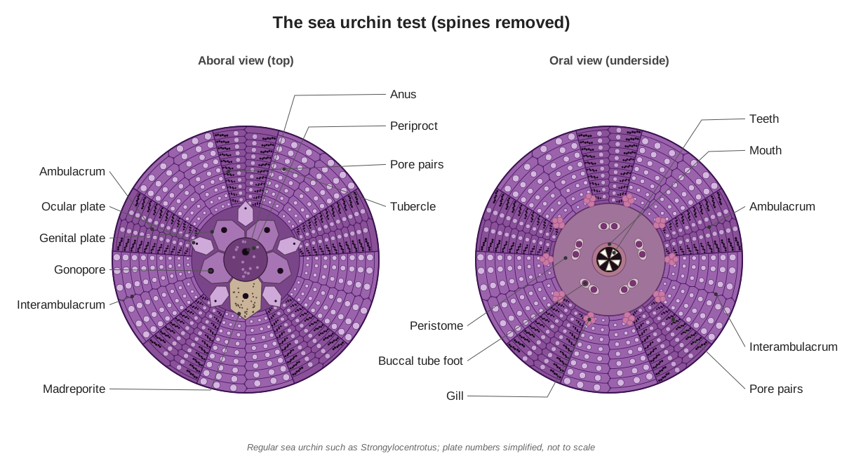
At the top, a ring of ten plates called the apical disc surrounds the periproct, the flexible membrane that holds the anus. Five large genital plates sit on the interambulacra, each with a gonopore; one of them, genital plate 2, is enlarged and sieve-like and forms the madreporite. Five small ocular plates sit at the tops of the ambulacra; each has a tiny pore for the end of a radial canal (NHM Echinoid Directory). New ambulacral plates are added just below the ocular plates as the urchin grows (Turner, 2026).[6, 19]
On the underside, the peristome is a soft membrane covering a large hole in the test, with the mouth at its centre. It carries five pairs of small buccal plates, each with a tube foot. Around its edge, inside the test, the perignathic girdle forms five tall arches called auricles that anchor the muscles of the lantern (NHM Echinoid Directory; Fox).[7, 1]
What are sea urchin spines and pedicellariae for?
Sea urchin spines defend the animal and help it move and wedge itself into rock; pedicellariae are tiny stalked pincers between the spines that keep the surface clean and attack small intruders. Each spine sits on a ball-shaped tubercle and is moved by a ring of muscle, while collagen fibres can lock it rigidly in place (Fox; NHM Echinoid Directory).[1, 7]
Purple urchin spines are short, rarely more than 2.5 cm long (Walla Walla University). Adults have bright purple spines, while juveniles under about 30 mm are often pale green (UCSC MARINe). Broken spines regrow (ASU Ask A Biologist). Pedicellariae have three jaws on a stalk. Urchins have four main types: globiferous pedicellariae with venom glands, ophicephalous, tridentate and triphyllous; all four can occur on the same animal (Ehlert-Flaskämper et al., 2025). Small glossy sphaeridia on the ambulacral plates are thought to sense balance (Thompson et al., 2021).[26, 28, 24, 14, 18]
How do tube feet and the water vascular system work?
Sea urchin tube feet are hydraulic. Fluid thought to enter through the madreporite runs down the stone canal to a ring canal on top of Aristotle's lantern, then along five radial canals up the ambulacra. Squeezing the flat ampulla inside the test pushes fluid into its tube foot and extends it (Fox; University of Washington).[1, 21]
| Part | Position | What it does |
|---|---|---|
| Madreporite | Genital plate 2, top of test | Sieve-like entrance; helps keep pressure right[6, 21] |
| Stone canal | From madreporite down to the lantern | Links madreporite and ring canal[1] |
| Ring canal | Top of the lantern, around the oesophagus | Hub that feeds the radial canals[1] |
| Tiedemann's bodies | Five pouches on the ring canal | Clear particles from the fluid[1] |
| Radial canals | Down the lantern, through the auricles, up each ambulacrum | Supply the tube feet[1] |
| Ampullae | Inside the test, under each tube foot | Extend the tube foot; exchange gases[1] |
| Tube feet | Ten rows outside the test | Grip, walk, breathe, sense light[1, 11] |
| Terminal tentacles | Through the ocular plates | End of each radial canal[6, 1] |
Each tube foot connects to its ampulla through two pores in the test, which is why the pores come in pairs. Urchin ampullae are flat, standing on edge like the pages of a book, rather than round bulbs as in a sea star (Fox). The suckers are stiffened by tiny calcite plates, and extended tube feet are usually longer than the spines. Strongylocentrotus has Tiedemann's bodies on its ring canal but no Polian vesicles (Fox).[1, 2]
What is Aristotle's lantern?
Aristotle's lantern is the sea urchin's jaw apparatus: five interlocking jaws called pyramids, each holding one long tooth, surrounding the mouth inside the test. Muscles push the lantern out to scrape algae off rock and pull it back in. Aristotle described it in his History of Animals, and its shape recalls a five-sided horn lantern (ASU Ask A Biologist).[25]
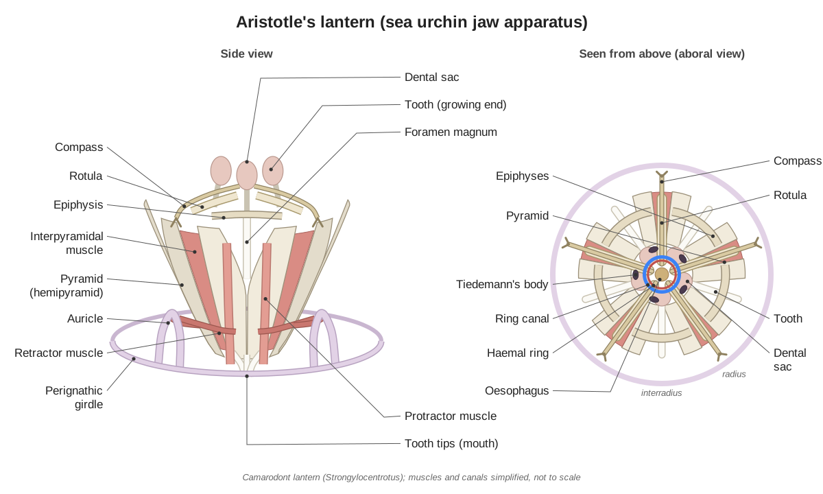
Each pyramid is two wedge-shaped hemipyramids joined in the middle, with a notch, the foramen magnum, at the top of their joint. Small axe-shaped epiphyses cap each pyramid, a tongue-shaped rotula lies between neighbouring pyramids, and a narrow compass rests on each rotula (NHM Echinoid Directory). Sources count the pieces differently: the NHM gives about 50 skeletal elements and 60 muscles, while Fox gives 40 ossicles (five jaws of eight) and 60 muscles.[4, 1]
| Part | Number | Job |
|---|---|---|
| Pyramids (10 hemipyramids) | 5 | The jaws; they close on each other[4, 1] |
| Teeth | 5 | Scrape and bite; grow continuously[1, 12] |
| Epiphyses | 10 | Cap the pyramids; carry the rotulae[4] |
| Rotulae | 5 | Brace neighbouring jaws[4] |
| Compasses | 5 | Raise and lower the lantern coelom wall[4] |
| Interpyramidal muscles | 5 | Close the jaws[1] |
| Protractor and retractor muscles | Paired | Push the lantern out and pull it in[1] |
The teeth wear down at the tip and grow continuously from a soft upper end held in a dental sac (Fox). In camarodont urchins, the group that includes the purple urchin, about 1.5 mm of tooth is replaced each week; the tooth is self-sharpening, and its "stone" part has the most magnesium (Stock, 2014). Purple urchins use their teeth, and to a lesser extent their spines, to grind shelter pits in rock (Troha et al., 2024). Strongylocentrotus has a camarodont lantern, with a deep V-shaped foramen magnum, enlarged epiphyses that meet above it, and T-shaped teeth (NHM Echinoid Directory).[1, 12, 13, 5]
Sea urchin internal anatomy
Inside a sea urchin, the large body cavity (the perivisceral coelom) is filled with fluid. Aristotle's lantern sits in the centre of the floor, the stomach and intestine loop around the inner wall, five gonads hang under the top of the test, and the axial complex runs from the madreporite down to the lantern (Ziegler et al., 2008; Fox).[8, 1]
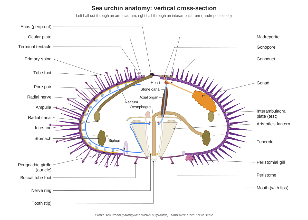
Food takes a long route. It passes from the mouth up the pharynx inside the lantern, then along the oesophagus to the wall of the test. There the gut widens into the stomach, which makes one full circle around the lower part of the test, counterclockwise when seen from above. At its end the gut reverses as the intestine, which circles back clockwise, higher on the wall, and finally the rectum rises to the anus (Fox). A thin tube, the siphon, bypasses the stomach and probably carries off swallowed water so it does not dilute the digestive enzymes (Fox; Holland & Ghiselin, 2009).[1, 10]
Does a sea urchin have a heart, blood or kidneys?
A sea urchin has no heart like ours, but it has a small pulsating vessel at the top of its axial organ, just under the madreporite. Colourless haemal fluid runs in a ring around the top of the lantern and in vessels along the gut; most transport is done by the body-cavity fluid (Fox; Smith & Hill, 2025).[1, 16]
The axial organ is a soft brown spindle wrapped around the stone canal. Its function has long been debated: it has been called a heart, a contractile vessel and a source of immune cells. Echinoderms are generally said to have no excretory organs (Fox), but genes of the filtering cells of vertebrate kidneys are active only in the urchin axial organ, so Smith & Hill (2025) propose that it filters waste, which then leaves through the gut. The pulsating vessel beats about five times a minute according to Fox; in S. purpuratus it has been described as two contractile chambers (cited by Ziegler et al., 2009).[1, 16, 9]
Does a sea urchin have a brain or eyes?
A sea urchin has no brain. Its nervous system is a nerve ring around the pharynx at the base of Aristotle's lantern, from which five ribbon-like radial nerves run up the inside of the ambulacra, between the radial canals and the test (Fox; University of Washington). It has no eyes either, but its tube feet sense light.[1, 21, 11]
In the purple urchin, each tube foot has up to 140 light-sensitive cells at the rim of its sucker and at its base. The basal cells sit in a cup of the opaque skeleton, which shades them from one side, so Ullrich-Lüter et al. (2011) proposed that the whole urchin works like a huge compound eye. A newer gene-expression study of juvenile urchins found that most of the body has a "head-like" identity and describes sea urchins as all-brain rather than no-brain (Paganos et al., 2025).[11, 15]
How do sea urchins breathe?
Sea urchins breathe mainly through their tube feet. Oxygen crosses the thin walls of the feet into the water vascular system, and the flat ampullae inside the test pass it on to the body-cavity fluid (Fox; Walla Walla University). Regular urchins also have five pairs of bushy gills around the peristome.[1, 26]
Sources stress different jobs for these peristomial gills. Fox describes them as the respiratory organs of the lantern coelom, supplying oxygen to the hard-working lantern muscles. The NHM Echinoid Directory explains them as outpocketings that buffer the change in volume inside the rigid test when the lantern moves in and out; pencil urchins (cidaroids) lack them.[1, 7]
Male vs female sea urchins and reproduction
Male and female purple sea urchins look the same from outside (UCSC MARINe). Each has five gonads, ovaries or testes, against the interambulacra, and each gonad opens through a gonoduct to a gonopore on top of the test. Fertilisation happens in the water: females release orange eggs and males release milky white sperm (Fox; University of Washington).[28, 1, 23]
In Strongylocentrotus both ovaries and testes look orangish; only the gametes inside differ (Fox). In the breeding season the gonads can fill up to 80% of the space inside the test (University of Washington), and most purple urchins spawn from January to March (Walla Walla University; Animal Diversity Web). Each gonad is a mass of small sacs filled with nutritive cells that store food reserves before the eggs or sperm develop; these gonads are the seafood sold as uni (Rodríguez & Olmos, 2026). The larva, the echinopluteus, is bilaterally symmetrical and turns into a five-sided juvenile at metamorphosis (University of Washington; Fox).[1, 22, 21, 26, 27, 17]
Sea urchin vs starfish: pentaradial symmetry compared
Sea urchins and starfish share the echinoderm body plan: five-part symmetry, a water vascular system with a madreporite, tube feet and a calcite skeleton in the skin. In an urchin the ambulacra have spread up over the body until they enclose almost all of it, leaving only the small periproct at the top (Fox).[1, 2]
The urchin's plates are also fused into a rigid test instead of a flexible grid (UCMP). Compare with our 3D starfish model, or see how other body plans differ in comparative anatomy.[30]
| Feature | Sea urchin (Strongylocentrotus) | Starfish (Asterias) |
|---|---|---|
| Body | Rigid globe, no arms | Central disc with five flexible arms |
| Skeleton | Fused plates forming a test | Rod-like ossicles forming a flexible grid |
| Feeding | Scrapes algae with Aristotle's lantern | Everts its cardiac stomach over prey |
| Gut | Long stomach and intestine looping round the test | Large stomach with a pair of pyloric caeca in each arm |
| Gonads | Five, one per interambulacrum | Ten, a pair in each arm |
| Ampullae | Flat, like pages of a book | Round bulbs |
| Gas exchange | Tube feet and peristomial gills | Papulae (skin gills) and tube feet |
Sea urchin facts in this table come from Fox's Strongylocentrotus exercise, starfish facts from his Asterias exercise.[1, 2]
Purple sea urchin facts: size, habitat and diet
The purple sea urchin's test is usually about 5 cm across and at most about 10 cm (Walla Walla University; Animal Diversity Web; UCSC MARINe); the Monterey Bay Aquarium gives up to 7 cm. It lives from Alaska to Baja California, from the low intertidal to 160 m (Walla Walla University; UCSC MARINe), often in pits ground into rock.[26, 27, 28, 29]
Purple urchins eat algae, especially drifting giant kelp, but hungry urchins graze living kelp down to the holdfast and can turn a kelp forest into an "urchin barren" (UCSC MARINe; Monterey Bay Aquarium). Sea otters, sunflower stars and California sheephead eat them (Monterey Bay Aquarium). They grow slowly, reaching large size in about 10 years (Walla Walla University); Animal Diversity Web gives an average of 20 years in captivity, and the related red sea urchin can live for more than a century (Amir et al., 2020).[28, 29, 26, 27, 20]
Sea urchin anatomy labeled: every structure
The text version of the 3D model: every labeled structure of the purple sea urchin, grouped by body system, with what it does.
Test (skeleton)
- Ambulacral plates (Ambulacra, ambulacral zones of the test)
- The five narrower double columns of test plates that run from the mouth to the top, each plate pierced by pore pairs for the tube feet. Carry the tube feet. Each pore pair links one tube foot outside to its ampulla and the radial canal inside.
- Interambulacral plates (Interambulacra)
- The five broader double columns of plates between the ambulacra. They have no pores and carry the biggest spines. Make up most of the rigid wall of the test; the five gonads lie against their inner surface.
- Pore pairs (Podial pores)
- Pairs of tiny holes through the ambulacral plates, one pair for each tube foot. Two podial ducts pass through the two pores and connect one ampulla inside the test to one tube foot outside.
- Tubercles
- Shiny, rounded knobs on the plates where the spines sit. Form the ball of a ball-and-socket joint with the base of a spine.
- Genital plates
- Five large plates around the top of the test, each with a gonopore. Carry the five gonopores. One of them, genital plate 2, is the madreporite.
- Ocular plates
- Five small plates of the apical disc, one at the top of each ambulacrum. Each has a tiny pore through which the end of a radial canal emerges as the terminal tentacle.
- Periproct
- The small, flexible membrane at the very top of the test, with the anus in it. Closes the top opening of the test while letting the anus open.
- Peristome (Peristomial membrane)
- The large, soft, spine-free area on the underside, with the mouth at its centre. A flexible floor around the mouth that lets the lantern move in and out.
- Perignathic girdle (Auricles)
- A ring of calcite projections on the inside of the test around the peristome, with five tall arches called auricles. Anchors the muscles that move Aristotle's lantern. The radial canals and radial nerves pass through the auricle arches.
Spines & pedicellariae
- Primary spines
- The large, pointed purple spines that cover the test. Defence, walking (with the tube feet) and wedging the urchin into holes in rock.
- Secondary spines
- Shorter, thinner spines between the primary spines. Fill the gaps between the big spines and protect the skin and tube feet.
- Pedicellariae
- Tiny pincers on flexible stalks, scattered among the spines. Keep the surface clean and fight off small animals; some types inject venom.
- Sphaeridia
- Tiny glossy knobs on the ambulacral plates, like miniature spines. Thought to sense orientation and balance.
Aristotle's lantern
- Aristotle's lantern (pyramids) (Jaw apparatus; five pyramids, each a pair of hemipyramids)
- The urchin's jaw apparatus: five wedge-shaped jaws (pyramids) arranged like a five-sided lantern around the mouth. Bites and scrapes algae from rock. Muscles push the lantern out of the mouth and pull it back in.
- Teeth (Tooth bands)
- Five long, curved calcite teeth, one inside each pyramid; their hard tips meet at the mouth. Scrape algae and other food off rock; in the purple urchin they also grind out shelter pits in rock.
- Epiphyses
- Small, axe-shaped pieces on the top shoulders of each pyramid. Support the rotulae and brace the top of each jaw.
- Rotulae
- Five tongue-shaped bars at the top of the lantern, one between each pair of jaws. Brace neighbouring jaws together and act as pivots when the jaws open and close.
- Compasses
- Five narrow, spatula-shaped rods lying on top of the rotulae. Raise and lower the walls of the lantern coelom as the lantern moves in and out while feeding.
- Dental sacs
- Bulging, thin-walled sacs around the soft, growing upper end of each tooth. Enclose the place where new tooth material is laid down.
- Interpyramidal muscles
- Blocks of muscle joining each pyramid to its neighbours. Close the jaws and give the strong biting action.
- Protractor muscles
- Paired muscles from the perignathic girdle to the upper outer edge of the lantern. Push the lantern out, so the teeth protrude from the mouth.
- Retractor muscles
- Paired muscles from the auricles to the lower outer part of the pyramids. Pull the lantern back into the test.
Digestive
- Mouth
- The opening in the centre of the peristome, ringed by the five tooth tips. Takes in algae scraped up by the teeth.
- Pharynx
- The first part of the gut, a wide tube running straight up through the core of the lantern. Carries food from the mouth up to the oesophagus.
- Oesophagus (Esophagus)
- A narrow tube from the top of the lantern to the start of the stomach. Carries food from the pharynx to the stomach.
- Stomach (Lower gut loop)
- A wide, flat, sacculated tube that makes one full loop around the inside of the test. Main site of digestion.
- Siphon
- A narrow tube along the inner curve of the stomach that bypasses it. Probably carries water past the stomach so it does not dilute the digestive enzymes.
- Intestine (Upper gut loop)
- The second loop of the gut, paler than the stomach, running the opposite way around the test. Further digestion and absorption; forms faecal pellets.
- Rectum
- The last part of the gut, rising straight up to the anus. Carries faeces to the anus.
- Anus
- The opening at the top of the urchin, in the periproct. Releases faecal pellets.
- Mesenteries
- Thin sheets and strands that hang the gut from the inside of the test. Hold the delicate gut loops in place.
Water vascular
- Madreporite (Madreporic plate, genital plate 2)
- The largest plate of the apical disc, finely perforated like a sieve. Connects the water vascular system with the seawater and helps keep its pressure right.
- Stone canal
- A thin tube from the madreporite down to the ring canal on top of the lantern. Links the madreporite to the ring canal.
- Ring canal (Water ring)
- A small, transparent ring of canal at the top of the lantern, around the oesophagus. The hub of the water vascular system: it feeds the five radial canals.
- Tiedemann's bodies
- Five small, flat pouches of the ring canal, like dark smudges on the inside of the lantern. Remove foreign particles from the water vascular fluid by phagocytosis.
- Radial canals
- Five canals from the ring canal to the ambulacra, each running up the inside of the test. Supply water to the ampullae and tube feet through many short lateral canals.
- Ampullae
- Flat fluid-filled sacs inside the test, one for each tube foot, stacked like the pages of a book. Squeeze fluid into the tube feet to extend them, and exchange gases with the body cavity.
- Tube feet (Podia)
- Long, slender, extendable tubes ending in suckers, in ten rows from top to bottom. Walking, gripping, passing food to the mouth, breathing and sensing light and touch.
- Buccal tube feet (Buccal podia, on the buccal plates)
- Ten short, thick tube feet in a ring around the mouth, each on its own small buccal plate. Touch and taste food at the mouth.
- Terminal tentacles
- The tip of each radial canal, poking out of the ocular plate as a tiny tentacle. End of the radial canal; a small sensory tentacle.
Haemal & axial complex
- Axial organ (Axial gland, axial hemal vessel)
- A soft brown spindle wrapped around the stone canal. Part of the haemal system; it may filter body fluid like a simple kidney.
- Heart (pulsating vessel) (Dorsal sac, 'echinoid heart')
- A small contractile sac at the top end of the axial organ, just under the madreporite. Contracts slowly and is thought to help move haemal fluid; whether it is a true heart is debated.
- Haemal ring (Hemal ring)
- A small ring vessel lying on top of the ring canal at the top of the lantern. Hub of the haemal system: vessels run from it to the gut, the axial organ, the Tiedemann's bodies and the ambulacra.
- Marginal sinuses (Intestinal haemal vessels)
- Haemal vessels running along the inner and outer edges of the gut. Thought to carry nutrients from the gut to the rest of the body.
Nervous
- Nerve ring (Circumoral nerve ring)
- A ring of nerve tissue around the pharynx at the base of the lantern; the urchin has no brain. Links the five radial nerves and coordinates the spines, tube feet and lantern.
- Radial nerves (Radial nerve cords)
- Five ribbon-like nerves running from the nerve ring up the inside of each ambulacrum. Carry signals to and from the tube feet, spines and pedicellariae.
Reproductive
- Gonads (Ovaries or testes ('uni'))
- Five large orange lobed organs, ovaries in females and testes in males. Make eggs or sperm and store nutrients.
- Gonoducts
- Short ducts from the top of each gonad to its gonopore. Carry eggs or sperm to the outside.
- Gonopores (Genital pores)
- Five small pores on top of the urchin, one in each genital plate. Release eggs or sperm into the sea, where fertilisation happens.
Gills
- Peristomial gills (Buccal sacs)
- Five pairs of small, bushy sacs around the edge of the peristome. Supply oxygen to the busy lantern muscles, and buffer volume changes inside the test as the lantern moves.
Frequently asked questions
Do sea urchins have a brain?
No. A sea urchin has no brain, only a nerve ring around its mouth inside Aristotle's lantern, with five radial nerves running up the inside of the test along the ambulacra. These control the spines, tube feet and pedicellariae. Recent gene-expression studies suggest so much of the body has a 'head-like' identity that some biologists describe urchins as all-brain rather than no-brain.
Do sea urchins have eyes?
Not true eyes, but they can sense light. In the purple sea urchin, each tube foot has up to 140 light-sensitive cells at the rim of its sucker and at its base. The opaque test shades the basal cells from one side, so researchers proposed that the whole urchin works like a huge compound eye that can tell where light comes from.
Where is a sea urchin's mouth?
On the underside. The mouth is in the centre of the peristome, a soft membrane on the flat oral surface, and you can see the tips of five white teeth sticking out of it. The anus is at the opposite pole, on top of the urchin, in the small periproct surrounded by the genital and ocular plates.
What is Aristotle's lantern?
Aristotle's lantern is the sea urchin's jaw apparatus: five pyramids (jaws), each holding one continuously growing tooth, plus epiphyses, rotulae and compasses, worked by about 60 muscles. Sources count about 40 to 50 skeletal pieces. It scrapes algae off rock and can grind pits into stone. Aristotle described it, and it looks like a five-sided horn lantern.
How do sea urchins breathe?
Mainly through their tube feet. Oxygen passes across the thin walls of the tube feet into the water vascular system, and the flat ampullae inside the test hand it on to the body cavity fluid. Regular urchins also have five pairs of bushy peristomial gills around the mouth, which supply the hard-working lantern muscles.
Do sea urchins have a heart?
Not a heart like ours. Sea urchins have a small pulsating vessel at the top of the axial organ, just below the madreporite, inside a sac called the dorsal sac. It contracts slowly, about five times a minute in Fox's description, and some texts call it the heart. Most transport is done by the fluid in the body cavity.
What part of a sea urchin do people eat?
The gonads. The five orange gonads, ovaries or testes, are sold as uni or sea urchin roe and valued for their colour, texture and flavour. The purple sea urchin is one of the commercially harvested species, but on the Pacific coast divers mostly take the larger red sea urchin.
How can you tell a male sea urchin from a female?
From the outside you cannot: male and female purple sea urchins look the same. The sex shows only when they spawn. Eggs are orange and sperm are milky white, released through the five gonopores on top of the test. Inside, both ovaries and testes look orangish, but squeezing a gonad shows eggs or white sperm.
References
Every source below was opened and checked on 25 October 2026.
- Fox, R. Invertebrate Anatomy OnLine: Strongylocentrotus droebachiensis, green sea urchin (laboratory exercise). Lander University.
- Fox, R. Invertebrate Anatomy OnLine: Asterias forbesi, sea star (laboratory exercise). Lander University.
- Natural History Museum, London. The Echinoid Directory: skeletal morphology of regular echinoids (archived page).
- Natural History Museum, London. The Echinoid Directory: Aristotle's lantern and lantern structure (archived pages).
- Natural History Museum, London. The Echinoid Directory: major lantern types (archived page).
- Natural History Museum, London. The Echinoid Directory: apical disc and periproct (archived page).
- Natural History Museum, London. The Echinoid Directory: peristome, perignathic girdle and tubercles (archived pages).
- Ziegler, A., Faber, C., Mueller, S. & Bartolomaeus, T. (2008). Systematic comparison and reconstruction of sea urchin (Echinoidea) internal anatomy: a novel approach using magnetic resonance imaging. BMC Biology 6: 33.
- Ziegler, A., Faber, C. & Bartolomaeus, T. (2009). Comparative morphology of the axial complex and interdependence of internal organ systems in sea urchins. Frontiers in Zoology 6: 10.
- Holland, N. D. & Ghiselin, M. T. (2009). Magnetic resonance imaging (MRI) has failed to distinguish between smaller gut regions and larger haemal sinuses in sea urchins. BMC Biology 7: 39.
- Ullrich-Lüter, E. M., Dupont, S., Arboleda, E., Hausen, H. & Arnone, M. I. (2011). Unique system of photoreceptors in sea urchin tube feet. PNAS 108: 8367–8372.
- Stock, S. R. (2014). Sea urchins have teeth? A review of their microstructure, biomineralization, development and mechanical properties. Connective Tissue Research 55: 41–51.
- Troha, L. U., Narvaez, C. A. & Russell, M. P. (2024). Effects of rock type and food availability on bioerosion by the purple sea urchin, Strongylocentrotus purpuratus. Integrative and Comparative Biology 64: 1527–1535.
- Ehlert-Flaskämper, S., Motti, C. A. & Harris, R. J. (2025). Prickly defenders: a review of venomous sea urchins (Echinoidea). Marine Drugs 23: 253.
- Paganos, P. et al. (2025). Single-nucleus profiling highlights the all-brain echinoderm nervous system. Science Advances 11: eadx7753.
- Smith, L. C. & Hill, T. M. (2025). Ultrafiltration and fluid excretion in echinoids involves the axial organ with elimination via the intestine. Life 15: 767.
- Rodríguez, A. & Olmos, J. (2026). From genes to gonads: molecular control points of reproduction in the sea urchin as a framework for aquaculture. International Journal of Molecular Sciences.
- Thompson, J. R., Paganos, P., Benvenuto, G., Arnone, M. I. & Oliveri, P. (2021). Post-metamorphic skeletal growth in the sea urchin Paracentrotus lividus. EvoDevo 12: 3.
- Turner, R. L. (2026). The tagmatized echinoderm. Integrative Organismal Biology 8: obag040.
- Amir, Y., Insler, M., Giller, A., Gutman, D. & Atzmon, G. (2020). Senescence and longevity of sea urchins. Genes 11: 573.
- University of Washington, Virtual Embryo: Anatomy & physiology of the sea urchin.
- University of Washington, Virtual Embryo: Internal anatomy.
- University of Washington, Virtual Embryo: Spawning a sea urchin.
- Arizona State University, Ask A Biologist: Urchin anatomy.
- Arizona State University, Ask A Biologist: Aristotle's lantern.
- Cowles, D. Strongylocentrotus purpuratus (Stimpson, 1857). Invertebrates of the Salish Sea, Walla Walla University.
- Worley, A. Strongylocentrotus purpuratus, purple sea urchin. Animal Diversity Web, University of Michigan.
- MARINe, Multi-Agency Rocky Intertidal Network (UC Santa Cruz). Strongylocentrotus purpuratus.
- Monterey Bay Aquarium. Purple sea urchin.
- University of California Museum of Paleontology. Introduction to the Echinoidea.
More interactive anatomy models
- Starfish anatomyWater vascular system, tube feet, two stomachs and pyloric caeca of a sea star.
- Starfish dissectionGuided sea star dissection: aboral wall, digestive glands, gonads and ampullae.
- Human anatomy1,000+ bones, muscles, organs, vessels and nerves from real anatomical data.
- Octopus anatomyThree hearts, a ring-shaped brain, eight arms and a beak, in 3D.
- Squid anatomyPen, eight arms, two tentacles, giant axons and three hearts of the longfin squid.
- Squid dissectionStep-by-step guided squid dissection with every structure labeled.