

What is anatomy and physiology?
Human anatomy is the scientific study of the body's structures. Human physiology is the scientific study of the chemistry and physics of those structures and of how they work together to support life. Anatomy asks what a part is and where it lies; physiology asks how it works. Physiologists specialise too: neurophysiology, for example, studies the brain, spinal cord and nerves, from the organ level down to molecules. (OpenStax Anatomy & Physiology 2e, 1.1 Overview of Anatomy and Physiology.)
In the wider life sciences, physiology is the biological science concerned with the life-supporting properties, functions and processes of living organisms or their parts. Its recognised specialties include electrophysiology, endocrinology, neurophysiology, comparative physiology and psychophysiology. (NLM Medical Subject Headings (MeSH), Physiology.)
The word physiology was first recorded in English between 1555 and 1565, from Latin physiologia and Greek physiología, 'the science of natural causes and phenomena'. Anatomy, by contrast, means 'cutting up'. (Dictionary.com, physiology.)
For the definition, word origin and every branch of anatomy, from gross anatomy to histology, see what is anatomy.
Anatomy vs physiology: the difference
The difference between anatomy and physiology is the difference between structure and function. Anatomy describes what a body part is, its shape and where it sits; physiology explains how it works and why. The heart's four chambers and valves are anatomy; the pressure changes that open and close those valves are physiology.
| Anatomy | Physiology | |
|---|---|---|
| Studies | Structure: the parts of the body, their shape and position | Function: the chemistry and physics of those parts and how they work together |
| Key question | What is it, and where is it? | How does it work, and why? |
| Word origin | Greek anatomē, “dissection” | Greek physiología, “science of natural causes and phenomena” |
| Typical methods | Dissection, microscopy, X-ray, CT, MRI, ultrasound | Measuring living processes; functional imaging such as PET and fMRI shows metabolism, blood flow and brain activity |
| Heart example | Four chambers, four valves, a left ventricle wall much thicker than the right | Pressure differences open and close the valves; the left ventricle generates higher pressure |
| Lung example | Alveoli about 200 µm across with a 0.5 µm respiratory membrane | Oxygen and carbon dioxide cross that membrane by simple diffusion |
| Sub-fields | Gross, microscopic (histology, cytology), developmental, comparative, neuroanatomy | Neurophysiology, endocrinology, electrophysiology, comparative physiology |
A quick way to remember it: anatomy is the map, physiology is the traffic. Most A&P exam questions ask for both at once: name the structure, then explain what it does.
Structure determines function
The idea that links the two subjects is that form is closely related to function in all living things. OpenStax gives two everyday examples: your eyelid can close and reopen in a blink to clear dust because of how its nerves and muscles are arranged, and your thumb can be opposed to the four fingers, which is what lets the hand pinch, grasp and use a pen or phone. Its advice to students is to keep relating the form of each structure to its function. (OpenStax Anatomy & Physiology 2e, 1.1 Overview of Anatomy and Physiology.)
The four examples below are the ones that come up again and again in A&P courses. Each shows a structure whose shape only makes sense once you know its job.

Example 1: alveoli, a huge, thin surface for gas exchange
Gas exchange happens in the alveoli, air sacs about 200 µm across. Most of their surface (up to 97%) is covered by type I alveolar cells, flat cells that are highly permeable to gases. With the capillary wall they form the respiratory membrane, only about 0.5 µm thick, across which oxygen and carbon dioxide move by simple diffusion. Type II cells secrete surfactant, which lowers surface tension and helps keep the alveoli open. (OpenStax Anatomy & Physiology 2e, 22.1 Organs and Structures of the Respiratory System.)
Add up all the alveoli and the lungs exchange gases across a very large, highly permeable surface of about 70 square metres, wrapped in capillaries with very thin walls and no smooth muscle. (OpenStax Anatomy & Physiology 2e, 22.2 The Lungs.)
Kenhub's figures agree closely: alveoli 0.2–0.5 mm across, a respiratory membrane of only 0.5 µm (a red blood cell is about 7.5 µm wide), type I cells over about 95% of the surface and a total area of around 75 square metres. Structure → function: a thin barrier keeps the diffusion distance short, and a vast area moves enough gas for the whole body. (Kenhub, Alveoli (Ferng, reviewed by Mytilinaios).)
Example 2: intestinal villi, folds on folds for absorption
The small intestine is about 3.05 m long in a living person, and its lining is folded at three scales: circular folds of the mucosa and submucosa; villi 0.5–1 mm long, about 20 to 40 per square millimetre; and microvilli about 1 µm long, an estimated 200 million per square millimetre, forming the brush border. Each villus holds a capillary bed and a lymphatic capillary, the lacteal, which carry absorbed nutrients away. OpenStax says these folds increase the absorptive surface more than 600-fold, to about 200 m². (OpenStax Anatomy & Physiology 2e, 23.5 The Small and Large Intestines.)
That textbook total is disputed. Helander and Fändriks (2014) recalculated the area from an average small-intestine diameter of 2.5 cm, with villi enlarging the surface about 6.5 times and microvilli about 13 times, and arrived at about 30 m² for the small intestine and about 32 m² for the whole digestive tract. Whichever figure you use, the principle is the same: folding packs a large absorptive surface into a narrow tube. (BioNumbers (Harvard), Average surface area of parts of the gastrointestinal tract, from Helander & Fändriks, Scand J Gastroenterol 49:681 (2014).)
Example 3: heart valves, one-way doors driven by pressure
The heart has four valves. The atrioventricular valves (the tricuspid on the right and the mitral, or bicuspid, on the left) are anchored by chordae tendineae to papillary muscles, which stop the flaps being forced back into the atria when the ventricles contract. The semilunar valves (pulmonary and aortic) have no cords; their pocket-like cusps fill with blood and seal the opening when the ventricles relax. The valves open and close passively: blood flows towards lower pressure, and the pressure difference across each valve opens or seals it. (OpenStax Anatomy & Physiology 2e, 19.1 Heart Anatomy.)
The chambers show the same principle. The muscle of the left ventricle is much thicker and better developed than that of the right, because the left ventricle must generate enough pressure to drive blood through the long, high-resistance systemic circuit, while the right ventricle pumps only through the pulmonary circuit to the lungs. (OpenStax Anatomy & Physiology 2e, 19.1 Heart Anatomy.)
Example 4: neurons, built to collect and send signals
A neuron's shape follows its job. Branched dendrites receive information from other neurons at synapses; the cell body holds the nucleus and most organelles; and one axon carries the nerve impulse to target cells, starting at the trigger zone by the axon hillock and ending in synaptic end bulbs. Many axons are wrapped in myelin, made by glial cells, which insulates them much like the plastic around an electrical wire, with bare gaps called nodes of Ranvier. (OpenStax Anatomy & Physiology 2e, 12.2 Nervous Tissue.)
That insulation changes the physiology. In an unmyelinated axon the signal spreads continuously, which is slow; in a myelinated axon it effectively jumps from node to node (saltatory conduction), which is faster. A wide axon also conducts faster than a narrow one. (OpenStax Anatomy & Physiology 2e, 12.4 The Action Potential.)
| Structure | Structural feature | Function it makes possible | Learn more |
|---|---|---|---|
| Alveoli | Walls about 0.5 µm thick; about 70–75 m² in total | Fast diffusion of oxygen and carbon dioxide | Lungs, Respiratory system |
| Villi and microvilli | Folds on folds: circular folds, villi, brush border | A far larger surface for absorbing nutrients | Small intestine, Digestive system |
| Heart valves | Leaflets tethered by chordae tendineae to papillary muscles | One-way flow, driven by pressure differences | Blood flow through the heart, Heart |
| Neurons | Branched dendrites; a single axon wrapped in myelin with nodes of Ranvier | Many inputs collected; fast, saltatory conduction | Action potential, Neuron anatomy |
Use it in exams. For any structure, ask three questions: what is it made of, what shape is it, and what does that shape let it do? Thin and flat usually means diffusion, folded means surface area, thick and muscular means pressure, and branched means collecting or distributing.
Levels of organisation: from chemicals to the whole body
The body is built in levels of increasing complexity, each made from the one below. OpenStax's figure uses six: chemical, cellular, tissue, organ, organ system and organism. At the chemical level, atoms of elements such as hydrogen, oxygen, carbon, nitrogen, calcium and iron bond into molecules such as water, proteins and sugars, the building blocks of every structure. (OpenStax Anatomy & Physiology 2e, 1.2 Structural Organization of the Human Body.)
A cell is the smallest independently functioning unit of a living organism; inside it, organelles are tiny functioning units suspended in water-based cytoplasm. A tissue is a group of many similar cells that work together for a specific function. An organ is an anatomically distinct structure made of two or more tissue types. An organ system is a group of organs that work together to perform major functions, and an organism is a living being that can carry out all the functions of life on its own. (OpenStax Anatomy & Physiology 2e, 1.2 Structural Organization of the Human Body.)
There are four primary tissue types: epithelial tissue covers surfaces and forms glands, connective tissue binds and supports, muscle tissue contracts to produce movement, and nervous tissue carries electrochemical signals. Every organ is a combination of them. (OpenStax Anatomy & Physiology 2e, 4.1 Types of Tissues.)
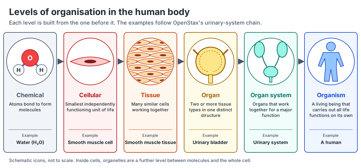
| Level | What it is | Example | Explore |
|---|---|---|---|
| Chemical | Atoms bonded into molecules | Water, proteins, sugars | Cellular respiration |
| Cellular | The smallest independently functioning unit of life | Smooth muscle cell | Cell anatomy |
| Tissue | Many similar cells working together | Smooth muscle tissue | Tissue types, Skin layers |
| Organ | Two or more tissue types in one distinct structure | Urinary bladder | Organs of the body |
| Organ system | Organs that work together for a major function | Urinary system | Urinary system |
| Organism | A living being that carries out all life functions on its own | A human | 3D human body |
The 11 organ systems of the human body
OpenStax groups the organs into 11 organ systems. It warns that assigning organs to systems can be imprecise, because most organs contribute to more than one: the testes and ovaries, for example, are listed both as endocrine glands, which secrete hormones, and as reproductive organs, which make gametes. (OpenStax Anatomy & Physiology 2e, 1.2 Structural Organization of the Human Body.)
The count depends on the source. The National Cancer Institute's SEER training module lists ten major systems (skeletal, muscular, nervous, endocrine, cardiovascular, lymphatic, respiratory, digestive, urinary and reproductive) with no separate integumentary system; OpenStax adds the skin, hair and nails as the integumentary system to make 11. (National Cancer Institute SEER Training Modules, Introduction to the Human Body.)
| System | Main organs | Main functions | Guides |
|---|---|---|---|
| Integumentary | Skin, hair, nails | Encloses internal structures; contains many sensory receptors | Skin layers |
| Skeletal | Bones, cartilage, joints | Supports the body; with the muscles, enables movement | Skeletal system, Joints |
| Muscular | Skeletal muscles, tendons | Enables movement; helps maintain body temperature | Muscular system, Sarcomere |
| Nervous | Brain, spinal cord, peripheral nerves | Detects and processes sensory information; activates responses | Brain, Spinal cord, Autonomic nervous system |
| Endocrine | Pituitary, thyroid, pancreas, adrenal glands, testes, ovaries | Secretes hormones; regulates bodily processes | Pituitary, Thyroid, Adrenal glands |
| Cardiovascular | Heart, blood vessels | Delivers oxygen and nutrients to tissues; helps equalise body temperature | Circulatory system, Heart blood flow |
| Lymphatic | Thymus, spleen, lymph nodes, lymphatic vessels | Returns fluid to the blood; defends against pathogens | Lymphatic system, Spleen |
| Respiratory | Nasal passages, trachea, lungs | Delivers oxygen to the blood; removes carbon dioxide | Respiratory system, Lungs |
| Digestive | Stomach, liver, gallbladder, small and large intestines | Processes food for use by the body; removes undigested waste | Digestive system, Stomach, Liver |
| Urinary | Kidneys, urinary bladder | Controls water balance; removes and excretes wastes from the blood | Urinary system, Nephron |
| Reproductive | Testes and epididymis; ovaries, uterus and mammary glands | Produces sex hormones and gametes; the female system supports the embryo and fetus and produces milk | Male, Female |
See every system in place, switch them on and off and peel back skin and muscle in the interactive 3D human anatomy model.
What the organ systems do together: the functions of life
OpenStax lists six functions that keep a human alive: organisation (trillions of cells kept in distinct internal compartments), metabolism (the sum of all anabolic, building, and catabolic, breaking-down, reactions in the body), responsiveness (adjusting to internal and external change, such as sweating to cool down), movement (from skeletal muscle down to single organs and cells), development (all the changes the body goes through in life, including differentiation, growth and repair) and reproduction. (OpenStax Anatomy & Physiology 2e, 1.3 Functions of Human Life.)
Homeostasis and feedback loops
Homeostasis is the state of steady internal conditions that living things maintain, and much of physiology is about how the body achieves it. Every regulated value has a set point, the value around which its normal range fluctuates, and a normal range, the restricted set of values that is optimally healthful and stable. Keeping inside that range requires the body to monitor its internal conditions continuously. (OpenStax Anatomy & Physiology 2e, 1.5 Homeostasis.)
Body temperature is the classic example. The average normal body temperature is generally accepted as 37 °C (98.6 °F), but studies have found a normal range from about 36.1 °C (97 °F) to 37.2 °C (99 °F). (MedlinePlus, Body temperature norms.)
Negative feedback reverses a deviation from the set point. It needs three parts: a sensor (receptor) that monitors the value, a control centre that compares it with the normal range, and an effector that changes it back. When body temperature rises above 37 °C, nerve cells in the skin and brain (sensors) signal the brain's temperature-regulatory centre (control centre); it drives the sweat glands (effectors) and dilates the blood vessels of the skin, so heat is lost. In the cold, the heat-gain centre cuts blood flow to the skin, and if heat loss is severe the brain triggers shivering, whose muscle contractions release heat. (OpenStax Anatomy & Physiology 2e, 1.5 Homeostasis.)
Blood glucose is regulated the same way. When endocrine cells in the pancreas detect excess glucose in the blood (the stimulus), the beta cells release the hormone insulin; as glucose falls back to normal, insulin release stops, which keeps blood sugar from dropping below the normal range. (OpenStax Anatomy & Physiology 2e, 1.5 Homeostasis.)
Positive feedback intensifies a change instead of reversing it. In childbirth, the baby's head stretching the cervix triggers the release of oxytocin, which makes the uterine muscle contract harder, which pushes the head harder against the cervix; the loop stops only when the baby is born. Blood clotting is another positive feedback cascade: each step stimulates the release of more clotting substances. (OpenStax Anatomy & Physiology 2e, 1.5 Homeostasis.)
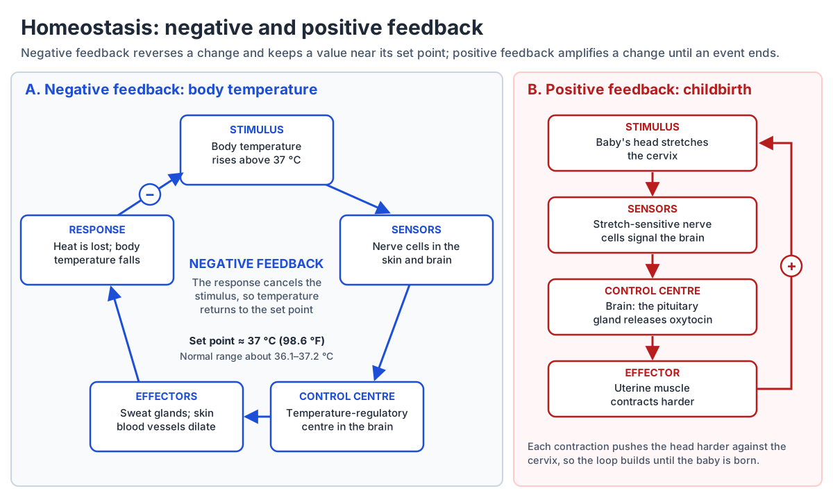
| Negative feedback | Positive feedback | |
|---|---|---|
| Effect on a change | Reverses it, back towards the set point | Intensifies it |
| Parts | Sensor, control centre, effector | The same parts, but the response strengthens the stimulus |
| Stops when | The value is back in its normal range | The triggering event is over (the baby is born, the clot is formed) |
| Examples | Body temperature; blood glucose and insulin | Childbirth and oxytocin; blood clotting |
How anatomy and physiology courses are structured
Anatomy and physiology is usually taught as a two-semester sequence, A&P 1 and A&P 2. The Human Anatomy & Physiology Society (HAPS) publishes more than 800 learning outcomes for it, organised into lettered modules; it notes that most first-semester courses cover modules A to J and second-semester courses the rest, and it takes no position on the order. (Human Anatomy & Physiology Society, HAPS A&P Learning Outcomes.)
HAPS's end-of-course exams show the usual split. The A&P 1 exam covers the body plan, homeostasis, chemistry and cell biology, histology, and the integumentary, skeletal, muscular and nervous systems with the special senses. The A&P 2 exam covers the endocrine, cardiovascular, lymphatic and immune, respiratory, digestive, urinary and reproductive systems, plus metabolism and fluid, electrolyte and acid–base balance. The nervous and cardiovascular systems carry the most questions, 14 each. (Human Anatomy & Physiology Society, HAPS A&P I and A&P II Exams.)
The endocrine system is the swing topic: the exam places it in A&P 2, while the learning-outcomes guidance counts module J in the first semester. Check your own syllabus. (Human Anatomy & Physiology Society, HAPS A&P Learning Outcomes.)
Labs bring the anatomy to life with anatomical models, skeletons, diagrams and photographs, and with dissection and the inspection of cadavers. (LibreTexts Medicine, 1.1A Defining Anatomy (A and P for STEM Educators, James Madison University).)
| A&P 1 | Questions | A&P 2 | Questions |
|---|---|---|---|
| A. Body plan and organisation | 2 | J. Endocrine system | 6 |
| B. Homeostasis | 1 | K. Cardiovascular system | 14 |
| C. Chemistry and cell biology | 6 | L. Lymphatic system and immunity | 4 |
| D. Histology | 3 | M. Respiratory system | 7 |
| E. Integumentary system | 2 | N. Digestive system | 6 |
| F. Skeletal system and articulations | 5 | O. Metabolism | 3 |
| G. Muscular system | 6 | P. Urinary system | 5 |
| H. Nervous system | 14 | Q. Fluid, electrolyte and acid–base balance | 7 |
| I. Special senses | 4 | R. Reproductive system | 5 |
How to study anatomy and physiology
A 2013 review in Psychological Science in the Public Interest by John Dunlosky and colleagues rated ten common study techniques. Only two received the highest utility rating: practice testing (quizzing yourself with flash cards or end-of-chapter questions) and distributed practice (spreading study out over time). Summarising, highlighting and underlining, and rereading were among the five rated low. (Association for Psychological Science, Which Study Strategies Make the Grade? (Dunlosky et al. 2013).)
The effect holds for anatomy. Dobson, Perez and Linderholm (2017) had students learn skeletal muscle anatomy with the same 24 minutes of study arranged in five ways. Retrieval beat restudying, spacing beat cramming, and the combination was best: a week later, students who studied and retrieved across seven days recalled about 33.9 items on average, against about 12.9 for one massed session with retrieval. (Dobson, Perez & Linderholm, Distributed Retrieval Practice Promotes Superior Recall of Anatomy Information, Anatomical Sciences Education 10:339 (2017).)
An A&P study plan that uses the evidence
- Learn the language first. Anatomical terms come from Greek and Latin and their meanings are fixed, so learning common roots makes each new name easier to decode. Start with the anatomical terms, directional terms, body planes.
- Pair every structure with its function. Write the function next to each name, and ask what the shape lets it do.
- Test, don't reread. Cover the labels on a diagram and name each part from memory, for example the label-it-yourself diagram on the heart blood flow page or the anatomy quiz.
- Space it out. Revisit each system on several days spread over the week instead of in one long session before the exam.
- Draw the loop. For every hormone or reflex, sketch stimulus, sensor, control centre, effector and response, and mark whether the feedback is negative or positive.
- Study in 3D. Rotate the 3D human body, fade the skin and muscles, and find each organ in place before you learn its details.
Anatomy and physiology: frequently asked questions
What is anatomy and physiology in simple terms?
Anatomy is the study of the body's structure: what the parts are, what they look like and where they are. Physiology is the study of how those parts work, from the chemistry inside cells to whole organ systems. Together, A&P explains how the body is built and how it keeps itself alive.
What is the difference between anatomy and physiology?
Anatomy describes structure and physiology explains function. The lungs' 70 m² of thin-walled alveoli are anatomy; the diffusion of oxygen into the blood across that 0.5 µm membrane is physiology. The heart's four valves are anatomy; the pressure changes that open and close them are physiology. Each makes sense only with the other.
Why are anatomy and physiology taught together?
Because form and function are closely related in all living things, it is hard to understand one without the other. Knowing that the left ventricle's wall is much thicker than the right's is anatomy; knowing it is thicker because it must pump blood around the whole body at higher pressure is physiology, and that is what makes the fact stick.
What are the 11 organ systems?
Integumentary, skeletal, muscular, nervous, endocrine, cardiovascular, lymphatic, respiratory, digestive, urinary and reproductive. That is the OpenStax list; some sources count ten by leaving out the integumentary system, and the lymphatic system is often taught together with immunity. Many organs belong to more than one system.
What is homeostasis in anatomy and physiology?
Homeostasis is the body's ability to keep internal conditions, such as temperature and blood glucose, within a narrow normal range around a set point. It mostly works by negative feedback: a sensor detects a change, a control centre compares it with the set point, and an effector reverses it, as when sweating cools you down after your temperature rises.
What is covered in A&P 1 vs A&P 2?
A&P 1 usually covers body organisation, homeostasis, chemistry, cells, tissues, skin, bones and joints, muscles, the nervous system and the special senses. A&P 2 usually covers the endocrine, cardiovascular, lymphatic and immune, respiratory, digestive, urinary and reproductive systems, metabolism, and fluid, electrolyte and acid–base balance. The endocrine system moves between the two depending on the course.
Is anatomy and physiology hard?
It is demanding mainly because of volume: HAPS lists more than 800 learning outcomes for the two-semester course, and there are thousands of names to learn. It becomes manageable when you learn the terminology first, link every structure to its function, and test yourself on spaced days instead of rereading notes.
How do you study for anatomy and physiology?
Quiz yourself and spread your study out: practice testing and distributed practice are the two techniques rated most useful by learning researchers, and in an anatomy experiment their combination more than doubled recall a week later compared with a single massed session. Label blank diagrams from memory, rotate 3D models, draw feedback loops for each system, and always ask what a structure's shape lets it do.
References
- OpenStax Anatomy & Physiology 2e, 1.1 Overview of Anatomy and Physiology
- OpenStax Anatomy & Physiology 2e, 1.2 Structural Organization of the Human Body
- OpenStax Anatomy & Physiology 2e, 1.3 Functions of Human Life
- OpenStax Anatomy & Physiology 2e, 1.5 Homeostasis
- OpenStax Anatomy & Physiology 2e, 1.7 Medical Imaging
- OpenStax Anatomy & Physiology 2e, 4.1 Types of Tissues
- OpenStax Anatomy & Physiology 2e, 12.2 Nervous Tissue
- OpenStax Anatomy & Physiology 2e, 12.4 The Action Potential
- OpenStax Anatomy & Physiology 2e, 19.1 Heart Anatomy
- OpenStax Anatomy & Physiology 2e, 22.1 Organs and Structures of the Respiratory System
- OpenStax Anatomy & Physiology 2e, 22.2 The Lungs
- OpenStax Anatomy & Physiology 2e, 23.5 The Small and Large Intestines
- NLM Medical Subject Headings (MeSH), Physiology
- Dictionary.com, physiology
- Kenhub, Alveoli (Ferng, reviewed by Mytilinaios)
- BioNumbers (Harvard), Average surface area of parts of the gastrointestinal tract, from Helander & Fändriks, Scand J Gastroenterol 49:681 (2014)
- National Cancer Institute SEER Training Modules, Introduction to the Human Body
- MedlinePlus, Body temperature norms
- Human Anatomy & Physiology Society, HAPS A&P I and A&P II Exams
- Human Anatomy & Physiology Society, HAPS A&P Learning Outcomes
- LibreTexts Medicine, 1.1A Defining Anatomy (A and P for STEM Educators, James Madison University)
- Association for Psychological Science, Which Study Strategies Make the Grade? (Dunlosky et al. 2013)
- Dobson, Perez & Linderholm, Distributed Retrieval Practice Promotes Superior Recall of Anatomy Information, Anatomical Sciences Education 10:339 (2017)
- OpenStax Anatomy & Physiology 2e, 1.6 Anatomical Terminology
More interactive anatomy models
- What is anatomy?What anatomy studies, its branches, how anatomists name and describe the body, and how to learn it.
- Comparative anatomyHomologous, analogous and vestigial structures compared across vertebrates, with diagrams.
- Animal anatomyEvery animal on the site by group, with what each body plan teaches.
- Anatomy quizFree anatomy quizzes and games: find-the-structure quizzes on 3D models and labelling practice.
- Human anatomy1,000+ bones, muscles, organs, vessels and nerves from real anatomical data.
- Octopus anatomyThree hearts, a ring-shaped brain, eight arms and a beak, in 3D.