

What is a tissue?
In anatomy, a tissue is a group of cells found together in the body. All the tissues of the body fall into four primary types: epithelial, connective, muscle and nervous tissue. (OpenStax Anatomy and Physiology 2e, 4.1: Types of Tissues.)
Cleveland Clinic describes tissues as groups of cells with similar or related jobs and places them in the body's levels of organisation: cells form tissues, tissues form organs, organs form organ systems and organ systems form the body. Every organ is made of at least two tissue types, and the body contains at least 200 different types of cells. (Cleveland Clinic: Body Tissues.)
The tissues develop from the three germ layers of the embryo. The ectoderm gives rise to the epidermis, the skin glands and the nervous system; the mesoderm to the connective tissues, including bone, cartilage and blood, and to muscle; and the endoderm to the lining of the airways and of most of the digestive system, and to the digestive glands. (OpenStax Anatomy and Physiology 2e, 4.1: Types of Tissues.)
How tissues come together as the internal organs, shown in 3D. Organs of the body.
The four types of tissue
Epithelial tissue is made of sheets of cells that cover the outside of the body, line internal cavities and passageways, and form certain glands. Connective tissue binds the body together and supports, protects and integrates its parts. Muscle tissue is excitable: it responds to stimulation by contracting, which produces movement. Nervous tissue carries electrical signals, nerve impulses, from one region of the body to another. (OpenStax Anatomy and Physiology 2e, 4.1: Types of Tissues.)
One simple way to tell them apart under the microscope is the amount of material between the cells. Epithelial, muscle and nervous tissues consist of closely packed cells with little extracellular material, whereas connective tissue has scattered cells in an abundant extracellular matrix of ground substance and fibres. (Digital Histology: Tissue basics.)
Connective tissue is the most abundant and widespread of the four, found almost everywhere in the body. Informal terms such as soft tissue or brain tissue are not separate tissue types. (Cleveland Clinic: Body Tissues.)
The four tissue types compared
| Tissue | Main job | Cells and matrix | Examples |
|---|---|---|---|
| Epithelial | Covers surfaces, lines cavities and passageways, forms glands | Tightly packed cells on a basement membrane; no blood vessels | Epidermis, lining of the gut and airways, kidney tubules, glands |
| Connective | Binds, supports, protects, transports, stores fat | Scattered cells in an abundant matrix of fibres and ground substance | Tendons, ligaments, fat, cartilage, bone, blood, lymph |
| Muscle | Contracts to produce movement | Elongated, excitable cells (fibres) | Skeletal muscles, heart, walls of hollow organs |
| Nervous | Carries electrical signals | Neurons and supporting glial cells | Brain, spinal cord, nerves |
Sources: OpenStax §4.1 to 4.5, Digital Histology and Cleveland Clinic.
Epithelial tissue: types, locations and functions
Epithelium is highly cellular, with little material between its cells. It is almost completely avascular, so it gets its nutrients by diffusion or absorption from below. Each epithelium is polarised, with a free (apical) surface and a basal surface, and it is anchored to the underlying connective tissue by a basement membrane. Many epithelia can replace damaged and dead cells quickly. (OpenStax Anatomy and Physiology 2e, 4.2: Epithelial Tissue.)
Epithelia are named by the shape of their cells (squamous, flat; cuboidal, as tall as wide; columnar, taller than wide) and by the number of layers: simple (one layer), stratified (several layers), pseudostratified (one layer that looks layered because the nuclei sit at different heights) and transitional (a stratified epithelium whose cells change shape). (OpenStax Anatomy and Physiology 2e, 4.2: Epithelial Tissue.)
Simple squamous epithelium lines the air sacs (alveoli) of the lungs, the blood vessels (where it is called endothelium) and the serous membranes (mesothelium); its thin cells allow rapid diffusion. Simple cuboidal epithelium lines the kidney tubules and gland ducts and secretes and absorbs. Simple columnar epithelium lines much of the digestive tract and parts of the female reproductive tract, absorbing and secreting mucus and enzymes; ciliated forms line the uterine tubes and parts of the airways. (OpenStax Anatomy and Physiology 2e, 4.2: Epithelial Tissue.)
Pseudostratified columnar epithelium lines the trachea, the bronchi and much of the upper respiratory tract, where its cilia sweep mucus along. Stratified squamous epithelium is the most common stratified type and resists abrasion: keratinised, it forms the epidermis of the skin; non-keratinised, it lines the mouth, oesophagus (esophagus) and vagina. Transitional epithelium is found only in the urinary system, in the ureters and bladder, where it stretches as they fill. (OpenStax Anatomy and Physiology 2e, 4.2: Epithelial Tissue.)
Cells in an epithelium are tied together by junctions. Tight junctions seal neighbouring cells so substances cannot leak between them; anchoring junctions (desmosomes, hemidesmosomes and adherens junctions) give strong, flexible attachment to each other and to the basement membrane; and gap junctions are channels that let ions and small molecules pass from cell to cell. (OpenStax Anatomy and Physiology 2e, 4.2: Epithelial Tissue.)
Cleveland Clinic sums up the job of epithelium as forming boundaries: it separates and protects the tissues beneath and controls what passes through. Its examples are the epidermis of the skin and the cornea of the eye. (Cleveland Clinic: Body Tissues.)
The epidermis, an epithelium, with the dermis and hypodermis beneath it, in 3D. Skin layers.
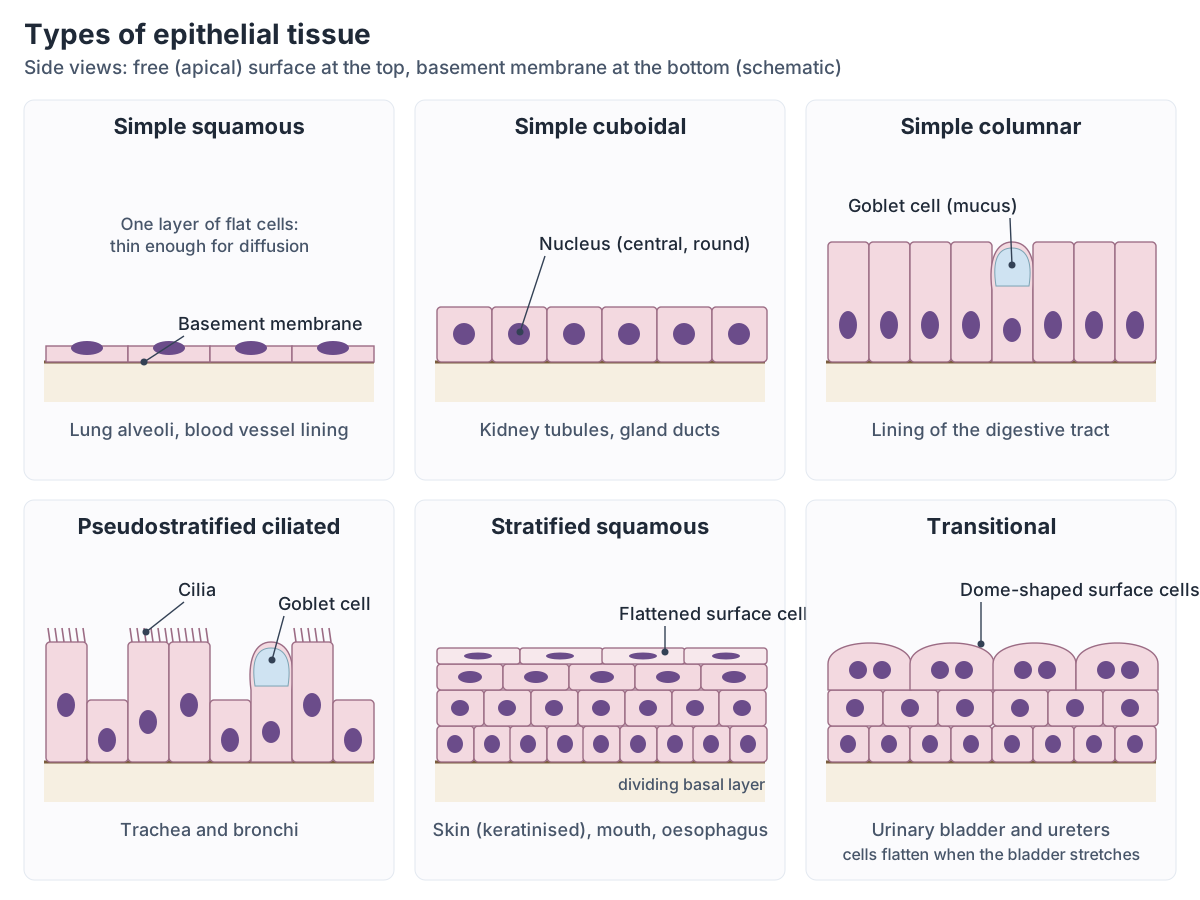
Types of epithelium and where they are found
| Epithelium | Layers and cell shape | Function | Where it is found |
|---|---|---|---|
| Simple squamous | One layer of flat cells | Diffusion, smooth lining | Lung alveoli, blood vessels (endothelium), serous membranes (mesothelium) |
| Simple cuboidal | One layer of cube-shaped cells | Secretion and absorption | Kidney tubules, gland ducts |
| Simple columnar | One layer of tall cells (some ciliated) | Absorption, secretion of mucus and enzymes | Digestive tract, uterine tubes, parts of the airways |
| Pseudostratified columnar | One layer that looks layered; ciliated | Secretes and moves mucus | Trachea, bronchi, upper respiratory tract |
| Stratified squamous | Many layers, flat at the surface | Protection against abrasion | Skin (keratinised); mouth, oesophagus, vagina (non-keratinised) |
| Stratified cuboidal and columnar | Several layers | Uncommon; lines some ducts | Some glands and ducts, parts of the urethra |
| Transitional | Many layers of cells that change shape | Stretching | Ureters and urinary bladder |
Source: OpenStax §4.2 Epithelial Tissue.
Glandular epithelium: exocrine and endocrine glands
Glands are made of epithelial cells. Endocrine glands have no ducts and release hormones into the interstitial fluid and the blood. Exocrine glands release their secretions through ducts onto an epithelial surface; mucus, sweat, saliva and milk are exocrine secretions. (OpenStax Anatomy and Physiology 2e, 4.2: Epithelial Tissue.)
Exocrine glands are also classed by how their cells release the secretion. In merocrine secretion the product leaves by exocytosis and the cell stays intact; in apocrine secretion part of the top of the cell is shed with it; and in holocrine secretion the whole cell breaks down and becomes the secretion, as in the sebaceous (oil) glands of the skin. (OpenStax Anatomy and Physiology 2e, 4.2: Epithelial Tissue.)
Connective tissue: proper, supportive and fluid
Connective tissue consists of cells scattered in a matrix of ground substance and protein fibres. It supports and connects other tissues, protects organs, defends the body against microbes, transports fluids and nutrients, stores fat and insulates the body. (OpenStax Anatomy and Physiology 2e, 4.3: Connective Tissue Supports and Protects.)
Three kinds of fibre give connective tissues their properties. Collagen fibres are strong and flexible and dominate tendons and ligaments; elastic fibres return to their original shape after stretching, as in the skin and some ligaments; and reticular fibres form fine, branching networks, abundant in the liver and spleen. The most common cell is the fibroblast, which makes the fibres and ground substance; others include adipocytes, which store fat, mesenchymal stem cells, macrophages, which engulf pathogens and debris, and mast cells, which release histamine. (OpenStax Anatomy and Physiology 2e, 4.3: Connective Tissue Supports and Protects.)
Connective tissue proper is loose or dense. Loose connective tissue includes areolar tissue, which surrounds blood vessels and fills spaces between muscle fibres, adipose tissue, which stores fat and cushions organs such as the kidneys, and reticular tissue, which forms the framework of lymphatic tissue, the spleen and the liver. Dense connective tissue is regular in tendons and ligaments, irregular in the dermis of the skin, and elastic in the walls of arteries and the ligaments of the vocal folds. (OpenStax Anatomy and Physiology 2e, 4.3: Connective Tissue Supports and Protects.)
Supportive connective tissue is cartilage and bone. Hyaline cartilage is found in the rib cage, the nose and on joint surfaces, fibrocartilage in the menisci of the knee and the intervertebral discs, and elastic cartilage in the external ear. Cartilage has no blood vessels, so nutrients must diffuse through its matrix, which is why it heals slowly. Bone is either compact (solid and strong) or spongy (cancellous, lighter, inside bones and at the ends of long bones). Blood and lymph are fluid connective tissues. (OpenStax Anatomy and Physiology 2e, 4.3: Connective Tissue Supports and Protects.)
Cleveland Clinic gives bone with its marrow, blood, lymph and the skin below its outermost layer as examples of connective tissue, and notes that the proteins of the matrix decide how dense and hard each connective tissue is. (Cleveland Clinic: Body Tissues.)
Compact and spongy bone, osteons and bone cells in the anatomy of a long bone. Bone structure.
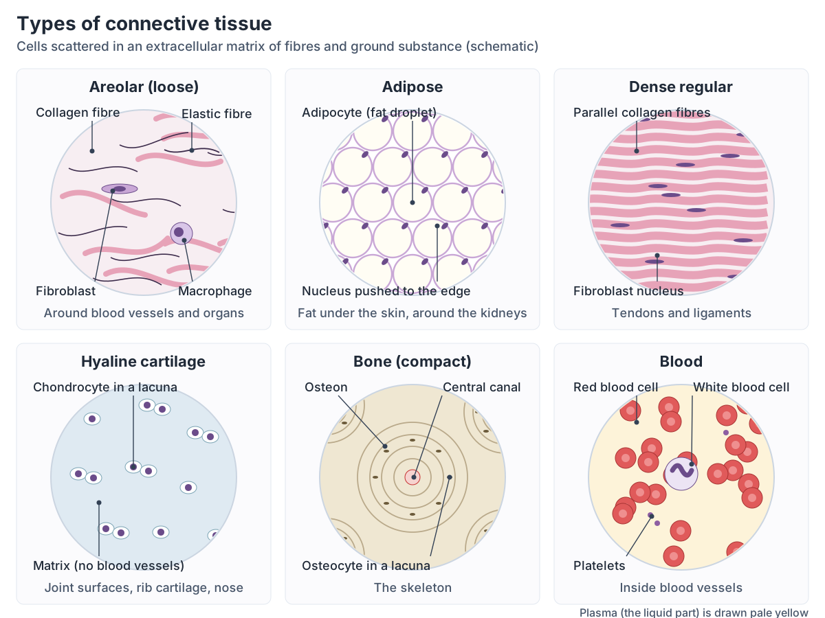
Types of connective tissue
| Group | Type | Where it is found |
|---|---|---|
| Loose connective tissue proper | Areolar | Around blood vessels, between muscle fibres |
| Loose connective tissue proper | Adipose | Fat stores; around the kidneys |
| Loose connective tissue proper | Reticular | Framework of lymphatic tissue, spleen and liver |
| Dense connective tissue proper | Regular | Tendons and ligaments |
| Dense connective tissue proper | Irregular | Dermis of the skin |
| Dense connective tissue proper | Elastic | Walls of arteries, vocal fold ligaments |
| Supportive | Hyaline cartilage | Rib cage, nose, joint surfaces |
| Supportive | Fibrocartilage | Menisci of the knee, intervertebral discs |
| Supportive | Elastic cartilage | External ear |
| Supportive | Bone (compact and spongy) | The skeleton |
| Fluid | Blood and lymph | Blood vessels and lymphatic vessels |
Source: OpenStax §4.3 Connective Tissue Supports and Protects.
Muscle tissue: skeletal, cardiac and smooth
Skeletal muscle is made of long, cylindrical fibres with striations (stripes) and many nuclei at the edge of each fibre. It is under voluntary control, attaches to bones and guards the entrances of the body such as the mouth and anus, and it moves the body, produces heat and protects organs. (OpenStax Anatomy and Physiology 2e, 4.4: Muscle Tissue and Motion.)
Cardiac muscle is found only in the heart, where it pumps blood. Its cells are short, branched and striated, each with a single central nucleus, and they are joined end to end by intercalated discs, which contain anchoring junctions and gap junctions. Smooth muscle has short, spindle-shaped cells with one nucleus and no visible striations; it lies in the walls of hollow organs and passageways, where it moves food, secretions and blood. Cardiac and smooth muscle are both involuntary. (OpenStax Anatomy and Physiology 2e, 4.4: Muscle Tissue and Motion.)
Cleveland Clinic describes muscle tissue as electrically reactive fibres that bundle together for strength and lists heart muscle, the muscle of the digestive tract and skeletal muscle as its three examples. (Cleveland Clinic: Body Tissues.)
The contractile unit that gives skeletal and cardiac muscle their stripes, in 3D. Sarcomere diagram.
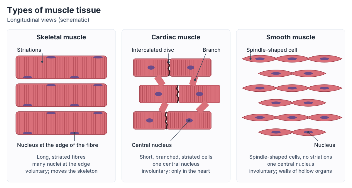
Skeletal vs cardiac vs smooth muscle
| Feature | Skeletal | Cardiac | Smooth |
|---|---|---|---|
| Cell shape | Long cylindrical fibres | Short, branched | Short, spindle-shaped |
| Striations | Yes | Yes | No |
| Nuclei | Many, at the edge | One, central | One per cell |
| Control | Voluntary | Involuntary | Involuntary |
| Special features | Attached to bones | Intercalated discs | In walls of hollow organs |
| Main job | Movement, heat, protection | Pumps blood | Moves food, secretions and blood |
Source: OpenStax §4.4 Muscle Tissue and Motion.
Nervous tissue: neurons and glial cells
Nervous tissue contains two kinds of cell: neurons, which carry signals, and neuroglia (glial cells), which support them. A neuron has a cell body holding the nucleus and most organelles, branching dendrites that receive signals, and a single long axon, often wrapped in myelin, that carries the action potential away to a synapse with the next cell. (OpenStax Anatomy and Physiology 2e, 4.5: Nervous Tissue Mediates Perception and Response.)
Neurons are grouped by shape. A multipolar neuron has several dendrites and one prominent axon; a bipolar neuron has one dendrite and one axon; and a unipolar neuron has a single process that divides into a functional dendrite and axon. (OpenStax Anatomy and Physiology 2e, 4.5: Nervous Tissue Mediates Perception and Response.)
In the brain and spinal cord, astrocytes regulate the ions around neurons, clear some neurotransmitters and help form the blood-brain barrier; oligodendrocytes make the myelin; and microglia protect against infection (OpenStax notes they are related to macrophages rather than true nervous tissue). In the peripheral nerves, Schwann cells make the myelin. (OpenStax Anatomy and Physiology 2e, 4.5: Nervous Tissue Mediates Perception and Response.)
Clusters of neuron cell bodies form the grey matter of the brain and spinal cord and clusters of axons form the white matter. Most nervous tissue passes signals on, but some areas act like circuit breakers that stop them. (Cleveland Clinic: Body Tissues.)
How a neuron fires: depolarisation, repolarisation and the ion channels involved. Action potential diagram.
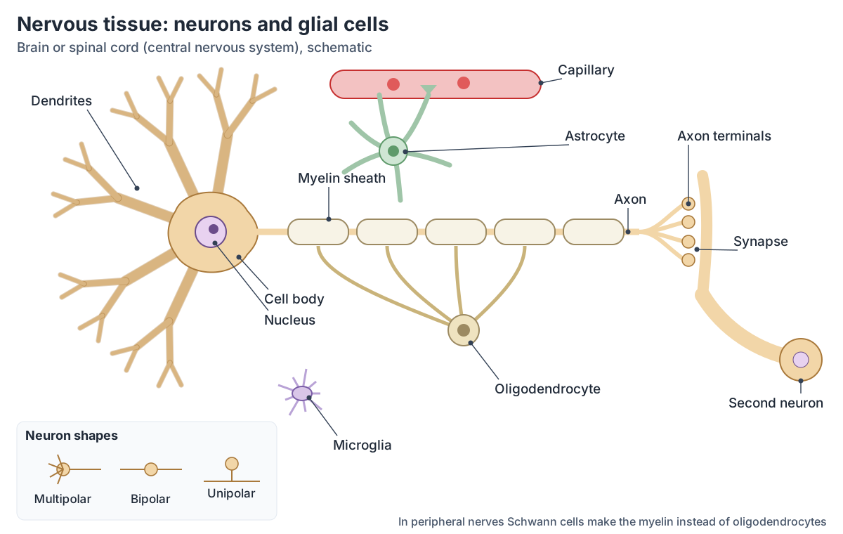
Tissue membranes: mucous, serous, cutaneous and synovial
Tissues also form thin sheets called membranes. Mucous membranes are epithelial membranes that line the cavities and passageways open to the outside: the digestive, respiratory, excretory and reproductive tracts. Serous membranes are epithelial membranes of mesothelium that line closed body cavities: the pleural, pericardial and peritoneal cavities. (OpenStax Anatomy and Physiology 2e, 4.1: Types of Tissues.)
The skin is the cutaneous membrane, an epithelial membrane that covers the surface of the body. Synovial membranes are made of connective tissue rather than epithelium and line the cavities of freely movable joints such as the shoulder, elbow and knee. (OpenStax Anatomy and Physiology 2e, 4.1: Types of Tissues.)
Synovial joints and the membrane that lines them. Types of joints.
How tissues heal and age
Injured tissue becomes inflamed, showing the four cardinal signs of redness, swelling, pain and local heat, sometimes with loss of function. Repair then happens by regeneration, the addition of new cells of the same type as those lost, or by scarring, in which fibroblasts lay down collagen; once the new capillaries disappear, a pale scar is often left. (OpenStax Anatomy and Physiology 2e, 4.6: Tissue Injury and Aging.)
With age, wound healing slows and immune defences weaken. The skin and other tissues become thinner and drier as elastic and collagen fibres decrease, bones lose minerals and cells divide less as their telomeres shorten. Cancers are also named after their tissue of origin: carcinomas arise from epithelial cells and sarcomas from connective tissue. (OpenStax Anatomy and Physiology 2e, 4.6: Tissue Injury and Aging.)
Frequently asked questions
What are the four types of tissue in the human body?
The four tissue types are epithelial tissue, which covers surfaces, lines cavities and forms glands; connective tissue, which binds, supports and protects and includes bone, cartilage, fat and blood; muscle tissue, which contracts to move the body and its organs; and nervous tissue, which carries electrical signals through the brain, spinal cord and nerves.
What is the most abundant tissue in the body?
Connective tissue is the most abundant and widespread tissue type. It is found almost everywhere, from the bones, cartilage, tendons and ligaments to fat, the dermis of the skin and the blood. Its cells are scattered in a large amount of extracellular matrix made of protein fibres and ground substance.
Is blood a connective tissue?
Yes. Blood, together with lymph, is classed as a fluid connective tissue. It develops, with the other connective tissues, from the mesoderm of the embryo. The other connective tissues are connective tissue proper (loose and dense), cartilage and bone, and all of them have cells scattered in an extracellular matrix rather than packed tightly together.
What are the types of epithelial tissue?
Epithelia are named by cell shape and layers. The main types are simple squamous (alveoli, blood vessels), simple cuboidal (kidney tubules), simple columnar (gut lining), pseudostratified columnar (trachea), stratified squamous (skin, mouth, oesophagus), stratified cuboidal and columnar (some ducts) and transitional epithelium (bladder and ureters).
What is the difference between skeletal, cardiac and smooth muscle?
Skeletal muscle has long, striated fibres with many nuclei and is under voluntary control. Cardiac muscle, found only in the heart, has short, branched, striated cells with one nucleus, joined by intercalated discs, and is involuntary. Smooth muscle has spindle-shaped cells without striations and lines the walls of hollow organs; it is also involuntary.
What cells make up nervous tissue?
Nervous tissue is made of neurons, which carry electrical signals, and glial cells, which support them. In the brain and spinal cord the glia are astrocytes, oligodendrocytes, which make myelin, and microglia, which fight infection; in the peripheral nerves, Schwann cells make the myelin.
References
- OpenStax. Anatomy and Physiology 2e, §4.1 Types of Tissues. Rice University, 2022.
- OpenStax. Anatomy and Physiology 2e, §4.2 Epithelial Tissue. Rice University, 2022.
- OpenStax. Anatomy and Physiology 2e, §4.3 Connective Tissue Supports and Protects. Rice University, 2022.
- OpenStax. Anatomy and Physiology 2e, §4.4 Muscle Tissue and Motion. Rice University, 2022.
- OpenStax. Anatomy and Physiology 2e, §4.5 Nervous Tissue Mediates Perception and Response. Rice University, 2022.
- OpenStax. Anatomy and Physiology 2e, §4.6 Tissue Injury and Aging. Rice University, 2022.
- Cleveland Clinic. Body Tissues. Last updated 2025-04-01.
- Digital Histology. Tissue basics.
Related anatomy
- Bone structureLong bone anatomy with a labeled section and 3D femur: diaphysis, epiphyses, growth plate, periosteum, marrow, compact…
- Muscular systemExplore an interactive 3D muscle model: how muscles move bones, how they are named, the upper body and leg muscles…
- Skeletal systemExplore a realistic 3D human skeleton: the 206 bones, axial vs appendicular skeleton, bone types, bony landmarks and…
- Brain anatomyBrain anatomy explained: cerebrum, lobes, gyri, insula, corpus callosum, thalamus, hippocampus, brainstem and…
- Organs of the bodyHuman anatomy organs explained with a labelled internal organs diagram: organs by body system and location, left vs…
Explore the whole body in the interactive 3D human anatomy model, or browse the A–Z list of human structures.