

Sheep anatomy diagram: the parts of a sheep (labeled)
The external parts of a sheep are the named regions that farmers, show judges and vets use: the poll, forehead, face and muzzle on the head; the neck, top of the shoulder, back, loin, hip, rump and dock along the top line; the ribs, flanks and belly on the side; and the forearm, knee, cannon, pastern, dewclaw, stifle, hock and leg on the limbs.[12, 11]
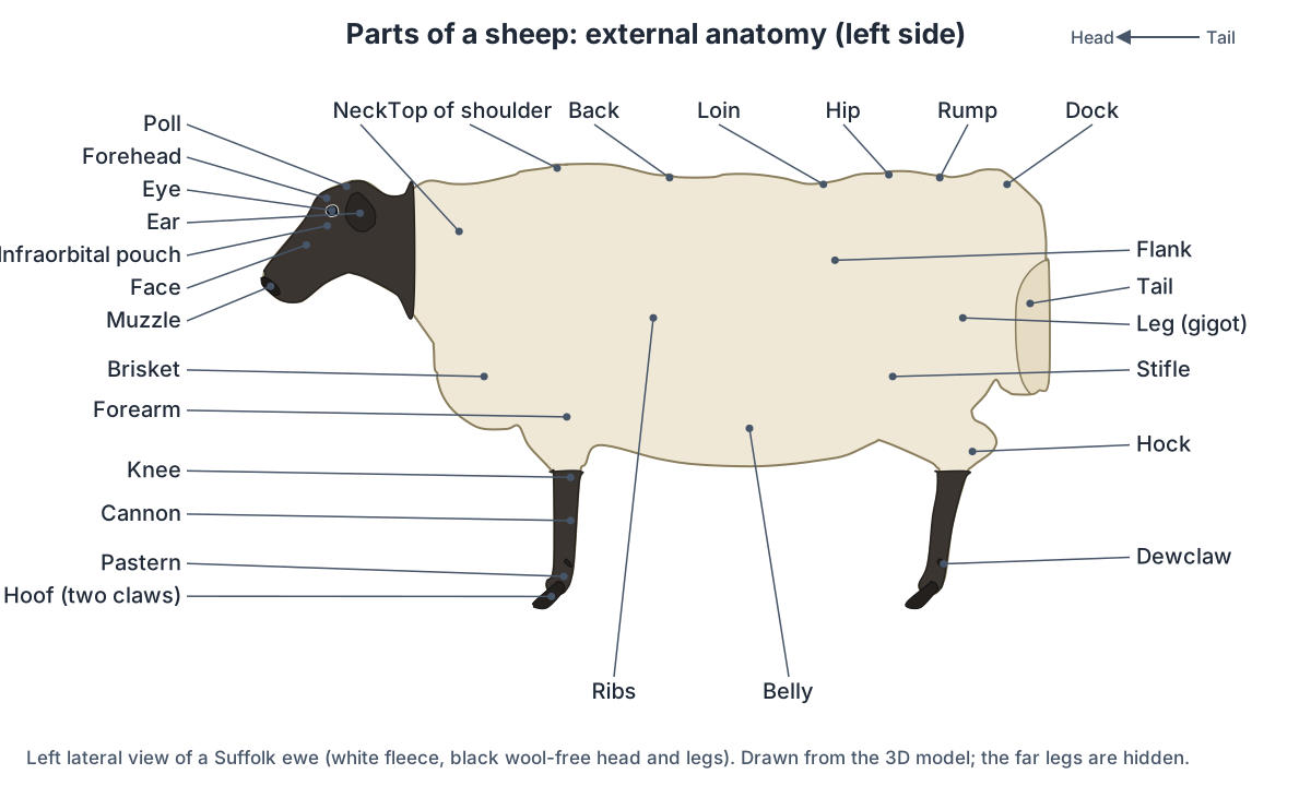
| Part | Where it is | What lies beneath |
|---|---|---|
| Poll | Top of the head, between and behind the ears or horns | Top of the skull[11] |
| Muzzle | Lower front of the head: nostrils, mouth and chin | Nose, lips and the front of both jaws[11] |
| Brisket | Lower chest between the forelegs | Lower chest wall[11] |
| Loin | Each side of the spine between the last rib and the hip | Lumbar vertebrae[11] |
| Rump | Top of the hindquarters | Sacrum[11] |
| Dock | Base of the tail; on docked lambs, where the tail was removed | Base of the tail[11, 13] |
| Flank | Hollow between the last rib and the hip | Abdominal wall; the rumen on the left[11, 4] |
| Leg (gigot) | The thigh of a sheep | Femur and the thigh muscles[11] |
| Knee | Front leg, above the cannon | Carpus (wrist): radius, carpal bones and metacarpus[11] |
| Stifle and hock | Hind-leg joints | Stifle: femur, patella and tibia; hock: tibia, tarsus and metatarsus[11] |
| Cannon and pastern | Below the knee or hock, then below the fetlock | Fused metacarpal or metatarsal bones; then the phalanges[11, 24] |
| Hoof and dewclaws | Two claws on each foot; two small horny dewclaws behind the fetlock | Third and fourth digits; reduced digits[11, 24] |
Sheep also have three kinds of skin gland that goats lack: an infraorbital pouch below the inner corner of each eye, an interdigital gland shaped like a tobacco pipe between the two claws of every foot, and a pair of inguinal pouches beside the udder.[26, 11, 28] The interdigital gland stores a thick, oily secretion and empties it onto the skin through a duct. The duct can become blocked, swelling the pouch, which rarely causes lameness; the gland can also become infected.[30] A ram has a raised "knob" on the nose just above the nostrils.[11]
Sheep anatomy at a glance
A sheep has the same organ systems as other mammals, built around a stomach of four compartments that fills most of the left side of the abdomen.[4, 3] This page describes an adult Suffolk, a meat breed with a white fleece and a black, wool-free head and legs; Suffolks of both sexes are naturally polled (hornless).[14, 15, 16] Other breeds differ in size, fleece, horns and tail, but have the same organs. Vets use four direction words for a standing animal: cranial (towards the head), caudal (towards the tail), dorsal (the back) and ventral (the belly).
Sheep body by the numbers
| Feature | Adult sheep | Source |
|---|---|---|
| Vertebrae | 7 neck, 13 chest, 6 lumbar (sometimes 7), 4 fused sacral, about 16–24 tail | [17, 18, 19] |
| Ribs | 13 pairs; a 14th pair is common | [20, 19] |
| Teeth | 32 permanent, no upper incisors; 20 milk teeth | [2, 7] |
| Toes | 2 claws and 2 dewclaws per foot | [11, 24] |
| Stomach | 1 stomach with 4 compartments | [3, 6] |
| Udder | 2 glands, each with 1 teat | [49, 50] |
| Resting heart rate | 70–80 a minute (MSD); 60–90 (Virginia Tech) | [61, 64] |
| Resting breathing rate | 16–34 a minute (MSD); 12–20 (Virginia Tech) | [62, 64] |
| Rectal temperature | 38.3–39.9 °C (MSD); 102.2–104.9 °F, about 39.0–40.5 °C (Virginia Tech) | [63, 64] |
| Oestrous cycle | About 17 days (14–20), autumn and winter | [67, 68] |
| Pregnancy | About 147 days (142–152), about 5 months | [69, 2] |
Normal ranges differ between sources because they measure different flocks and conditions; a sheep that seems unwell should be seen by a vet.
Sheep internal organs: where everything is
Inside a sheep, the chest holds the heart and lungs in front of the diaphragm, and the abdomen behind it is dominated by the stomach: the rumen lies against the left body wall, while the liver lies on the right against the diaphragm and the intestines fill much of the right side.[1, 4] The bladder and the reproductive tract sit at the entrance to the pelvis, with the bladder below the uterus in ewes.[1]
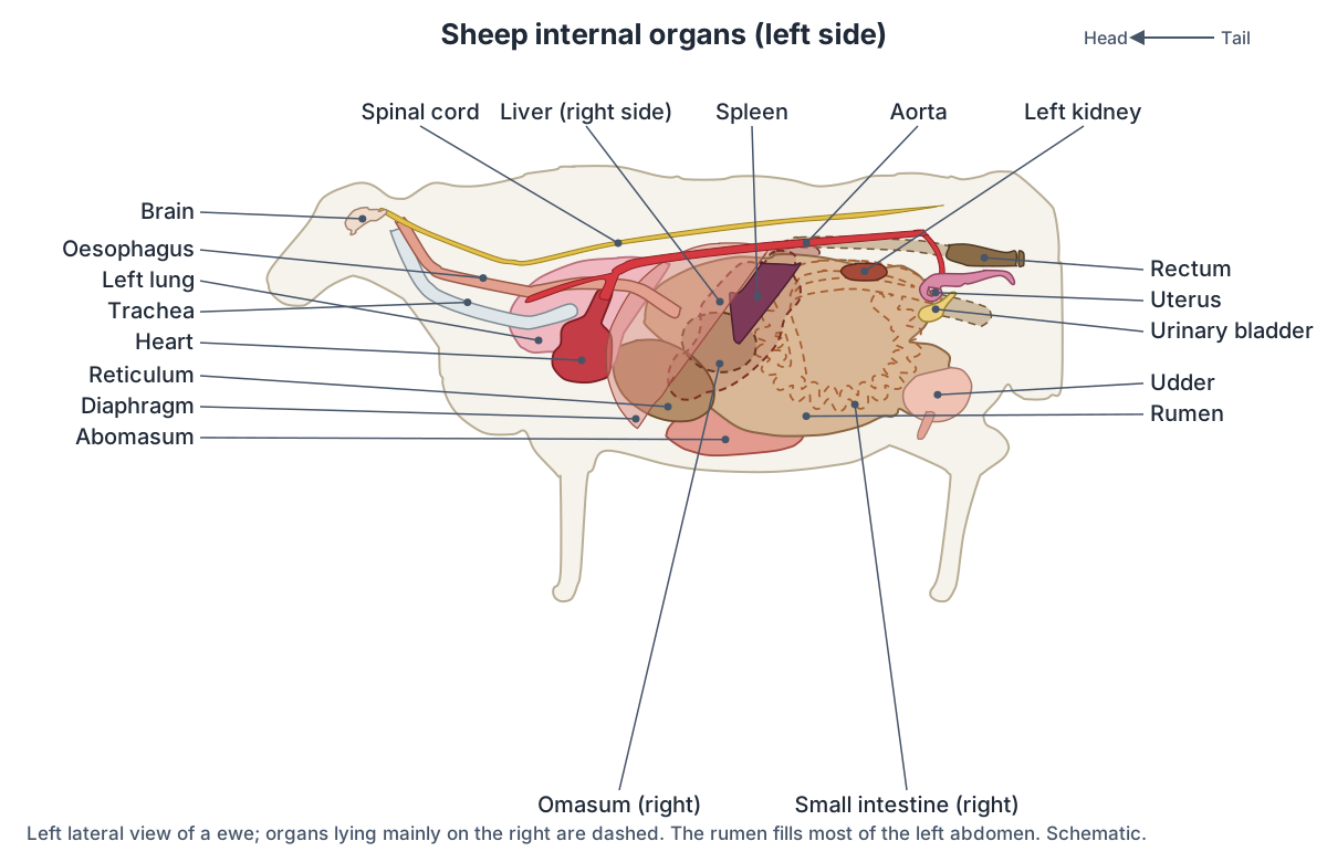
| Organ | Where it is |
|---|---|
| Heart | Low in the chest, roughly from the 3rd to the 6th rib; its caudal edge may touch the diaphragm[1, 36] |
| Lungs | Chest; the left lung has 2 lobes and the right lung 4[34, 1] |
| Rumen | Most of the left side of the abdomen, seen from the left flank[4, 1] |
| Reticulum | Front of the abdomen, right against the diaphragm[1] |
| Omasum | Right of the rumen and reticulum, higher than in cattle[5] |
| Abomasum | Floor of the abdomen on both sides, against the belly wall[1] |
| Spleen | Left side, between the rumen and the diaphragm, under the last ribs[46, 1] |
| Liver and gallbladder | Right front of the abdomen; the gallbladder reaches below the liver's lower edge[1] |
| Intestines | Mainly the right side; the spiral colon lies in the right flank[1, 48] |
| Kidneys | Roof of the abdomen; the rumen can push the left kidney to the right of the midline[1, 32] |
| Bladder | Pelvic floor, reaching forwards into the abdomen when full[1] |
The ruminant stomach of a sheep
A sheep has one stomach with four compartments, not four stomachs: the rumen, reticulum and omasum make up the forestomach, and the abomasum is the true stomach.[3, 6, 2] Swallowed grass goes into the rumen and reticulum, where microbes ferment the fibre; the sheep brings it back up as cud, chews it again and swallows it, and the gas from fermentation is burped up.[3, 6]
- Rumen (paunch): the largest compartment, a fermentation vat filling most of the left abdomen. In sheep its papillae are large and tongue-like. The dorsal sac is the longer of the two sacs, but the ventral sac is larger, reaches further to the right than in cattle, and its caudal blind sac extends further back than the dorsal one.[4, 5]
- Reticulum (honeycomb): the front compartment, lined with honeycomb ridges and lying against the diaphragm.[3, 1] In lambs the reticular groove sends milk straight to the omasum and abomasum, past the rumen.[3]
- Omasum ("many piles"): a round compartment packed with leaves of tissue. The leaves give it a large surface that absorbs water from the digesta before it passes to the abomasum; relatively little digestion happens here.[3, 6]
- Abomasum (true stomach): secretes enzymes and acid like the stomach of a pig or a person.[6, 3]
In a mature sheep the rumen holds about 5 to 10 gallons (roughly 19 to 38 litres), far more than the other three compartments, and the small intestine is about 80 feet (24 m) long.[6] In smaller ruminants such as sheep, the rumen makes up about 75% of the stomach's volume, the reticulum 8%, the omasum 4% and the abomasum 13%.[4]
The proportions differ from a cow's: in sheep the reticulum and abomasum are relatively larger and the omasum relatively smaller.[4, 5] A young lamb lives on milk, which the reticular groove sends past the rumen until the forestomach develops.[3, 6] For the inside of each compartment, the rumen pillars and how rumination works in detail, see the cow digestive system guide, which covers the same four compartments in cattle.
Sheep teeth and the dental pad
An adult sheep has 32 teeth and no upper front teeth: the front of the upper jaw is a hard dental pad, and the eight lower front teeth (three pairs of incisors and a pair of canines shaped like incisors) clip grass against it.[9, 10, 7] Further back, each side of each jaw has three premolars and three molars that grind the food and cud.[10, 2] A lamb has 20 temporary teeth, including four pairs of milk teeth at the front.[7, 2]
| Permanent front teeth | New Mexico State University | Sheep 101 (Maryland) |
|---|---|---|
| 1st (central) pair | 1–1½ years | About 1 year |
| 2nd pair | 2 years | 2 years |
| 3rd pair | About 3 years | Not given |
| 4th (corner) pair, full mouth | 4 years | By 4 years |
Ages from New Mexico State University and Sheep 101; eruption can vary by about six months with breed and diet.[9, 8] A sheep that has lost or broken front teeth is "broken mouthed", and one with none is a "gummer".[7]
Sheep skeleton
A sheep's spine usually has 7 cervical, 13 thoracic, 6 lumbar and 4 fused sacral vertebrae, followed by about 16 to 24 tail vertebrae.[17, 18, 19] Extra vertebrae are fairly common: a 14th pair of ribs is common, and a 2024 study notes that extra thoracic or lumbar vertebrae are common in Mongolian sheep.[20, 19]
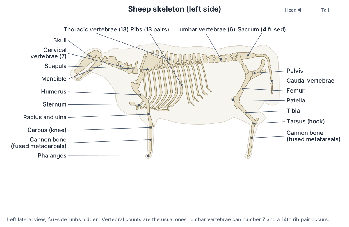
Each leg ends in two weight-bearing digits. The cannon bone of the foreleg is the fused third and fourth metacarpals, and in the hind leg the fused third and fourth metatarsals; the lower ends stay separate, one for each claw.[24, 25] The "knee" of a sheep is its carpus (wrist), and the hock is the joint of the tibia, tarsal and metatarsal bones.[11] Sheep nasal bones are pointed at the front, unlike the notched nasal bones of cattle.[22]
Horns, where present, grow on a bony core from the frontal bone covered by a keratin sheath, and in sheep they sit close to the eye socket.[23, 21] Whether a sheep is horned depends on breed, sex and genes at a single locus: in breeds of Central and Western European origin a sheep with one copy of the horned variant is horned if it is a ram and polled if it is a ewe, while in southern European and African breeds the pattern is less consistent.[23] Tails also vary by breed, from thin to fat and from short to long, and many lambs are docked.[11, 13]
Sheep muscles
The 3D model does not show muscles, but the main muscles that move a sheep are in the limbs and along the spine. In the foreleg they include the brachiocephalicus, trapezius, latissimus dorsi, deltoid and triceps brachii.[70] The hind leg (the gigot, sold as leg of lamb) holds the biceps femoris, semitendinosus, semimembranosus and the quadriceps group, with the gluteal muscles over the hip and rump and the gastrocnemius at the back of the lower leg, above the hock.[70, 71] Epaxial muscles above the vertebrae steady the spine and move the neck and back up and down and from side to side, while hypaxial muscles below the spine flex the neck and back.[70]
Sheep heart and blood vessels
A sheep's heart lies low in the chest between the lungs, roughly from the 3rd to the 6th rib (the 2nd to 5th intercostal spaces in Akkaraman sheep), and its caudal edge can touch the diaphragm.[1, 36] In sheep the right coronary artery is much smaller than the left.[36] The aorta leaves the left ventricle and runs back under the spine, the pulmonary trunk carries blood from the right ventricle to the lungs, and the caudal vena cava returns blood from the back of the body to the right atrium.[37] For the chambers, valves and vessels on a real heart, follow the sheep heart dissection guide.
Sheep lungs and breathing
A sheep's left lung has two lobes, a cranial lobe in two parts and a caudal lobe, and its right lung has four: cranial (in two parts), middle, caudal and accessory.[34, 1] As in cattle, the bronchus to the right cranial lobe leaves the trachea itself, before it divides; this tracheal bronchus is absent in cats, dogs and horses.[35, 34] Lung abscesses are a common internal form of caseous lymphadenitis, which in sheep more often affects the internal organs than the skin nodes.[60]
Kidneys and urinary system
Sheep kidneys are smooth and bean-shaped, like a dog's, not divided into lobes like a cow's, and inside each has a single fused, crest-like papilla instead of many.[32, 1, 33] The left kidney hangs against the rumen and a full rumen can push it right of the midline.[1, 32] The ureters run to the ovoid bladder and pass obliquely through its wall.[1] In rams the urethra continues past the tip of the penis as the urethral process, where urinary stones often lodge, especially in castrated males.[1, 2]
Sheep brain and spinal cord
The sheep brain is remarkably similar to the human brain, which is why it is the standard brain dissected in anatomy labs; its cerebrum is proportionately smaller and its brainstem lies further back.[59] The cerebrum handles the higher functions, the cerebellum coordinates movement and balance, and the brainstem links the brain to the spinal cord and helps regulate breathing and the heart.[40] For every structure a lab sheet asks you to label, use the sheep brain dissection guide.
Sheep eyes and sense of smell
Sheep eyes sit on the sides of the head and have large, horizontal, roughly rectangular pupils. Together these give a very wide field of view, commonly about 270 to 320 degrees and less in sheep with heavy wool on the face, so a sheep can scan its surroundings with only slight head movement.[72, 73] Both eyes see the same scene only in a narrower zone in front, and that is where sheep judge depth well; they often stop to look closely at something unfamiliar before moving on.[73, 72] Sheep also have an excellent sense of smell, which helps rams find ewes in heat and ewes find their own lambs.[72]
Fleece, skin and lymph nodes
Wool fibres are secondary (undercoat) hairs: the woolly skin has mostly compound follicle groups, while the face, lower legs and ears have mostly simple follicles.[31] Lanolin is the yellow grease in the fleece, made by the skin.[13] Thanks to the fleece, adult sheep tolerate heat and cold well, but newborn lambs and newly shorn sheep can easily get too cold.[2]
The spleen is a flat, triangular, bluish-red organ on the left side, attached to the rumen; a goat's is roughly four-sided.[45, 46] The submandibular, parotid, prescapular and prefemoral lymph nodes are the usual sites of caseous lymphadenitis abscesses.[60]
Ewe vs ram anatomy
A ewe has an udder of two glands with one teat each, and a uterus with two horns; a ram has two testes in a woolly scrotum in the groin and a penis with an S-shaped bend.[49, 51, 56]
| Ewe | Ram |
|---|---|
| Udder of 2 glands, each with 1 teat, held up by medial and lateral suspensory ligaments[49, 50] | Testes oval and elongated, long axis vertical, in a thin, woolly scrotum[56, 57] |
| Vulva just below the anus; vagina; a cervix whose canal is crossed by rings, their number varying by breed[2, 53, 54] | Epididymis on each testis; ductus deferens widening into an ampulla[56] |
| Bicornuate uterus with button-like caruncles, which are concave in sheep[51, 2, 52] | Vesicular and bulbourethral glands; the prostate is only a thin layer in the urethral wall[55, 56] |
| Ovaries and oviducts; cycles about every 17 days in autumn and winter[67, 68] | Fibroelastic penis with a sigmoid flexure behind the scrotum, ending in the urethral process[56, 1] |
| Inguinal pouches beside the udder; their wax seems to help lambs find the teats[27] | Muscles and sweat glands in the scrotum raise, lower and cool the testes[58] |
Ewe lambs reach puberty at about 6 to 9 months, depending on season and nutrition.[68, 67] Scrotal size is measured in a ram's breeding soundness exam because testis size is closely linked to sperm output.[58] Where horns occur, they are usually a feature of rams.[11, 23] In the 3D model, the Female/Male switch shows each set of organs on the same Suffolk body.
Sheep vs cow anatomy
Sheep and cattle are both ruminants with the same four-compartment stomach, the same dental formula and no upper incisors, so most of their anatomy matches; the differences are in proportions, the kidneys, the udder, the placenta and the male tract.[2, 3]
| Feature | Sheep | Cow |
|---|---|---|
| Vertebral formula | C7 T13 L6 (–7) S4 Cd16–24 | C7 T13 L6 S5 Cd18–20[17, 18] |
| Rib pairs | 13, often 14 | 13[20] |
| Adult teeth | 32, no upper incisors | 32, no upper incisors[2, 10] |
| Stomach proportions | Reticulum and abomasum relatively larger, omasum smaller | Larger omasum[4, 5] |
| Kidney surface | Smooth, bean-shaped, 1 fused papilla | Lobulated, many papillae[32, 33, 66] |
| Udder | 2 glands, 2 teats | 4 quarters, 4 teats[49, 50] |
| Placentome caruncles | Concave | Convex[2, 52] |
| Prostate | Disseminate part only | Body and disseminate part[55, 56] |
| Horn position (horned animals) | Close to the eye socket | Further back on the skull[21] |
| Oestrous cycle | About 17 days, autumn to winter | About 21 days, all year[67] |
| Pregnancy | About 147 days (5 months) | About 9 months (270–292 days)[2, 65] |
Explore the cow in 3D on the cow anatomy page.
Sheep anatomy labeled: every structure in the 3D model
This is the text version of the 3D sheep: all 82 labeled structures, grouped by body system, with where each is and what it does. Structures marked "females only" or "males only" appear when you switch sex in the viewer.
External
- Fleece and skin (Wool coat)
- The wool coat over the skin. A Suffolk has a white fleece and a black, wool-free head and legs (USSA; WikiVet breeds). Insulates the body: thanks to their wool, sheep tolerate both heat and cold well, while newborn lambs and newly shorn sheep can get dangerously cold (Underwood et al.).
- Muzzle (Nose, lips and chin)
- The lower front of the head: the nostrils, mouth and chin (BASU). The lips gather plants and the lower front teeth cut them against the dental pad (MSU Extension; NMSU).
- Eyes
- Two eyes on the sides of the head, set in bony orbits. Vision. The eyes on the sides of the head and the large, roughly rectangular pupils give a very wide field of view, so a sheep can scan its surroundings with only slight head movement (Sheep 201 behavior).
- Ears
- Suffolks have long, black, hair-covered ears that hang below the horizontal and tip slightly forward (USSA; WikiVet breeds). Collect sound for hearing.
- Infraorbital pouches (Lacrimal (lachrymal) pouches, infraorbital sinuses)
- A pair of skin pockets just below the inner corner of each eye, lined with glands (BASU; Sebaceous gland review 2026). Their glands make a waxy secretion that dries to a yellow, sticky film (BASU); the smell is one of the body odours that attract rams (Hamdani sheep study).
- Hooves (claws) (Cloven hoof)
- Each foot ends in a hoof divided into two claws; the gap between them is the cleft of the hoof (BASU). Carry the sheep's weight on two digits per foot.
- Dewclaws
- Two small horny digits behind each fetlock (BASU). Reduced digits that do not carry weight.
- Interdigital glands (Interdigital sinuses or pouches)
- A skin gland shaped like a tobacco pipe between the two digits of every foot, opening just above the cleft of the hoof (Vembur sheep study; MSD). Stores a thick, oily, translucent secretion in a small pouch and releases it through a duct onto the skin (MSD).
- Inguinal pouches (Inguinal sinuses)
- A pair of skin pouches beside the udder in ewes, also described in rams, that make a strong-smelling wax (Inguinal sinus study; BASU). The wax seems to help newborn lambs find the udder, along with the ewe's calls and touch (Inguinal sinus study).
Skeleton
- Skull
- The bones of the head. Suffolks are naturally polled (hornless) (WikiVet breeds; Farmer's Weekly). Protects the brain and carries the eyes, upper cheek teeth and air sinuses.
- Mandible (Lower jaw)
- The lower jaw, which carries all the front teeth, because sheep have no upper incisors (NMSU; WikiVet dental). Holds the eight lower front teeth, which bite against the dental pad, and the lower cheek teeth (NMSU; Sheep 201).
- Cervical vertebrae (Neck bones (7))
- Seven neck vertebrae (IMAIOS; Vertebrae study 2024). Support the head and neck and protect the spinal cord.
- Thoracic vertebrae (Chest vertebrae (13))
- Usually 13 thoracic vertebrae, one for each pair of ribs (IMAIOS; Vertebrae study 2024). Carry the ribs.
- Lumbar vertebrae (Loin vertebrae (6))
- Six lumbar vertebrae, sometimes seven (IMAIOS; Mongolian sheep study). Form the spine of the loin (BASU).
- Sacrum
- Four fused sacral vertebrae; cattle have five (IMAIOS; Vertebrae study 2024). Joins the spine to the pelvis.
- Caudal vertebrae (Tail bones)
- About 16 to 24 tail vertebrae, depending on the source: 16–20 (IMAIOS) or 16–24 (Vertebrae study 2024). Support the tail.
- Ribs (13 pairs)
- Usually 13 pairs of ribs, but a 14th pair is common in sheep (Vetscraft ribs; IMAIOS). Protect the heart and lungs, and the front of the abdomen, and move with breathing.
- Sternum (Breastbone)
- The breastbone, forming the floor of the chest. The ribs' costal cartilages join it.
- Scapula (Shoulder blade)
- The flat shoulder blade on the side of the chest. Joins the foreleg to the trunk through muscles.
- Forelimb bones (Humerus, radius and ulna, carpus, cannon bone, phalanges)
- The bones of the foreleg. The 'knee' of a sheep is its carpus (wrist), and the cannon bone below it is the fused third and fourth metacarpals (BASU; VetFlow). Carry the front of the body.
- Pelvis (Hip bones)
- The two hip bones, joined to the sacrum. The hook bones and pin bones are points of the pelvis (BASU). Carries the hind legs and surrounds the birth canal.
- Hindlimb bones (Femur, patella, tibia, tarsus, cannon bone, phalanges)
- The bones of the hind leg. The stifle is the joint of femur, patella and tibia; the hock is the joint of the tibia, tarsal and metatarsal bones (BASU). Carry and push the body.
Digestive system
- Dental pad
- A tough pad of tissue where upper front teeth would be (WikiVet dental; NMSU). The lower incisors press grass against it to cut it (WikiVet dental; MSU Extension).
- Lower incisors (Front teeth (8))
- Eight lower front teeth: three pairs of incisors and a pair of canines shaped like incisors (NMSU; WikiVet dental). Clip grass and other plants against the dental pad (NMSU).
- Cheek teeth (Premolars and molars)
- Three premolars and three molars on each side of each jaw, 24 in all (WikiVet dental; Underwood et al.). Grind food and cud. Damaged molars make it hard for a sheep to keep its condition (Sheep 201).
- Tongue
- The muscular tongue on the floor of the mouth, part of the ruminant digestive tract (MSU Extension). Moves food and cud between the teeth.
- Salivary glands (Parotid and mandibular glands)
- Glands that make saliva, which helps with chewing and swallowing (MSU Extension). Saliva's most important job in ruminants is to buffer the pH of the rumen and reticulum (MSU Extension).
- Pharynx (Throat)
- The shared passage behind the mouth and nose where food and air cross. A passage for both air and food (OpenStax respiratory); in ruminants cud also passes back up through it (MSU Extension).
- Oesophagus (Esophagus, gullet)
- The muscular tube from the throat to the stomach, which works in both directions in ruminants (MSU Extension). Carries food down to the reticulum and brings cud back up to be chewed again (MSU Extension).
- Rumen (Paunch)
- The largest compartment of the stomach, a fermentation vat full of microbes that fills most of the left side of the abdomen (WikiVet; Sheep 101). Microbes break down fibre that the sheep cannot digest itself; the gas they make is burped up (Sheep 101; MSU Extension).
- Reticulum (Honeycomb)
- The front compartment, lined with a honeycomb pattern of ridges (MSU Extension; Sheep 101). Mixes food with the rumen and sorts it; the reticular groove in young lambs sends milk straight to the omasum and abomasum (MSU Extension).
- Omasum (Many piles, butcher's bible)
- A round compartment packed with leaves (laminae) of tissue (MSU Extension; Sheep 101). Absorbs water through its many leaves and passes food on to the abomasum; relatively little digestion happens here (MSU Extension; Sheep 101).
- Abomasum (True stomach)
- The fourth compartment, which works like the stomach of a pig or a person, with enzymes and acid (Sheep 101; MSU Extension). Digests food chemically before it passes to the small intestine (MSU Extension).
- Small intestine (Duodenum, jejunum, ileum)
- The long tube after the abomasum: duodenum, jejunum and ileum (MSU Extension). Digests food with bile and pancreatic juice and absorbs nutrients through finger-like villi (MSU Extension).
- Caecum (Cecum)
- A blind pouch at the start of the large intestine (MSU Extension). Plays a small part in digestion in ruminants, unlike in horses (MSU Extension).
- Colon (spiral colon) (Large intestine)
- The large intestine after the caecum; its ascending part coils into a flat spiral of centripetal and centrifugal turns (Duro et al.; Marzok & Tharwat). Absorbs water: the colon is where most water is taken up in the large intestine (MSU Extension).
- Rectum and anus
- The last part of the large intestine and its opening under the tail. Passes faeces out of the body (MSU Extension).
- Liver
- The large gland with four lobes: right, left, quadrate and caudate (Marzok & Tharwat; Santos et al.). Makes bile and sends it to the duodenum (OpenStax digestion).
- Gallbladder
- A pear-shaped sac that usually reaches beyond the lower edge of the liver (Marzok & Tharwat). Stores, concentrates and releases bile (OpenStax digestion).
- Pancreas
- A gland near the duodenum with digestive and hormone functions (MSU Extension; OpenStax digestion). Makes enzymes that digest sugars, proteins and fats, and hormones such as insulin and glucagon (OpenStax digestion).
Heart & vessels
- Heart
- The four-chambered heart, low in the chest from about the 3rd to the 6th rib (Marzok & Tharwat; Tıpırdamaz). The right ventricle pumps blood to the lungs and the left ventricle to the body (OpenStax). For its chambers and valves, see the sheep heart dissection guide.
- Aorta
- The main artery, leaving the left ventricle (OpenStax). Its branches carry blood to almost every tissue of the body (OpenStax circulation).
- Caudal vena cava
- The large vein returning blood from the back half of the body to the right atrium (OpenStax). Returns blood from the body to the right atrium (OpenStax).
- Pulmonary trunk
- The vessel leaving the right ventricle for the lungs (OpenStax). Carries oxygen-poor blood from the right ventricle towards the lungs (OpenStax).
- Jugular veins
- The large veins draining the head down each side of the neck. Drain blood from the head and neck back towards the heart (OpenStax circulation).
- Carotid arteries
- The arteries supplying the head, running up the neck beside the trachea. Supply the head and neck, including the brain (OpenStax circulation).
Lungs & airway
- Larynx (Voice box)
- The cartilage box at the top of the trachea. The epiglottis stops food entering the trachea, and the vocal folds make the voice (OpenStax respiratory).
- Trachea (Windpipe)
- The windpipe from the larynx to the lungs. Carries air from the larynx towards the lungs (OpenStax respiratory).
- Bronchi (Tracheal bronchus, principal and lobar bronchi)
- The airways branching from the trachea into each lung lobe, including the tracheal bronchus to the right cranial lobe (Özüdoğru & Özdemir; IMAIOS bronchus). Carry air to every lobe.
- Left lung
- Two lobes: a cranial lobe in two parts and a caudal lobe (Özüdoğru & Özdemir; Marzok & Tharwat). Gas exchange.
- Right lung
- Four lobes: cranial (in two parts), middle, caudal and accessory (Özüdoğru & Özdemir; Marzok & Tharwat). Gas exchange.
- Diaphragm
- The dome of muscle between the chest and the abdomen. The main breathing muscle.
Kidneys & bladder
- Left kidney
- Smooth and bean-shaped, not lobulated like a cow's (Diyala; Marzok & Tharwat). Cleans the blood and balances its contents, making urine (OpenStax urinary).
- Right kidney
- Smooth and bean-shaped, like a dog's kidney (Diyala; Marzok & Tharwat). Cleans the blood and balances its contents, making urine (OpenStax urinary).
- Ureters
- Two tubes carrying urine from the kidneys to the bladder. Carry urine; they pass obliquely through the bladder wall (Marzok & Tharwat).
- Urinary bladder
- An ovoid bag that reaches forward into the abdomen when full (Marzok & Tharwat). Stores urine.
- Urethra · females only
- The ewe's urethra, which carries urine out of the bladder. Rams have a urethra too, but the ram view does not draw it as a separate part: it runs along the pelvic floor and through the penis, and continues beyond the glans as the urethral process (Marzok & Tharwat). Carries urine out of the bladder.
Reproductive organs & udder
- Udder (Mammary glands) · females only
- Two mammary glands, the left and right halves of the udder (Anderson; Tikrit). A cow has four quarters. Makes milk for the lambs. Medial and lateral suspensory ligaments hold it up (Anderson).
- Teats · females only
- Two teats, one per gland (Anderson; Tikrit). Lambs suck milk from them.
- Vulva · females only
- The external opening of the reproductive tract, just below the anus (Underwood et al.). Opening for mating, urine and birth.
- Vagina · females only
- The muscular canal from the vulva to the cervix (OpenStax reproduction). Stretches to allow mating and the birth of lambs (OpenStax reproduction).
- Cervix · females only
- The neck of the uterus, whose canal is crossed by a series of rings (Cruz Júnior et al.). Links the vagina and the uterus. In cervical artificial insemination, semen is left at the cervical opening because the canal cannot be passed (Abril-Parreño et al.).
- Uterine body · females only
- The short body of the uterus before it divides into two horns (WikiVet uterus). Joins the cervix to the horns.
- Uterine horns · females only
- The two horns of the bicornuate uterus, where the lambs grow (WikiVet uterus). House the lambs; the lining has button-like caruncles where the placenta attaches (WikiVet uterus; Underwood et al.).
- Oviducts (Uterine tubes) · females only
- Two thin tubes from the tips of the uterine horns to the ovaries, each opening as a funnel (infundibulum) (OpenStax reproduction). Carry the egg to the uterus; fertilisation happens here (OpenStax reproduction).
- Ovaries · females only
- The two small ovaries near the tips of the uterine horns. Release eggs and make reproductive hormones (OpenStax reproduction).
- Scrotum · males only
- The pouch of skin holding the testes, in the groin (Underwood et al.); thin and woolly in the ram (Boukhliq et al.). Keeps the testes cooler than the body: sweat glands in its skin and muscles that raise or lower the testes control their temperature (Sheep 201 ram).
- Testes (Testicles) · males only
- Two oval, elongated testes with their long axis vertical (Boukhliq et al.; Diyala male). Make sperm in the seminiferous tubules and testosterone in the Leydig cells between them (Boukhliq et al.).
- Epididymis · males only
- The coiled duct on each testis: head at the top, tail at the bottom (Boukhliq et al.). Stores sperm while they mature and carries them on (Boukhliq et al.; Sheep 201 ram).
- Ductus deferens (Vas deferens) · males only
- The tube carrying sperm from the epididymis up the spermatic cord to the urethra; it widens into an ampulla near the bladder (Boukhliq et al.). Carries sperm.
- Vesicular glands (Seminal vesicles) · males only
- Paired, multilobulated accessory sex glands beside the ends of the ductus deferens (Boukhliq et al.). Add fluid to the semen; in bulls and boars they make a large part of the ejaculate (WikiVet glands).
- Bulbourethral glands · males only
- Small, round or oval paired glands at the back of the pelvic urethra (Boukhliq et al.; WikiVet glands). Add fluid to the semen.
- Penis · males only
- A fibroelastic penis with an S-shaped sigmoid flexure just behind the scrotum (Boukhliq et al.; Marzok & Tharwat). Erection mainly straightens the sigmoid flexure, pushing the free end out of the prepuce (Boukhliq et al.).
- Urethral process (Vermiform appendage, pizzle) · males only
- A worm-like extension of the urethra beyond the glans, attached on its left side in sheep (Marzok & Tharwat; Underwood et al.). Thought to spray semen at the cervix during mating (Marzok & Tharwat).
- Prepuce (Sheath) · males only
- The fold of skin that covers the penis when it is not erect (Boukhliq et al.). Protects the penis.
Brain & spinal cord
- Cerebrum
- The two folded cerebral hemispheres; proportionately smaller than in a human (Lumen / LibreTexts). Its wrinkled cortex carries out the higher functions of the nervous system (OpenStax nervous system). Full labels are in the sheep brain dissection guide.
- Cerebellum
- The folded 'little brain' behind the cerebrum (Lumen / LibreTexts). Receives balance signals and corrects movements so they match what the cerebrum intended (OpenStax nervous system).
- Brainstem
- The stalk joining the brain to the spinal cord; it lies further back than in a human (Lumen / LibreTexts). Links the brain to the spinal cord; its pons and medulla regulate breathing and the heart (OpenStax nervous system).
- Spinal cord
- The cord of nerve tissue in the vertebral canal. Its tracts carry sensory signals up to the brain and motor commands back out (OpenStax nervous system).
Spleen & lymph nodes
- Spleen
- A flat, triangular, bluish-red to purple organ (Yadav et al.; Thanvi). Filters the blood, removing microbes and dying red blood cells (OpenStax lymphatic).
- Prescapular lymph nodes (Superficial cervical lymph nodes)
- Lymph nodes in front of each shoulder blade, under the skin. Filter debris and germs out of the lymph (OpenStax lymphatic).
- Prefemoral lymph nodes (Subiliac lymph nodes)
- Lymph nodes in front of each thigh, under the skin of the flank. Filter debris and germs out of the lymph (OpenStax lymphatic).
Frequently asked questions
How many stomachs does a sheep have?
A sheep has one stomach with four compartments, not four stomachs. The rumen, reticulum and omasum make up the forestomach, where microbes ferment grass and the sheep brings food back up as cud to chew again. The fourth compartment, the abomasum, is the true stomach that digests food with acid, much like a human stomach.
Do sheep have top teeth?
Sheep have no upper front teeth. The front of the upper jaw is a tough dental pad, and the lower front teeth cut grass against it. They do have upper back teeth: three premolars and three molars on each side of each jaw grind food and cud.
How many teeth does a sheep have?
An adult sheep has 32 teeth: eight lower front teeth (three pairs of incisors plus a pair of canines that look like incisors) and 24 cheek teeth, six in each half of each jaw. A lamb has 20 temporary teeth, including four pairs of milk teeth at the front.
How can you tell a sheep's age by its teeth?
Count the pairs of permanent front teeth. The middle pair of milk teeth is replaced at about one year, a second pair appears at about two years, a third at about three and the last at about four, when the sheep has a full (solid) mouth. Timing can vary by about six months with breed and diet.
What are the parts of a sheep called?
The external parts of a sheep include the muzzle, face, forehead and poll on the head; the neck, top of the shoulder, back, loin, hip and rump along the top; the dock at the base of the tail; the fore and rear flanks, ribs and belly on the side; and the knee, cannon, pastern, dewclaw, hock, stifle and leg on the limbs.
How many ribs does a sheep have?
Most sheep have 13 pairs of ribs, one pair for each of the 13 thoracic vertebrae. A 14th pair is fairly common, and a 2024 study notes that extra thoracic or lumbar vertebrae are common in Mongolian sheep. The full vertebral formula is usually 7 neck, 13 chest, 6 lumbar and 4 sacral vertebrae plus the tail.
Where is a sheep's heart located?
A sheep's heart lies low in the chest between the lungs, roughly from the 3rd to the 6th rib (the 2nd to 5th spaces between the ribs). Its caudal edge can touch the diaphragm, which separates it from the reticulum in the abdomen. For the chambers, valves and vessels, see the sheep heart dissection guide.
What is the difference between a ewe and a ram?
A ewe has an udder of two halves with one teat each, and a uterus with two horns. A ram has two testes in a woolly scrotum in the groin and a penis with an S-shaped bend and a worm-like urethral process at its tip. In some breeds only rams grow horns.
Is a sheep brain similar to a human brain?
Yes. A sheep brain has the same main parts as a human brain, which is why it is used in school and college dissections, but its cerebrum is smaller in proportion and its brainstem lies further back. The sheep brain dissection guide labels every structure students are asked to find.
How long is a sheep pregnant?
Pregnancy in sheep lasts about 147 days, roughly five months, with a normal range of about 142 to 152 days. Breed, the ewe's age and diet, and the number and sex of the lambs all affect the length. Ewes come into heat about every 17 days in the breeding season, mainly in autumn.
References
Every source below was opened and checked on 30 October 2026. In the 3D viewer, each structure's facts name their source in brackets by the short name shown after each entry.
- Marzok, M. & Tharwat, M. (2025). Fundamentals of diagnostic ultrasonography in sheep and goat medicine: a comprehensive illustrated overview. Frontiers in Veterinary Science 12 (Marzok & Tharwat)
- Underwood, W. J. et al. (2015). Biology and Diseases of Ruminants (Sheep, Goats, and Cattle). In Laboratory Animal Medicine, 3rd ed., Elsevier (Underwood et al.)
- Parish, J. A., Rivera, J. D. & Boland, H. T. Understanding the Ruminant Animal Digestive System. Mississippi State University Extension P2503 (MSU Extension)
- WikiVet: Ruminant Stomach, Anatomy and Physiology (WikiVet)
- Vetscraft: Stomach of Sheep and Goat (Vetscraft)
- Schoenian, S. Sheep 101: Ruminants (cud chewing and the four-part stomach). University of Maryland Extension small ruminant program (Sheep 101)
- Schoenian, S. Sheep 201: Teeth (Sheep 201)
- Schoenian, S. Sheep 101: Telling how old a sheep is (Sheep 101 (age))
- Pitassi, Hernandez Gifford, Gifford & Ward. Sheep Production: Age Identification. New Mexico State University Guide B-412 (NMSU)
- WikiVet: Dental Formula, Ruminants (WikiVet dental)
- Sahu, S. P. Identification of Body Parts of Farm Animals. Bihar Veterinary College, Bihar Animal Sciences University (BASU)
- University of Kentucky Animal & Food Sciences: Sheep Discovery, Parts (University of Kentucky)
- Oregon State University Extension (Clackamas County 4-H): Tips and Tricks to Succeeding with Sheep (OSU 4-H)
- United Suffolk Sheep Association: Breed information (USSA)
- WikiVet: Terminal Sire Breeds (Suffolk) (WikiVet breeds)
- Farmer's Weekly (2019). The basics of the Suffolk breed (Farmer's Weekly)
- IMAIOS vet-Anatomy: Vertebral numbering (IMAIOS)
- Transitional, fused and/or supernumerary vertebrae in the lumbosacrocaudal region in domestic mammals (2024) (Vertebrae study 2024)
- Vertebral number variation in Mongolian sheep (2024) (Mongolian sheep study)
- Vetscraft: Ribs (Vetscraft ribs)
- University of Minnesota College of Veterinary Medicine: Ungulate Dissection Lab 18, skull (UMN Lab 18)
- Ahmed, N. S. (2019). Comparative of the skull. College of Veterinary Medicine, University of Mosul (Mosul)
- OMIA 000483-9940: Horns, polled in sheep (Online Mendelian Inheritance in Animals) (OMIA)
- VetFlow: Ossa metacarpalia (metacarpal bones) (VetFlow)
- IMAIOS vet-Anatomy: Metatarsal III and IV (IMAIOS metatarsal)
- Decoding sebaceous gland functions and diseases: insights from domestic animals (2026) (Sebaceous gland review 2026)
- Understanding the Inguinal Sinus in Sheep (2017). Animals 7(8) (Inguinal sinus study)
- Interdigital gland of Vembur sheep (2022). Animals 12 (Vembur sheep study)
- Sinus interdigitalis in Hamdani crossbred sheep (2024) (Hamdani sheep study)
- MSD Veterinary Manual: Impacted or Infected Oil Gland in Sheep (MSD)
- Hargis, A. M. & Myers, S. The Integument. In Zachary, J. F. (ed.) Pathologic Basis of Veterinary Disease, Elsevier (PBVD)
- Ismail, A. Urinary system II. College of Veterinary Medicine, University of Diyala (Diyala)
- The Urinary System. In Zachary, J. F. (ed.) Pathologic Basis of Veterinary Disease, Elsevier (PBVD urinary)
- Özüdoğru, Z. & Özdemir, H. (2019). A macroanatomic study on broncho-pulmonary segmentation in Hasak sheep. Turkish Journal of Agriculture, Food Science and Technology 7(12) (Özüdoğru & Özdemir)
- IMAIOS vet-Anatomy: Lobar bronchus to right cranial lobe (IMAIOS bronchus)
- Tıpırdamaz, S. (1987). Comparative studies on the hearts and coronary arteries of Akkaraman sheep and ordinary goats. Selçuk Üniv. Vet. Fak. Derg. 3 (Tıpırdamaz)
- OpenStax Anatomy and Physiology 2e, 19.1: Heart Anatomy (OpenStax)
- OpenStax Anatomy and Physiology 2e, 20.5: Circulatory Pathways (OpenStax circulation)
- OpenStax Anatomy and Physiology 2e, 23.6: Accessory Organs in Digestion (OpenStax digestion)
- OpenStax Anatomy and Physiology 2e, 13.2: The Central Nervous System (OpenStax nervous system)
- OpenStax Anatomy and Physiology 2e, 22.1: Organs and Structures of the Respiratory System (OpenStax respiratory)
- OpenStax Anatomy and Physiology 2e, 21.1: Anatomy of the Lymphatic and Immune Systems (OpenStax lymphatic)
- OpenStax Anatomy and Physiology 2e, 25.3: Gross Anatomy of the Kidney (OpenStax urinary)
- OpenStax Anatomy and Physiology 2e, 27.2: Anatomy and Physiology of the Ovarian Reproductive System (OpenStax reproduction)
- Yadav, G. et al. (2026). Comparative morphometrical studies on spleen of adult sheep and goat. Indian Journal of Veterinary Anatomy 38(1) (Yadav et al.)
- Thanvi, P. K. (2021). Gross morphological peculiarities of spleen of sheep (Ovis aries). Indian Journal of Veterinary Anatomy 33(2) (Thanvi)
- Santos, R. M. B., Prada, I. L. S. & Didio, L. J. A. (1991). Hepatic veins and liver segmentation in ovine (Ovis aries). Brazilian Journal of Veterinary Research and Animal Science 28(1) (Santos et al.)
- Duro, S. et al. (2025). Ansa spiralis variation in the ascending colon of sheep. Anatomia, Histologia, Embryologia 54(3) (Duro et al.)
- Anderson, D. E. Urogenital surgery, including mastectomy (small ruminants). AABP Proceedings (Anderson)
- Tikrit University College of Veterinary Medicine: The mammary glands (lecture) (Tikrit)
- WikiVet: Uterus, Anatomy and Physiology (WikiVet uterus)
- Carter, A. M. (2018). Evolution of placentation in cattle and antelopes. Animal Reproduction 15 (Carter)
- Cruz Júnior, C. A. et al. (2014). Anatomical and histological characterization of the cervix in Santa Inês hair ewes. Animal Reproduction 11(1) (Cruz Júnior et al.)
- Abril-Parreño, L. et al. (2021). Ewe breed differences in cervical anatomy and cervicovaginal mucus properties: an international study. Theriogenology (Abril-Parreño et al.)
- WikiVet: Accessory Sex Glands, Anatomy and Physiology (WikiVet glands)
- Boukhliq, R., El Allali, K. & Tibary, A. (2018). Gross anatomy and ultrasonographic examination of the reproductive organs in rams and bucks. Rev. Mar. Sci. Agron. Vét. 6(2) (Boukhliq et al.)
- Ismail, A. Male reproductive system. College of Veterinary Medicine, University of Diyala (Diyala male)
- Schoenian, S. Sheep 201: Reproduction in the ram (Sheep 201 ram)
- Lumen Learning, Human Anatomy Laboratory Manual, 11.7: Sheep Brain Dissection (LibreTexts) (Lumen / LibreTexts)
- MSD Veterinary Manual: Caseous Lymphadenitis of Sheep and Goats (MSD lymph nodes)
- MSD Veterinary Manual: Table, resting heart rates (MSD heart rates)
- MSD Veterinary Manual: Table, resting respiratory rates (MSD breathing)
- MSD Veterinary Manual: Table, normal rectal temperature ranges (MSD temperature)
- Gregg, C., Clarke, T. & Greiner, S. P. (2025). Monitoring Livestock Vital Signs. Virginia Cooperative Extension APSC-169 (Virginia Tech)
- MSD Veterinary Manual: Table, approximate gestation periods (MSD gestation)
- Larsen, R. CVM Large Animal Anatomy, chapter 7: Abdomen 2, bovine and camelid. University of Minnesota open textbook (UMN abdomen)
- MSD Veterinary Manual: Table, features of the reproductive cycle (MSD cycle)
- Scully, C. M. Reproductive Physiology of Sheep. MSD Veterinary Manual (MSD reproduction)
- Maryland Small Ruminant Program: What to expect when you're expecting (sheep and goat gestation) (Maryland)
- AnatomyStuff: Sheep Anatomy (veterinary anatomy blog) (AnatomyStuff)
- Al-Moadhen, H., Lees, J. C., Pannier, L. & McGilchrist, P. (2023). Quantifying the effect of grilling and roasting on the eating quality of lamb leg muscles. Foods 12(19) (Al-Moadhen et al.)
- Schoenian, S. Sheep 201: Behavior (Sheep 201 behavior)
- Wikipedia: Sheep (Wikipedia)
More interactive anatomy models
- Cow digestive systemThe ruminant stomach: rumen, reticulum, omasum and abomasum, in 3D.
- Cow anatomyCattle skeleton, organs and the ruminant stomach in 3D.
- Human anatomy1,000+ bones, muscles, organs, vessels and nerves from real anatomical data.
- Octopus anatomyThree hearts, a ring-shaped brain, eight arms and a beak, in 3D.
- Squid anatomyPen, eight arms, two tentacles, giant axons and three hearts of the longfin squid.
- Squid dissectionStep-by-step guided squid dissection with every structure labeled.