

What is a long bone?
A long bone is cylindrical and longer than it is wide. The femur, humerus and phalanges are examples. Long bones act as levers: they move when the muscles attached to them contract. (OpenStax Anatomy and Physiology 2e, 6.2: Bone Classification.)
Bone, or osseous tissue, is a hard, dense connective tissue that makes up most of the adult skeleton. The skeleton supports the body, makes movement possible, protects internal organs, produces blood cells and stores minerals such as calcium and phosphorus, as well as fat. (OpenStax Anatomy and Physiology 2e, 6.1: The Functions of the Skeletal System.)
The adult skeleton has 206 bones, sorted into five classes by shape: long, short, flat, irregular and sesamoid. Only the long bones have the shaft-and-ends plan described on this page, but every bone is built from the same two kinds of tissue, compact and spongy bone. (OpenStax Anatomy and Physiology 2e, 6.2: Bone Classification.)
The whole 3D skeleton with the axial and appendicular bones labelled. Skeletal system.
Anatomy of a long bone: the parts
The diaphysis is the tubular shaft that runs between the two ends of the bone. Its walls are made of dense, hard compact bone, and its hollow centre is the medullary cavity, which in adults is filled with yellow marrow. (OpenStax Anatomy and Physiology 2e, 6.3: Bone Structure.)
Each end of the bone is an epiphysis. The epiphyses are wider than the shaft and are filled with spongy bone, and red marrow fills the small spaces in the spongy bone. Where an epiphysis forms part of a joint, its surface is covered by a thin layer of articular cartilage, which reduces friction and absorbs shock. (OpenStax Anatomy and Physiology 2e, 6.3: Bone Structure.)
The metaphysis is the narrow region that joins each epiphysis to the diaphysis. In a growing bone it contains the epiphyseal plate (growth plate), a layer of hyaline cartilage. When growth stops, at about 18 to 21 years of age, the cartilage is replaced by bone and the plate becomes the epiphyseal line. (OpenStax Anatomy and Physiology 2e, 6.3: Bone Structure.)
Radiopaedia describes the metaphyses as the wide parts of long bones where growth occurs, specifically the part next to the growth plate. Their rich blood supply, with slow blood flow, makes them the usual starting point of bone infection (osteomyelitis) spread through the blood in children. (Radiopaedia: Metaphysis.)
Long bones also have small openings in the diaphysis called nutrient foramina, through which nutrient arteries enter the bone. (OpenStax Anatomy and Physiology 2e, 6.3: Bone Structure.)
The thigh bone in 3D: head, neck, trochanters, shaft and condyles. Femur.
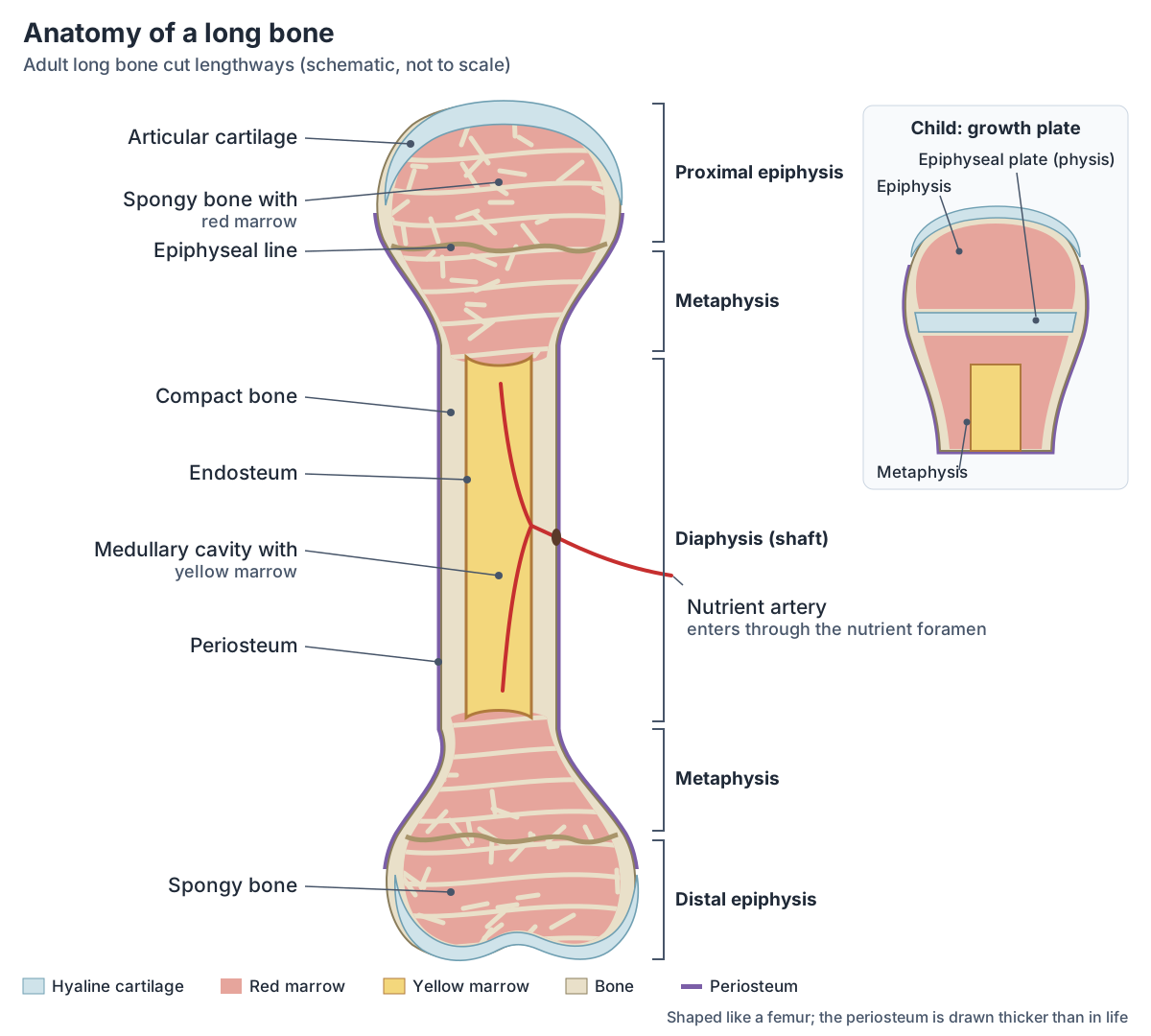
Parts of a long bone
| Part | Where | What it is made of or does |
|---|---|---|
| Diaphysis | Shaft | Tube of compact bone around the medullary cavity |
| Medullary cavity | Inside the diaphysis | Holds yellow marrow (fat) in adults; lined by endosteum |
| Epiphysis | Each end | Spongy bone filled with red marrow; covered by articular cartilage at joints |
| Metaphysis | Between diaphysis and epiphysis | Contains the growth plate in children; very vascular |
| Epiphyseal plate (physis) | In the metaphysis of a growing bone | Hyaline cartilage that lengthens the bone; becomes the epiphyseal line at about 18 to 21 years |
| Articular cartilage | Joint surfaces of the epiphyses | Hyaline cartilage that reduces friction and absorbs shock |
| Periosteum | Outer surface (not over articular cartilage) | Fibrous membrane with vessels and nerves; anchors tendons and ligaments |
| Endosteum | Lining of the medullary cavity | Thin membrane where growth, repair and remodelling occur |
| Nutrient foramen | Diaphysis | Opening for the nutrient artery |
Source: OpenStax Anatomy and Physiology 2e, §6.3 Bone Structure.
Periosteum and endosteum: the coverings of bone
The outer surface of a bone, except where it is covered by articular cartilage, is wrapped in the periosteum, a fibrous membrane that contains blood vessels, nerves and lymphatic vessels that nourish the compact bone. Tendons and ligaments attach to bone through the periosteum. (OpenStax Anatomy and Physiology 2e, 6.3: Bone Structure.)
The medullary cavity is lined by the endosteum, a delicate membrane where bone growth, repair and remodelling take place. (OpenStax Anatomy and Physiology 2e, 6.3: Bone Structure.)
The periosteum matters when a bone breaks. Its blood vessels tear and bleed, and its cells build the external callus of cartilage and bone that holds the broken ends together while they heal. (OpenStax Anatomy and Physiology 2e, 6.5: Fractures: Bone Repair.)
The growth plate (physis) and how long bones grow
Long bones form by endochondral ossification: a model made of hyaline cartilage is gradually replaced by bone. A primary ossification centre appears in the diaphysis by the second or third month of fetal life, and secondary ossification centres form in the epiphyses after birth. The flat bones of the face, most cranial bones and the clavicles form differently, directly from sheets of mesenchyme (intramembranous ossification). (OpenStax Anatomy and Physiology 2e, 6.4: Bone Formation and Development.)
The epiphyseal plate has four zones. The reserve zone anchors the plate to the epiphysis; in the proliferative zone chondrocytes divide; in the zone of maturation and hypertrophy they enlarge, which lengthens the bone; and in the zone of calcified matrix most chondrocytes have died and osteoblasts lay bone on the calcified cartilage, joining the plate to the diaphysis. (OpenStax Anatomy and Physiology 2e, 6.4: Bone Formation and Development.)
Growth in length continues until early adulthood, when chondrocytes stop dividing and bone replaces the cartilage, leaving only the epiphyseal line. A bone can still grow in diameter after that: osteoclasts remove bone lining the medullary cavity while osteoblasts add new bone under the periosteum, a process called appositional growth. (OpenStax Anatomy and Physiology 2e, 6.4: Bone Formation and Development.)
On an X-ray of a child, the physis shows as a dark (radiolucent) line between the metaphysis and the epiphysis. As the two fuse, the physis thins and disappears and endochondral ossification stops; a thin white line of bone, the physeal scar, can remain. Radiopaedia lists Salter-Harris fractures, which involve the growth plate, among the injuries of the physis. (Radiopaedia: Physis.)
Compact bone and the osteon (Haversian system)
The basic unit of compact bone is the osteon, or Haversian system. Each osteon is a column of concentric rings of bone matrix (lamellae) around a central (Haversian) canal, which carries blood vessels, nerves and lymphatic vessels along the bone. (OpenStax Anatomy and Physiology 2e, 6.3: Bone Structure.)
Perforating (Volkmann's) canals run at right angles to the central canals and connect them with the periosteum and the endosteum. Bone cells called osteocytes sit in small spaces (lacunae) between the lamellae, and tiny channels called canaliculi link the lacunae to each other and to the central canal, so nutrients can reach the cells and wastes can leave. (OpenStax Anatomy and Physiology 2e, 6.3: Bone Structure.)
Not all compact bone is in osteons. Circumferential lamellae run around the bone in flat layers under the periosteum and around the medullary cavity, and interstitial lamellae fill the gaps between osteons. (OpenStax Anatomy and Physiology 2e, 6.3: Bone Structure.)
Bone matrix has two parts that work together. Collagen fibres give it flexibility, so bone is not brittle, and crystals of hydroxyapatite, formed mainly from calcium phosphate and calcium carbonate, give it hardness and strength. (OpenStax Anatomy and Physiology 2e, 6.3: Bone Structure.)
Bone as one of the connective tissues, alongside epithelial, muscle and nervous tissue. Tissue types.
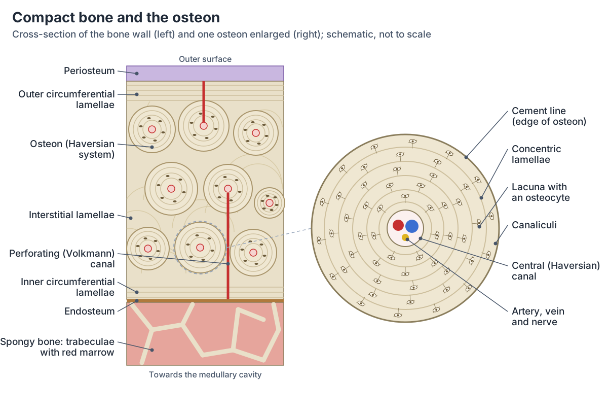
Spongy bone and bone marrow
Spongy (cancellous) bone is a lattice of thin bony struts called trabeculae. It contains osteocytes in lacunae, but they are not arranged in osteons. The trabeculae line up along the lines of stress in the bone, and the open spaces between them make the bone lighter. (OpenStax Anatomy and Physiology 2e, 6.3: Bone Structure.)
The spaces of spongy bone hold red marrow, where blood cells are made (haematopoiesis). Yellow marrow, the marrow of the medullary cavity in adult long bones, is mostly fat (adipose tissue), and its stored triglycerides can be used for energy. (OpenStax Anatomy and Physiology 2e, 6.1: The Functions of the Skeletal System.)
Compact vs spongy bone
| Feature | Compact bone | Spongy bone |
|---|---|---|
| Where in a long bone | Walls of the diaphysis, thin outer layer of the ends | Inside the epiphyses and metaphyses |
| Structure | Osteons (Haversian systems) with central and perforating canals | Lattice of trabeculae with no osteons |
| Marrow | Surrounds the medullary cavity with yellow marrow | Red marrow in the spaces between trabeculae |
| Strength | Dense and hard | Light; trabeculae follow lines of stress |
Source: OpenStax §6.3.
Bone cells: osteoblasts, osteocytes, osteoclasts and osteogenic cells
Osteogenic cells are undifferentiated cells in the deep layer of the periosteum and in the marrow. They are the only bone cells that divide, and they become osteoblasts. Osteoblasts build new bone by secreting collagen matrix and calcium salts; they do not divide, and once they are trapped in the matrix they become osteocytes. (OpenStax Anatomy and Physiology 2e, 6.3: Bone Structure.)
Osteocytes are the main cells of mature bone. Each lies in its own lacuna, keeps the mineral content of the surrounding matrix stable and connects with its neighbours through canaliculi. Osteoclasts are large cells with many nuclei that break down old or damaged bone; they come from monocytes and macrophages, not from osteogenic cells. (OpenStax Anatomy and Physiology 2e, 6.3: Bone Structure.)
Bonewald's review describes osteocytes as 90 to 95% of all bone cells in adult bone and as the longest-lived bone cells, surviving for decades. They sense mechanical loading, direct remodelling by controlling osteoblasts and osteoclasts, make sclerostin, which slows bone formation, and act as an endocrine organ: through FGF23 they help control how much phosphate the kidneys excrete. (Bonewald LF. The amazing osteocyte. J Bone Miner Res. 2011.)
Bone cells and what they do
| Cell | Where | Job |
|---|---|---|
| Osteogenic cell | Deep periosteum and marrow | Divides; becomes osteoblasts |
| Osteoblast | Bone surfaces where bone is forming | Lays down collagen matrix and calcium salts; becomes an osteocyte |
| Osteocyte | Lacunae inside the matrix | Maintains the matrix; senses load and directs remodelling; 90 to 95% of bone cells |
| Osteoclast | Bone surfaces where bone is being removed | Large multinucleated cell that resorbs bone; derived from monocytes and macrophages |
Sources: OpenStax §6.3 and Bonewald (2011).
Blood and nerve supply of a long bone
Nutrient arteries enter the diaphysis through the nutrient foramen and spread through the marrow cavity into the spongy bone. Vessels from the periosteum also penetrate the bone, metaphyseal arteries and veins enter the metaphyses from outside, and veins leave the bone through foramina. (OpenStax Anatomy and Physiology 2e, 6.3: Bone Structure.)
Nerves follow the same routes and are densest in the most active regions of the bone. They sense pain and help regulate blood flow and bone growth. (OpenStax Anatomy and Physiology 2e, 6.3: Bone Structure.)
Bone remodelling: bone is living tissue
Bone is reshaped throughout life. In modelling, which happens mainly during growth, bone is removed from one surface and laid down on another. In remodelling, old or damaged bone is resorbed and replaced at the same site; about 5 to 10% of the skeleton is remodelled every year. (OpenStax Anatomy and Physiology 2e, 6.4: Bone Formation and Development.)
A bone's inner and outer structure changes as the stress on it rises or falls: mechanical stress stimulates the deposition of mineral salts and collagen fibres. People who exercise regularly have denser bones than sedentary people, and resistance training has more effect than cardiovascular exercise. Bone density peaks at about 30 years of age. (OpenStax Anatomy and Physiology 2e, 6.6: Exercise, Nutrition, Hormones, and Bone Tissue.)
Osteoporosis is a loss of bone mass that happens when resorption outpaces formation. The compact bone thins and the trabeculae of spongy bone become fewer and smaller. It most often affects the upper end of the femur, the vertebrae and the wrist, and women lose bone faster than men from about 50 years of age, around menopause. (OpenStax Anatomy and Physiology 2e, 6.6: Exercise, Nutrition, Hormones, and Bone Tissue.)
How a broken long bone heals
Healing follows four steps. Torn vessels bleed and, about six to eight hours after the injury, form a clot called a fracture hematoma. Within about 48 hours, cells from the endosteum form an internal callus of fibrocartilage and cells from the periosteum form an external callus. Over several weeks osteoclasts clear dead bone and the cartilage of the calli is replaced by spongy bone, forming a bony callus. Finally remodelling restores compact bone at the edges and reshapes the bone. (OpenStax Anatomy and Physiology 2e, 6.5: Fractures: Bone Repair.)
Fractures are described by their pattern: transverse (straight across), oblique (at an angle), spiral (from twisting), comminuted (in several pieces), impacted (one fragment driven into another), greenstick (a partial break on one side of the bone) and open (compound) or closed (simple), depending on whether the bone breaks through the skin. (OpenStax Anatomy and Physiology 2e, 6.5: Fractures: Bone Repair.)
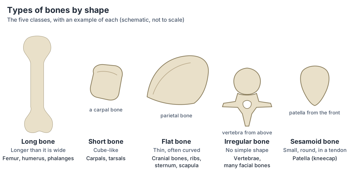
Types of bones by shape: long, short, flat, irregular and sesamoid
Short bones are cube-like, about equal in length, width and thickness; the carpals and tarsals give stability and support with limited movement. Flat bones are thin and often curved, such as the sternum, ribs, scapulae and cranial bones; they anchor muscles and often protect organs. Irregular bones, such as the vertebrae and many facial bones, have no simple shape. (OpenStax Anatomy and Physiology 2e, 6.2: Bone Classification.)
Sesamoid bones are small and round, like a sesame seed, and form inside tendons, which they protect from compressive forces. The patellae are the only sesamoid bones that every person has; the number of other sesamoid bones varies from person to person. (OpenStax Anatomy and Physiology 2e, 6.2: Bone Classification.)
How bones meet: fibrous, cartilaginous and synovial joints and their six shapes. Types of joints.
A long bone in 3D: the femur
The 3D model highlights the femur, the thigh bone, from BodyParts3D, a database built from MRI scans of an adult male volunteer. Rotate it to find the expanded upper and lower ends (the epiphyses), where the femur meets the hip and the knee, and the long, narrow shaft (the diaphysis) between them. (Mitsuhashi N et al. BodyParts3D: 3D structure database for anatomical concepts. Nucleic Acids Res. 2009.)
The model shows only the outer surface of the bone and does not include cartilage, marrow or the periosteum, and the femur of an adult has no growth plate left. Use the labeled section and osteon diagrams on this page to see what lies inside.
Bone structure in the 3D model
- Femur
- The thigh bone, the longest, heaviest and strongest bone in the body.
Frequently asked questions
What are the parts of a long bone?
A long bone has a shaft (diaphysis) of compact bone around a medullary cavity filled with yellow marrow, two wider ends (epiphyses) of spongy bone with red marrow, and a metaphysis between shaft and each end, which holds the growth plate in children. Articular cartilage covers the joint surfaces, the periosteum covers the rest, and the endosteum lines the cavity.
What is the difference between the diaphysis and the epiphysis?
The diaphysis is the long shaft of the bone, a tube of compact bone with the medullary cavity inside. The epiphyses are the wider ends; they are filled with spongy bone and red marrow and form joints with other bones, where they are covered by articular cartilage. The metaphysis joins the two.
What is the epiphyseal plate?
The epiphyseal plate, or growth plate (physis), is a layer of hyaline cartilage in the metaphysis of a growing long bone. Its cartilage cells divide and enlarge, and the cartilage is replaced by bone, which lengthens the bone. Growth stops in early adulthood, at about 18 to 21 years, when the plate turns into bone and leaves the epiphyseal line.
What is an osteon?
An osteon, or Haversian system, is the basic unit of compact bone: a column of concentric rings of bone matrix (lamellae) around a central canal that carries blood vessels and nerves. Osteocytes sit in lacunae between the rings and connect through tiny canaliculi, and perforating (Volkmann's) canals link neighbouring central canals with the bone surfaces.
What is the difference between compact and spongy bone?
Compact bone is dense and hard and is built from osteons; it forms the walls of the shaft and the outer layer of every bone. Spongy (cancellous) bone is a light lattice of trabeculae with no osteons; it fills the ends of long bones, follows the lines of stress and holds red marrow in its spaces.
What are the five types of bones?
Bones are classed by shape as long (longer than wide, such as the femur and humerus), short (cube-like, the carpals and tarsals), flat (thin and curved, such as the ribs, sternum and cranial bones), irregular (such as the vertebrae) and sesamoid (small bones inside tendons, such as the patella).
References
- OpenStax. Anatomy and Physiology 2e, §6.1 The Functions of the Skeletal System. Rice University, 2022.
- OpenStax. Anatomy and Physiology 2e, §6.2 Bone Classification. Rice University, 2022.
- OpenStax. Anatomy and Physiology 2e, §6.3 Bone Structure. Rice University, 2022.
- OpenStax. Anatomy and Physiology 2e, §6.4 Bone Formation and Development. Rice University, 2022.
- OpenStax. Anatomy and Physiology 2e, §6.5 Fractures: Bone Repair. Rice University, 2022.
- OpenStax. Anatomy and Physiology 2e, §6.6 Exercise, Nutrition, Hormones, and Bone Tissue. Rice University, 2022.
- Radiopaedia. Physis.
- Radiopaedia. Metaphysis.
- Bonewald LF. The amazing osteocyte. J Bone Miner Res. 2011;26(2):229-238.
- Mitsuhashi N, Fujieda K, Tamura T, et al. BodyParts3D: 3D structure database for anatomical concepts. Nucleic Acids Res. 2009;37:D782-D785.
- Mitsuhashi N. et al. (2009). BodyParts3D: 3D structure database for anatomical concepts. Nucleic Acids Research 37, D782–D785. Data © The Database Center for Life Science, CC BY 4.0.
Related anatomy
- Skeletal systemExplore a realistic 3D human skeleton: the 206 bones, axial vs appendicular skeleton, bone types, bony landmarks and…
- FemurFemur bone labelled: head, neck, trochanters, linea aspera, condyles and epicondyles, with muscle attachments, blood…
- Tissue typesThe four tissue types in human anatomy with labeled diagrams: epithelial, connective, muscle and nervous tissue, their…
- Types of jointsTypes of joints explained: fibrous, cartilaginous and synovial (diarthrosis) joints, a labelled synovial joint diagram…
- HumerusHumerus bone labeled: head, anatomical and surgical neck, tubercles, deltoid tuberosity, radial groove, epicondyles…
- TibiaTibia anatomy explained: where the shinbone is, the proximal tibia, tibial plateau, shaft and medial malleolus, its…
Explore the whole body in the interactive 3D human anatomy model, or browse the A–Z list of human structures.