

Internal oblique at a glance
- What it is
- The internal oblique is the middle of the three flat muscles of the side of the abdomen.
- Location
- It lies under the external oblique and over transversus abdominis, from the inguinal ligament, iliac crest and thoracolumbar fascia up to the lower ribs; in front it becomes an aponeurosis that joins the rectus sheath.
- Function
- It compresses the abdomen, flexes the trunk and rotates it to the same side.
- Body system
- Muscular · paired (left and right)
- FMA ID
- FMA13893 (Foundational Model of Anatomy)
Key facts
- TeachMeAnatomy: it 'originates from the inguinal ligament, iliac crest and lumbodorsal fascia' and 'inserts onto ribs 10-12'; its fibres run superomedially, at right angles to those of the external oblique.
- In the 3D model it is built as a layer under the external oblique; BodyParts3D has no internal oblique.
Where is the internal oblique?
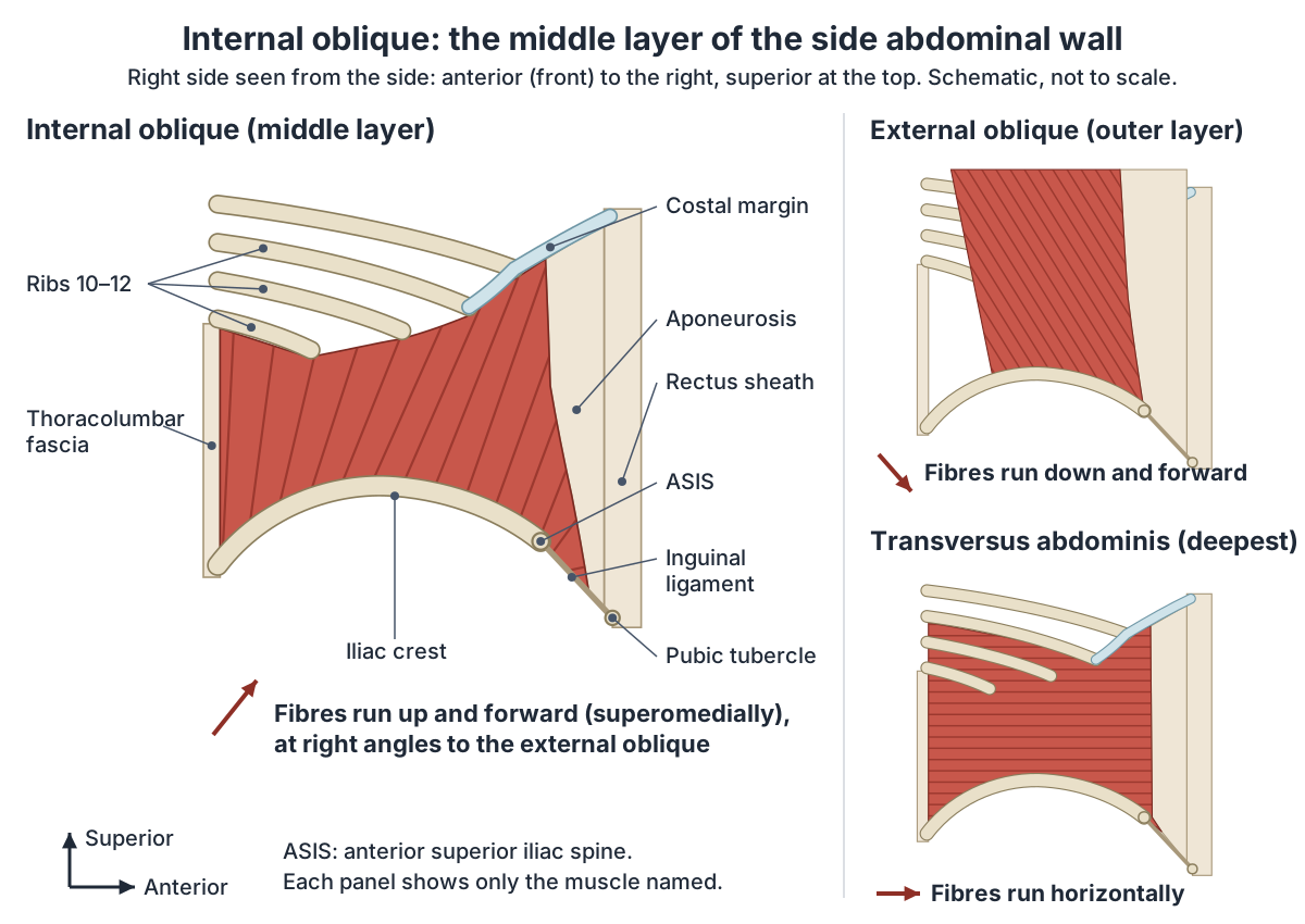
The internal oblique lies deep to the external oblique and superficial to transversus abdominis, so it is the middle of the three flat muscles of the side of the abdomen. It is fleshy at the side and becomes a flat tendon, an aponeurosis, towards the front, where it merges into the linea alba in the midline. (Radiopaedia: Internal oblique muscle.)
Its fibres run superomedially, up and towards the midline, at right angles to the fibres of the external oblique. Because the fibres of the three flat muscles run in different directions and cross each other, they strengthen the wall and lower the risk of abdominal contents pushing out through it. (TeachMeAnatomy: The Anterolateral Abdominal Wall.)
The fibres fan out from the hip region. Kenhub divides them into three groups: the anterior fibres run down and medially, the lateral fibres spread up and medially, and the posterior fibres climb to the lower ribs. (Kenhub: Internal abdominal oblique muscle.)
OpenStax gives a simple way to picture the two obliques: the external oblique fibres run down and in like four fingers sliding into the front pockets of your trousers, and the internal oblique fibres run up and in, the way the thumbs point. (OpenStax Anatomy and Physiology 2e, 11.4: Axial Muscles of the Abdominal Wall and Thorax.)
In the 3D model the internal oblique is an approximation, built along its textbook attachments, because BodyParts3D, the adult male data set the model is based on, has no internal oblique. It is drawn as a muscular sheet on each flank from the iliac crest and inguinal region up to the lower ribs, under the external oblique. The view opens on the left side of the body seen from the side, with the internal oblique, transversus abdominis, rectus abdominis, the twelfth rib and the hip bone labelled; the external oblique is hidden in this view so that the internal oblique can be seen. Its thickness is a typical value, not a measurement, and its aponeurosis to the rectus sheath is not modelled; the diagrams show it instead. Real thicknesses measured on ultrasound are given below.
All the muscles of the abdominal wall, the rectus sheath and the inguinal canal. Abdominal muscles.
Internal oblique origin and insertion
Radiopaedia gives the origin as the whole length of the lumbar (thoracolumbar) fascia, the anterior two-thirds of the intermediate line of the iliac crest and the lateral two-thirds of the grooved upper surface of the inguinal ligament. (Radiopaedia: Internal oblique muscle.)
For the inguinal ligament, other sources give a smaller share: Gray's Anatomy (1918) and a 2021 radiology case series on side strain both describe the lateral half of the ligament, so figures for the ligament range from a half to two-thirds. For the iliac crest, Radiopaedia, Kenhub and Gray's Anatomy all give the anterior two-thirds. (Mehta CP, Daftary A, Lawande M. Side Strain and Impact Injuries at the Iliac Crest Attachment of Lateral Abdominal Wall Musculature in Cricketers. Indian J Radiol Imaging. 2021.)
Kenhub adds a refinement: the anterior fibres were traditionally said to arise from the inguinal ligament, but it is now known that they arise from a deeper structure, the iliopectineal arch. The lateral fibres come from the anterior two-thirds of the iliac crest, and the posterior fibres from the iliac crest and thoracolumbar fascia. (Kenhub: Internal abdominal oblique muscle.)
It inserts on the inferior border of the costal cartilages of the lower three ribs (ribs 10 to 12). In front, the muscle becomes aponeurotic and the aponeurosis fuses in the midline at the linea alba. Lowest of all, it attaches to the pectineal line of the pubis together with the transversus abdominis aponeurosis, forming the conjoint tendon. (Radiopaedia: Internal oblique muscle.)
Kenhub describes the rib insertion as the inferior borders and tips of the lower three or four ribs and their cartilages, and gives the insertion of the conjoint tendon as the pubic crest and pecten pubis. (Kenhub: Internal abdominal oblique muscle.)
The conjoint tendon forms where the medial fibres of the internal oblique aponeurosis unite with the deeper fibres of the transversus abdominis aponeurosis. It attaches to the pubic crest and pecten pubis, reinforces the medial part of the back wall of the inguinal canal and lies directly behind the superficial inguinal ring. (Radiopaedia: Conjoint tendon.)
Internal oblique: origin, insertion, nerve, blood supply and action
| Feature | Internal oblique |
|---|---|
| Origin | Thoracolumbar fascia; anterior two-thirds of the iliac crest (intermediate line); lateral half to two-thirds of the inguinal ligament (or the iliopectineal arch behind it) |
| Insertion | Inferior borders of ribs 10 to 12 and their cartilages (some sources: lower three or four ribs); linea alba through its aponeurosis; pubic crest and pecten pubis through the conjoint tendon |
| Nerves | Lower intercostal (thoracoabdominal) nerves T7 to T11 and subcostal nerve T12; iliohypogastric and ilioinguinal nerves (L1) |
| Roots | T7 to T12 and L1 |
| Blood supply | Lower posterior intercostal and subcostal arteries; superior and inferior epigastric arteries; superficial and deep circumflex iliac arteries; posterior lumbar arteries |
| Actions | Both sides: flex the trunk and compress the abdomen (forced expiration, urination, defecation). One side: bends the trunk to the same side and rotates it to the same side, with the opposite external oblique |
| Layer | Middle flat muscle: external oblique outside, transversus abdominis inside; nerves run between it and transversus abdominis |
Compiled from Radiopaedia, Kenhub, TeachMeAnatomy, Mehta et al. (2021) and Gray's Anatomy (1918); ranges show where sources differ (see references).
The aponeurosis and the rectus sheath
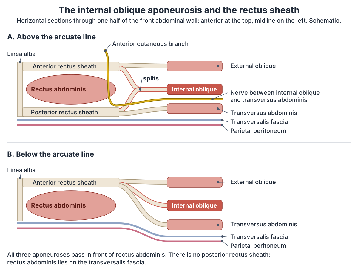
The aponeurosis of the internal oblique helps build the rectus sheath, the tough envelope around rectus abdominis. Above the arcuate line it splits into two layers around the muscle. The superficial layer joins the external oblique aponeurosis to form the anterior rectus sheath, and the deep layer joins the transversus abdominis aponeurosis to form the posterior rectus sheath. (Kenhub: Internal abdominal oblique muscle.)
Below the arcuate line all three aponeuroses, from the external oblique, internal oblique and transversus abdominis, pass in front of rectus abdominis. Radiopaedia places the arcuate line about 2.5 cm below the level of the umbilicus. (Radiopaedia: Internal oblique muscle.)
TeachMeAnatomy describes the same change happening about midway between the umbilicus and the pubic symphysis. Below this point the sheath has no posterior wall, so the back of rectus abdominis is in direct contact with the transversalis fascia; the line where the posterior layer ends is the arcuate line. (TeachMeAnatomy: The Anterolateral Abdominal Wall.)
The vertical 'six-pack' muscle that the rectus sheath encloses. Rectus abdominis.
Nerve and blood supply
The internal oblique is supplied mainly by the lower six thoracic spinal nerves (T7 to T12): the ends of the lower intercostal nerves and the subcostal nerve. The iliohypogastric and ilioinguinal nerves, both from L1, make smaller contributions. (Kenhub: Internal abdominal oblique muscle.)
TeachMeAnatomy lists the same nerves in a different way: the thoracoabdominal nerves (T7 to T11), the subcostal nerve (T12) and branches of the lumbar plexus. (TeachMeAnatomy: The Anterolateral Abdominal Wall.)
The two lumbar plexus branches both come from the L1 root. The iliohypogastric nerve crosses quadratus lumborum, pierces transversus abdominis and supplies the internal oblique and transversus abdominis. The ilioinguinal nerve follows the same course, supplies the same two muscles and then leaves the abdominal wall through the superficial inguinal ring. (TeachMeAnatomy: The Lumbar Plexus.)
These nerves travel in a definite layer. The anterior rami of T7 to T12 pass between the internal oblique and transversus abdominis, then pierce rectus abdominis and end as anterior cutaneous branches in the skin of the front of the abdomen. The L1 branches, the ilioinguinal and iliohypogastric nerves, also enter this plane. (Tsai HC et al. Transversus Abdominis Plane Block: An Updated Review of Anatomy and Techniques. Biomed Res Int. 2017.)
Its arteries are the lower posterior intercostal and subcostal arteries, the superior and inferior epigastric arteries, the superficial and deep circumflex iliac arteries and the posterior lumbar arteries. (TeachMeAnatomy: Internal Oblique.)
Surgeons make use of this blood supply. The internal oblique muscle flap, moved to repair the lower abdominal wall, groin and nearby areas, is based on the ascending branch of the deep circumflex iliac artery, which arises from the external iliac artery. (Watanabe S et al. The Internal Oblique Muscle Flap with Synthetic Mesh Reinforcement for a Large, Contaminated Abdominal Wall Defect. Plast Reconstr Surg Glob Open. 2020.)
The deepest flat muscle, which shares its nerves with the internal oblique. Transversus abdominis.
What does the internal oblique do?
When both internal obliques contract together, they flex the trunk, squeeze the abdominal organs and raise the pressure inside the abdomen. That pressure helps with forced breathing out, passing urine and emptying the bowels. When one side contracts on its own, it bends the trunk to that side and rotates it towards the same side. (Kenhub: Internal abdominal oblique muscle.)
Rotation needs a partner. The external oblique turns the trunk to the opposite side, working in synergy with the internal oblique of the other side. So the right internal oblique and the left external oblique together turn the trunk to the right, and the left internal oblique with the right external oblique turn it to the left. (Kenhub: External abdominal oblique muscle.)
TeachMeAnatomy gives the same pattern: bilateral contraction compresses the abdomen, while unilateral contraction rotates the torso to the same side (ipsilaterally). The external oblique, by contrast, rotates the torso to the opposite side. (TeachMeAnatomy: The Anterolateral Abdominal Wall.)
In breathing, normal breathing out is passive and needs no energy. In forced breathing out, the accessory muscles of the abdomen, including the obliques, contract and force the abdominal organs upwards against the diaphragm, which pushes more air out of the lungs. (OpenStax Anatomy and Physiology 2e, 22.3: The Process of Breathing.)
Radiopaedia sums up its actions as compressing the abdominal contents, assisting forced expiration by pulling down (depressing) the lower ribs, and flexing and bending the trunk. (Radiopaedia: Internal oblique muscle.)
The outer flat muscle, which twists the trunk to the opposite side. External oblique.
Its role in twisting, coughing and sitting
Twisting sports load the muscle heavily. In a baseball swing the trunk rotates towards the side opposite the batting side, and this movement needs a large contribution from the internal oblique on that opposite side. The same muscle is reported to help produce ball speed in pitching and bat speed in batting. (Komatsu S, Kaneko H, Nagashima M. Characteristics of internal oblique muscle strain in professional baseball players: a case series. BMC Sports Sci Med Rehabil. 2022.)
Coughing is another everyday task. A cough involves a quick, coordinated contraction of the muscles of the chest, abdomen and pelvis, which raises the pressure inside the chest and abdomen. (Kodama K et al. Cough-induced internal oblique hematoma. J Emerg Trauma Shock. 2013.)
The deep abdominal muscles also work in posture. In a study of 30 office workers who sat for 1 hour in one of three postures, only the slumped posture was linked with signs of fatigue in the internal oblique and transversus abdominis, appearing about 20 minutes into sitting. The authors suggest this fatigue may reduce the stability of the spine. (Waongenngarm P, Rajaratnam BS, Janwantanakul P. Internal Oblique and Transversus Abdominis Muscle Fatigue Induced by Slumped Sitting Posture. Saf Health Work. 2016.)
What lies next to the internal oblique?
Low down, the free lower border of the internal oblique lies in front of the deep inguinal ring and arches over the spermatic cord. Laterally its muscle fibres lie in front of the cord, while medially its tendinous fibres lie behind it. (Radiopaedia: Internal oblique muscle.)
This arch makes the internal oblique part of the inguinal canal. Kenhub's MALT summary puts the internal oblique and transversus abdominis in the roof (muscles) and the external and internal oblique aponeuroses in the anterior wall. The fibres travel up and then arch inferomedially as they cross the contents of the canal. (Kenhub: Inguinal canal.)
TeachMeAnatomy describes the anterior wall of the canal as reinforced laterally by the internal oblique, and notes that when the abdominal muscles contract, the walls clamp down on the canal. The ilioinguinal nerve runs through part of the canal and leaves through the superficial ring. (TeachMeAnatomy: The Inguinal Canal.)
At the back, the internal oblique borders two weak spots in the flank. The superior lumbar triangle is bounded by the twelfth rib above, quadratus lumborum medially and the internal oblique laterally. (Radiopaedia: Superior lumbar triangle.)
The inferior lumbar triangle (of Petit) lies between the iliac crest, the external oblique and latissimus dorsi, and the internal oblique forms its floor. (Radiopaedia: Inferior lumbar triangle.)
Higher up, the muscle continues into the chest wall: at the front, the lower internal intercostal muscles become continuous with the internal oblique. (Radiopaedia: Internal intercostal muscle.)
The inguinal ligament, inguinal canal and the region where the abdominal wall meets the thigh. Groin anatomy.
The cremaster muscle: a slip of the internal oblique
In males some of the anterior fibres of the internal oblique continue into the spermatic cord and form the cremaster muscle. (Kenhub: Internal abdominal oblique muscle.)
The cremasteric muscle and fascia form the middle covering of the spermatic cord, and both are continuations of the internal oblique muscle and its fascia. (Kenhub: Inguinal canal.)
Radiopaedia describes the cremaster as derived from the internal oblique muscle and aponeurosis, and notes that some authors include transversus abdominis and the transversalis fascia. It is supplied by the genital branch of the genitofemoral nerve and pulls the testis upwards, which matters for temperature control. (Radiopaedia: Cremaster muscle.)
How is the internal oblique examined? Ultrasound and real size
The internal oblique lies under the external oblique, but ultrasound shows it well. A probe placed at the midaxillary line, between the costal margin and the iliac crest, shows the typical three muscle layers of the side wall, one above the other. (Tsai HC et al. Transversus Abdominis Plane Block: An Updated Review of Anatomy and Techniques. Biomed Res Int. 2017.)
In 21 healthy young men scanned while seated, with the probe on the right side level with the umbilicus, the internal oblique measured about 7.1 to 7.2 mm thick at rest. When they pushed to turn to the right against resistance without moving the trunk (an isometric effort at half their maximum rotation strength), the right internal oblique thickened to about 9.6 mm, which fits its role in same-side rotation. (Sugaya T, Abe Y, Sakamoto M. Ultrasound evaluation of muscle thickness changes in the external oblique, internal oblique, and transversus abdominis muscles. J Phys Ther Sci. 2014.)
In a second study of 24 healthy men in their twenties, lying with the knees bent, the right internal oblique measured 0.85 cm (about 8.5 mm) thick, slightly thicker than the external oblique at 0.77 cm. (Kang KW, Kwon YH, Son SM. Ultrasound Measurement of the Transverse Abdominis, Internal Oblique, and External Oblique Muscles. Med Sci Monit. 2021.)
Variations
Descriptions of the attachments vary between sources: the rib insertion is given as the lower three or four ribs, and the anterior fibres may come from the iliopectineal arch rather than the inguinal ligament itself. (Kenhub: Internal abdominal oblique muscle.)
Descriptions of the layers that form the cremaster also differ. In a dissection study of 30 cadavers, Xiao and colleagues found that the internal oblique and its aponeurosis, and transversus abdominis, spread over the surface of the spermatic cord and continued as the cremaster. (Xiao Y, Zhou Z, Yan L, Tong C. Anatomical observations on 30 cadavers: the posterior wall of the inguinal canal and the cremaster. BMC Surg. 2024.)
Internal oblique strain, haematoma, hernias and nerve blocks
A side strain is an injury of the internal or external oblique or transversus abdominis, usually where they attach to the lower four ribs and only rarely at their pelvic attachments. Radiologists report that it typically involves the internal oblique at its lower rib attachments; their own series described four cricketers with injuries at the iliac crest instead. (Mehta CP, Daftary A, Lawande M. Side Strain and Impact Injuries at the Iliac Crest Attachment of Lateral Abdominal Wall Musculature in Cricketers. Indian J Radiol Imaging. 2021.)
In one Japanese professional baseball team with 188 players between 2012 and 2021, 23 players (12.2%) had 28 internal oblique strains. Most were at the insertion on the lower ribs, more often on the side opposite the batting or throwing side, and the mean time to return to play was 27.7 days. (Komatsu S, Kaneko H, Nagashima M. Characteristics of internal oblique muscle strain in professional baseball players: a case series. BMC Sports Sci Med Rehabil. 2022.)
Bleeding into the muscle, an internal oblique haematoma, is extremely rare. It has been reported after violent or long-lasting coughing and is usually caused by damage to the lower intercostal or lumbar arteries or the ascending branches of the deep circumflex iliac artery. (Kodama K et al. Cough-induced internal oblique hematoma. J Emerg Trauma Shock. 2013.)
The conjoint tendon, formed by the internal oblique and transversus abdominis aponeuroses, reinforces the back wall of the inguinal canal. If it weakens, it may predispose to a direct inguinal hernia. (Radiopaedia: Conjoint tendon.)
The nerves of the abdominal wall run in the plane between the internal oblique and transversus abdominis before piercing the rectus sheath to reach the midline. Anaesthetists use this: in abdominal wall blocks such as the transversus abdominis plane (TAP) block, local anaesthetic is injected into that plane so that it spreads to the nerves and relieves pain in the abdominal wall. (Fernandes HDS et al. Ultrasound-guided peripheral abdominal wall blocks. Clinics (Sao Paulo). 2021.)
Surgeons also use the fibre directions. The McBurney (gridiron) incision used in open appendicectomy splits the fibres of the abdominal wall muscles instead of cutting them, which allows excellent healing. (TeachMeAnatomy: The Anterolateral Abdominal Wall.)
Structures next to the internal oblique
The nearest named structures in the 3D model, measured from the centre of each part:
- Transversus abdominis · muscular
- External oblique · muscular
- Ascending colon · digestive
- Descending colon · digestive
- Taenia omentalis · digestive
- Rectus abdominis · muscular
- Musculophrenic artery · cardiovascular
- Lumbar artery · cardiovascular
- Internal intercostal muscle · muscular
- Lumbar vein · cardiovascular
Frequently asked questions
What does the internal oblique do?
Working together, the two internal obliques flex the trunk and squeeze the abdomen, raising the pressure inside it for forced breathing out, coughing, urination and defecation. One internal oblique on its own bends the trunk to its own side and rotates the trunk towards that side, working with the external oblique of the opposite side.
Where is the internal oblique located?
It forms the middle layer of the side wall of the abdomen, between the external oblique outside and transversus abdominis inside. It stretches from the iliac crest, the inguinal ligament and the thoracolumbar fascia of the lower back up to the lower ribs, and in front it becomes an aponeurosis that joins the rectus sheath.
What is the origin and insertion of the internal oblique?
It arises from the thoracolumbar fascia, the anterior two-thirds of the iliac crest and the lateral half to two-thirds of the inguinal ligament. It inserts on the lower borders of ribs 10 to 12 and their cartilages, on the linea alba through its aponeurosis, and on the pubic crest and pecten pubis through the conjoint tendon.
What is the difference between the internal and external oblique?
The external oblique is the outer layer and its fibres run down and towards the midline; the internal oblique lies under it, with fibres running up and towards the midline at right angles. The external oblique rotates the trunk to the opposite side and the internal oblique to the same side, so the two work as partners from opposite sides.
What nerve supplies the internal oblique?
It is supplied by the lower intercostal (thoracoabdominal) nerves T7 to T11, the subcostal nerve T12, and the iliohypogastric and ilioinguinal nerves from L1. These nerves run in the plane between the internal oblique and transversus abdominis, which is the layer targeted by a transversus abdominis plane block.
What is an internal oblique strain (side strain)?
A side strain is a tear of the side abdominal wall muscles, most often the internal oblique where it attaches to the lower ribs. It is seen in sports with forceful trunk rotation such as cricket, baseball and tennis. In one professional baseball team, 12.2% of players had an internal oblique strain over ten years, with a mean return to play of about four weeks.
How thick is the internal oblique?
Ultrasound studies in healthy young men measured it at about 7 to 8.5 mm thick at rest at the side of the abdomen: about 7.1 to 7.2 mm in 21 men sitting upright and 8.5 mm in 24 men lying with bent knees. It thickens when it contracts, for example to about 9.6 mm when pushing to turn towards the same side against resistance (an isometric effort).
References
- Kenhub. Internal abdominal oblique muscle.
- Kenhub. External abdominal oblique muscle.
- Kenhub. Inguinal canal.
- Internal oblique muscle. Radiopaedia reference article.
- Conjoint tendon. Radiopaedia reference article.
- Cremaster muscle. Radiopaedia reference article.
- Anterior abdominal wall. Radiopaedia reference article.
- Superior lumbar triangle. Radiopaedia reference article.
- Inferior lumbar triangle. Radiopaedia reference article.
- Internal intercostal muscle. Radiopaedia reference article.
- TeachMeAnatomy. The Anterolateral Abdominal Wall.
- TeachMeAnatomy. Internal Oblique (encyclopaedia entry).
- TeachMeAnatomy. The Inguinal Canal.
- TeachMeAnatomy. The Lumbar Plexus.
- OpenStax. Anatomy and Physiology 2e, §11.4 Axial Muscles of the Abdominal Wall and Thorax. Rice University, 2022.
- OpenStax. Anatomy and Physiology 2e, §22.3 The Process of Breathing. Rice University, 2022.
- Tsai HC, Yoshida T, Chuang TY, et al. Transversus abdominis plane block: an updated review of anatomy and techniques. Biomed Res Int. 2017;2017:8284363.
- Fernandes HDS, Azevedo AS, Ferreira TC, et al. Ultrasound-guided peripheral abdominal wall blocks. Clinics (Sao Paulo). 2021;76:e2170.
- Sugaya T, Abe Y, Sakamoto M. Ultrasound evaluation of muscle thickness changes in the external oblique, internal oblique, and transversus abdominis muscles considering the influence of posture and muscle contraction. J Phys Ther Sci. 2014;26(9):1399–1402.
- Kang KW, Kwon YH, Son SM. Ultrasound measurement of the transverse abdominis, internal oblique, and external oblique muscles associated with forward head posture and reduced cranio-vertebral angle. Med Sci Monit. 2021;27:e928987.
- Komatsu S, Kaneko H, Nagashima M. Characteristics of internal oblique muscle strain in professional baseball players: a case series. BMC Sports Sci Med Rehabil. 2022;14(1):118.
- Mehta CP, Daftary A, Lawande M. Side strain and impact injuries at the iliac crest attachment of lateral abdominal wall musculature in cricketers: an uncommon injury pattern. Indian J Radiol Imaging. 2021;31(2):373–377.
- Kodama K, Takase Y, Yamamoto H, Noda T. Cough-induced internal oblique hematoma. J Emerg Trauma Shock. 2013;6(2):132–134.
- Watanabe S, Sakuma H, Kono H, Shimono A, Matsuura N. The internal oblique muscle flap with synthetic mesh reinforcement for a large, contaminated abdominal wall defect. Plast Reconstr Surg Glob Open. 2020;8(9):e3083.
- Waongenngarm P, Rajaratnam BS, Janwantanakul P. Internal oblique and transversus abdominis muscle fatigue induced by slumped sitting posture after 1 hour of sitting in office workers. Saf Health Work. 2016;7(1):49–54.
- Gray H. Anatomy of the Human Body. 20th ed. Philadelphia: Lea & Febiger; 1918. The Muscles of the Abdomen: Obliquus internus abdominis (Internet Archive full text).
- Xiao Y, Zhou Z, Yan L, Tong C. Anatomical observations on 30 cadavers: new insights into the relationship between the posterior wall of the inguinal canal and the cremaster. BMC Surg. 2024;24(1):231.
- Foundational Model of Anatomy: Internal oblique (FMA13893). EMBL-EBI Ontology Lookup Service.
- Mitsuhashi N. et al. (2009). BodyParts3D: 3D structure database for anatomical concepts. Nucleic Acids Research 37, D782–D785. Data © The Database Center for Life Science, CC BY 4.0.
Related anatomy
- Abdominal musclesAbdominal muscles explained: the two oblique muscles, transversus abdominis, rectus abdominis, pyramidalis, the rectus…
- External obliqueExternal oblique: The largest and most superficial lateral abdominal wall muscle. See where it is in an interactive 3D…
- Transversus abdominisTransversus abdominis: Transversus abdominis is the deepest of the three flat muscles of the abdominal wall. See where…
- Rectus abdominisRectus abdominis: Rectus abdominis is the long, vertical strap muscle on each side of the midline of the front of the…
- GroinGroin anatomy with labelled diagrams and a 3D model: inguinal ligament and canal, femoral triangle, groin muscles…
- Internal intercostal muscleInternal intercostals: attachments, fibre direction, nerves, arteries, forced expiration, the parasternal part and the…
Explore the whole body in the interactive 3D human anatomy model, or browse the A–Z list of human structures.