

Where is the groin?
The groin, or inguinal region, is the lower part of the front of the abdominal wall on each side, just above the thigh, lateral to the pubic tubercle and below and medial to the anterior superior iliac spine (ASIS). In the nine-region map of the abdomen there are two inguinal regions, right and left; another name for them is the iliac regions. (Kenhub: Inguinal region.)
Inside the abdomen, the right inguinal region lies over part of the small intestine, the caecum, the appendix and the ascending colon, and the left over part of the small intestine and the descending and sigmoid colon. In females each also contains the ovary and uterine tube of that side. (Kenhub: Inguinal region.)
Below the inguinal ligament, at the top of the front of the thigh, lies the femoral triangle, the space through which the femoral nerve, artery and vein enter the leg. Its upper border is the inguinal ligament itself. (TeachMeAnatomy: The Femoral Triangle.)
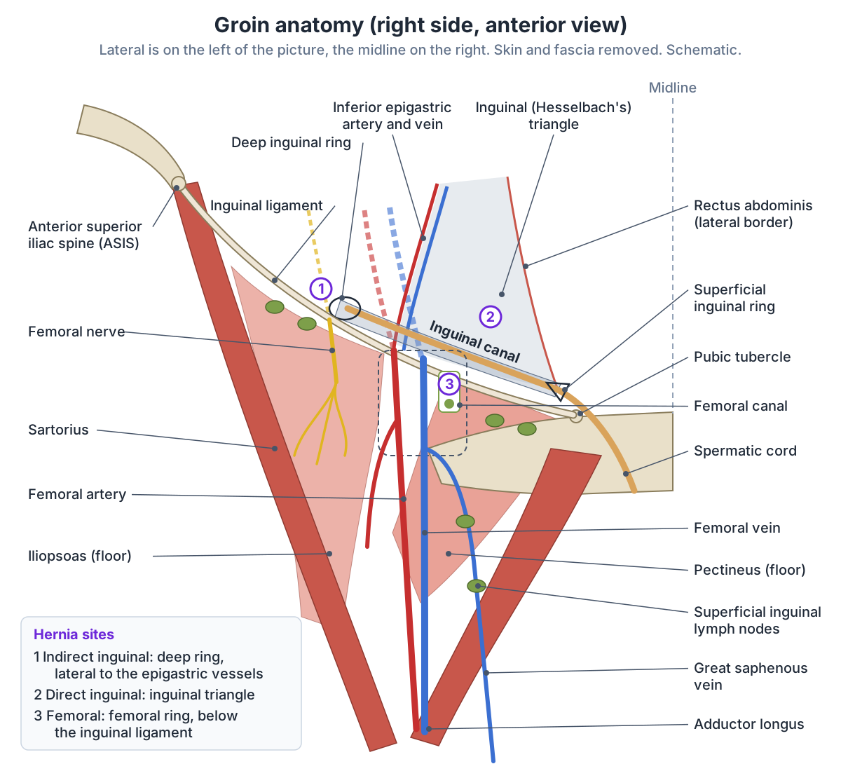
The inguinal ligament
The inguinal ligament is a fibrous band from the ASIS to the pubic tubercle. It is the lower border of the aponeurosis of the external oblique muscle, thickened and folded back on itself. Its lateral half runs obliquely and its medial half is almost horizontal; its grooved upper surface forms the floor of the inguinal canal, and below it is continuous with the fascia lata of the thigh. (Radiopaedia: Inguinal ligament.)
At its medial end, fibres turn back to the pectineal line of the superior pubic ramus as the lacunar ligament (of Gimbernat) and continue laterally along that line as the pectineal ligament (of Cooper). Internal oblique arises from the lateral two-thirds of the ligament and transversus abdominis from its lateral third. The ligament is named after François Poupart, who described it in 1705. (Radiopaedia: Inguinal ligament.)
The space beneath the ligament (the retroinguinal passage) carries iliopsoas, the femoral nerve, the femoral artery and vein, and lymphatic vessels from the inguinal nodes out of the abdomen and pelvis into the thigh. (Radiopaedia: Inguinal ligament.)
Two surface landmarks are easily confused. The mid-inguinal point lies exactly midway between the ASIS and the pubic symphysis, and the femoral pulse is felt just below it, where the femoral artery crosses the inguinal ligament. (TeachMeAnatomy: The Femoral Triangle.)
The midpoint of the inguinal ligament lies halfway between the ASIS and the pubic tubercle, and the deep inguinal ring sits about 1 cm above it. Kenhub's inguinal canal page places the ring at the midpoint itself, so sources vary by about a centimetre. (Kenhub: Inguinal region.)
Inguinal canal anatomy: walls, rings and contents
The inguinal canal is an oblique passage about 4 cm long through the lower abdominal wall, with an opening at each end. It begins at the deep (internal) inguinal ring, a round opening in the transversalis fascia about 1 cm above the inguinal ligament and lateral to the inferior epigastric vessels, and ends at the superficial (external) inguinal ring, a V-shaped gap in the external oblique aponeurosis above and lateral to the pubic tubercle. (Radiopaedia: Inguinal canal.)
Kenhub gives the adult length as 3 to 5 cm, places the superficial ring about 1 cm above and lateral to the pubic tubercle, and notes that both rings are larger in males, to allow the testis to pass. (Kenhub: Inguinal canal.)
The walls are remembered as MALT: the roof is muscle (internal oblique and transversus abdominis), the anterior wall is aponeurosis (external and internal oblique), the floor is ligament (inguinal and lacunar ligaments), and the posterior wall is transversalis fascia and the conjoint tendon. (Radiopaedia: Inguinal canal.)
TeachMeAnatomy describes the walls slightly differently: the anterior wall is the external oblique aponeurosis reinforced laterally by internal oblique, the posterior wall is the transversalis fascia, the roof is transversalis fascia, internal oblique and transversus abdominis, and the floor is the inguinal ligament thickened medially by the lacunar ligament. (TeachMeAnatomy: The Inguinal Canal.)
In males the canal carries the spermatic cord to the testis; in females it carries the round ligament of the uterus from the uterus to the labium majus. In both sexes it also carries the ilioinguinal nerve, which enters part-way along and leaves through the superficial ring and is the nerve most at risk in hernia repair, and the genital branch of the genitofemoral nerve. (TeachMeAnatomy: The Inguinal Canal.)
The canal exists because the gonads form on the back wall of the abdomen and descend, guided by a cord called the gubernaculum. In males the testis passes through the canal into the scrotum, and a pouch of peritoneum that goes with it, the processus vaginalis, normally closes; if it stays open it can lead to an indirect inguinal hernia or a hydrocele. In females the gubernaculum becomes the ovarian ligament and the round ligament of the uterus. (TeachMeAnatomy: The Inguinal Canal.)
The full inguinal canal guide: the abdominal wall layers that form its walls, with a labelled diagram. Abdominal muscles and the inguinal canal.
Femoral triangle: borders and contents
The femoral triangle is bounded above by the inguinal ligament, laterally by the medial border of sartorius and medially by the medial border of adductor longus (some sources use its lateral border). Its floor is formed by pectineus, iliopsoas and adductor longus, and its roof by the fascia lata. (TeachMeAnatomy: The Femoral Triangle.)
Kenhub remembers the borders as SAIL (sartorius, adductor longus, inguinal ligament) and the floor as APPI (adductor longus, pectineus, psoas major, iliacus). The apex points down towards the adductor canal, and the roof is fascia lata, superficial fascia and skin. (Kenhub: Femoral triangle.)
From lateral to medial the triangle contains the femoral nerve, femoral artery, femoral vein and femoral canal, remembered as NAVEL: nerve, artery, vein, empty space and lymphatics. The artery, vein and canal lie inside a fascial sleeve, the femoral sheath, and the great saphenous vein drains into the femoral vein within the triangle. (TeachMeAnatomy: The Femoral Triangle.)
The femoral sheath encloses everything except the femoral nerve and is continuous above with the transversalis and iliac fascia. The femoral nerve (L2 to L4) enters the thigh under the inguinal ligament and soon divides into anterior and posterior branches; its saphenous branch follows the femoral artery into the adductor canal. (Kenhub: Femoral triangle.)
The femoral canal is the smallest and most medial compartment of the sheath, about 1.3 cm long. Its borders are the lacunar ligament medially, the femoral vein laterally, the inguinal ligament in front, and the pectineal ligament, superior pubic ramus and pectineus behind. Its upper opening, the femoral ring, is closed by the femoral septum. It contains lymphatic vessels, a deep lymph node and loose tissue, and its empty space lets the femoral vein widen when venous return rises. (TeachMeAnatomy: The Femoral Canal.)
The artery of the femoral triangle, its branches and the femoral pulse. Femoral artery.
Groin muscles: the adductors and iliopsoas
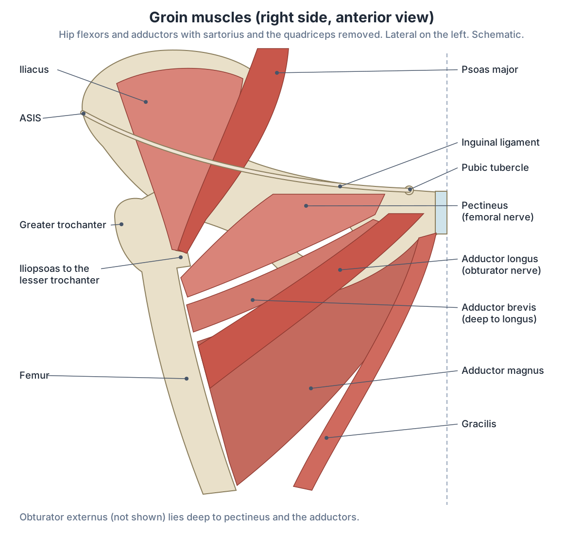
The adductor group has six muscles: adductor longus, adductor magnus, adductor brevis, gracilis, obturator externus and pectineus. All are supplied by the obturator nerve except pectineus, which is supplied by the femoral nerve. They pull the thigh towards the midline and steady the pelvis and leg when the foot is on the ground. (Tyler TF et al. Groin injuries in sports medicine. Sports Health. 2010.)
TeachMeAnatomy counts five medial thigh muscles and leaves pectineus out. Adductor longus fans out from the pubis to the linea aspera of the femur and forms the medial border of the femoral triangle. Adductor brevis lies deep to it and separates the anterior and posterior branches of the obturator nerve. Adductor magnus, the largest, has an adductor part supplied by the obturator nerve and a hamstring part supplied by the tibial part of the sciatic nerve, the only dual nerve supply in the group. Gracilis is the most superficial and medial and crosses both the hip and the knee. (TeachMeAnatomy: Muscles of the Medial Compartment of the Thigh.)
Pectineus arises from the pectineal line of the superior pubic ramus and inserts on the pectineal line and upper linea aspera of the femur. It is mainly supplied by the femoral nerve (L2, L3), but in some people its back part also receives a branch from the obturator or accessory obturator nerve. It flexes and adducts the thigh and forms the medial part of the floor of the femoral triangle. (Kenhub: Pectineus muscle.)
Iliopsoas flexes the hip. Psoas major arises from the lumbar vertebrae and is supplied by the anterior rami of L1 to L3; iliacus arises from the iliac fossa and is supplied by the femoral nerve. They insert together on the lesser trochanter of the femur. Sartorius, the longest muscle in the body, runs from the ASIS to the upper medial tibia and flexes, abducts and laterally rotates the hip. (TeachMeAnatomy: Muscles of the Anterior Compartment of the Thigh.)
Iliopsoas, the gluteal muscles and the deep hip rotators. Hip muscles.
Groin muscles: origin, insertion, nerve and action
| Muscle | Origin | Insertion | Nerve | Main action |
|---|---|---|---|---|
| Psoas major | Lumbar vertebrae | Lesser trochanter (with iliacus) | Anterior rami L1 to L3 | Flexes the hip |
| Iliacus | Iliac fossa | Lesser trochanter (with psoas major) | Femoral nerve | Flexes the hip |
| Sartorius | Anterior superior iliac spine | Upper medial surface of the tibia | Femoral nerve | Flexes, abducts and laterally rotates the hip; flexes the knee |
| Pectineus | Pectineal line of the superior pubic ramus | Pectineal line and upper linea aspera of the femur | Femoral nerve (L2, L3), sometimes also obturator or accessory obturator | Flexes and adducts the hip |
| Adductor longus | Body of the pubis | Linea aspera of the femur | Obturator nerve (L2 to L4) | Adducts the thigh |
| Adductor brevis | Body and inferior ramus of the pubis | Linea aspera, above adductor longus | Obturator nerve (L2 to L4) | Adducts the thigh |
| Adductor magnus | Inferior pubic and ischial rami (adductor part); ischial tuberosity (hamstring part) | Linea aspera; adductor tubercle and medial supracondylar line | Obturator nerve (L2 to L4); tibial part of sciatic nerve (L4 to S3) | Adducts; adductor part flexes, hamstring part extends the hip |
| Gracilis | Inferior pubic ramus and body of the pubis | Medial surface of the tibial shaft | Obturator nerve (L2 to L4) | Adducts the hip and flexes the knee |
| Obturator externus | Obturator membrane and surrounding bone | Back of the greater trochanter region | Obturator nerve (L2 to L4) | Adducts and laterally rotates the thigh |
Sources: TeachMeAnatomy (medial and anterior thigh compartments), Kenhub (pectineus) and Tyler et al. 2010.
Nerves of the groin
Six branches of the lumbar plexus supply the groin. The iliohypogastric nerve (T12, L1) supplies skin over the pubic region; the ilioinguinal nerve (L1) passes through the superficial inguinal ring to the skin of the upper inner thigh, the root of the penis and the front of the scrotum, or the mons pubis and labia majora; and the genitofemoral nerve (L1, L2) divides into a genital branch, which supplies the cremaster muscle and the front of the scrotum or the labia, and a femoral branch to the skin of the upper front of the thigh. (TeachMeAnatomy: The Lumbar Plexus.)
The lateral cutaneous nerve of the thigh (L2, L3) enters the thigh at the lateral end of the inguinal ligament and supplies the skin of the front and side of the thigh down to the knee. The femoral nerve (L2 to L4) supplies the anterior thigh muscles (iliacus, pectineus, sartorius and quadriceps femoris), and the obturator nerve (L2 to L4) supplies the medial thigh muscles and skin. (TeachMeAnatomy: The Lumbar Plexus.)
The genital branch of the genitofemoral nerve supplies the cremaster muscle in males. The ilioinguinal nerve passes only partly through the inguinal canal and leaves through the superficial ring, which is why it is the nerve most at risk during inguinal hernia repair. (TeachMeAnatomy: The Inguinal Canal.)
The femoral, obturator and sciatic nerves from the spine to the foot. Nerves of the leg.
Nerves of the groin
| Nerve | Roots | Supplies in the groin |
|---|---|---|
| Iliohypogastric | T12, L1 | Skin over the pubic region; internal oblique and transversus abdominis |
| Ilioinguinal | L1 | Skin of the upper inner thigh, root of the penis and front of the scrotum (mons pubis and labia majora in females) |
| Genitofemoral, genital branch | L1, L2 | Cremaster; skin of the front of the scrotum (mons pubis and labia majora) |
| Genitofemoral, femoral branch | L1, L2 | Skin of the upper front of the thigh |
| Lateral cutaneous nerve of the thigh | L2, L3 | Skin of the front and side of the thigh to the knee |
| Femoral | L2 to L4 | Iliacus, pectineus, sartorius, quadriceps; skin of the front of the thigh |
| Obturator | L2 to L4 | Adductor muscles and gracilis; skin of the medial thigh |
Source: TeachMeAnatomy, the lumbar plexus.
Groin blood vessels and inguinal lymph nodes
The femoral artery continues the external iliac artery below the inguinal ligament and lies between the femoral nerve and the femoral vein; in the triangle it gives off the deep femoral artery (profunda femoris). The femoral vein lies medial to the artery, receives the great saphenous vein through the saphenous opening in the fascia lata, and becomes the external iliac vein at the ligament. (Kenhub: Femoral triangle.)
The great saphenous vein pierces the saphenous opening 1 to 3 cm below the inguinal ligament and joins the femoral vein at the saphenofemoral junction in the femoral triangle. A valve 1 to 2 mm below the junction is present in about 99% of people, and near the groin the vein receives the superficial epigastric, superficial circumflex iliac and superficial external pudendal veins. (Radiopaedia: Great saphenous vein.)
Kenhub counts about 12 to 14 inguinal lymph nodes in superficial and deep groups. They drain the lower limb, the external genitalia, the perineum and the lowest part of the anterior abdominal wall, and the superficial nodes lie with the great saphenous vein in the superficial fascia. (Kenhub: Inguinal region.)
The superficial inguinal nodes form a line just below the inguinal ligament and receive lymph from the penis, scrotum, perineum, buttock and abdominal wall as far as the umbilicus. Further nodes lie on each side of the upper great saphenous vein, and one to three deep nodes lie on the medial side of the femoral vein. The testis is different: its lymph drains to the para-aortic nodes, so testicular disease rarely enlarges the inguinal nodes. (TeachMeAnatomy: Lymphatic Drainage of the Lower Limb.)
On imaging, an inguinal lymph node more than 15 mm across its short axis is considered abnormal. (Radiopaedia: Inguinal lymph nodes.)
Lymph nodes and lymphatic vessels throughout the body, including the inguinal nodes. Lymphatic system.
Groin hernias: indirect, direct and femoral
Inguinal hernias are the commonest abdominal wall hernias, up to 80% of them, and are up to seven times more common in males. An indirect inguinal hernia leaves the abdomen lateral to the inferior epigastric vessels through the deep inguinal ring, running in front of the spermatic cord in males or following the round ligament in females. A direct inguinal hernia pushes through a weak floor of the canal medial to those vessels, in Hesselbach's triangle; direct hernias are rare in women. (Radiopaedia: Inguinal hernia.)
Indirect hernias are the more common type and come from a processus vaginalis that failed to close; they can run the whole canal into the scrotum. Direct hernias are acquired, usually in adults, through weakness of the abdominal wall. Both can appear as a lump in the scrotum or labium majus. (TeachMeAnatomy: The Inguinal Canal.)
The international HerniaSurge guidelines put the lifetime risk of a groin hernia at 27 to 43% in men and 3 to 6% in women, and inguinal hernia repair at 8 to 10 times more common in males, a higher ratio than Radiopaedia's 7 to 1. Hernias peak at about 5 years of age, mainly indirect, and again at 70 to 80 years, mainly direct. (HerniaSurge Group. International guidelines for groin hernia management. Hernia. 2018.)
Hesselbach's (inguinal) triangle is bounded by the lateral border of rectus abdominis medially, the inferior epigastric vessels laterally and the medial third of the inguinal ligament below. (Kenhub: Inguinal canal.)
A femoral hernia passes through the femoral ring into the femoral canal, below the inguinal ligament. It appears as a lump below and lateral to the pubic tubercle, is more common in women, which TeachMeAnatomy links to their wider pelvis, and is prone to strangulation because the borders of the canal are tough and do not stretch. (TeachMeAnatomy: The Femoral Canal.)
Kenhub puts femoral hernias at 2 to 4% of hernias and describes them as a round, often tender bulge just below the pubic tubercle; because the femoral ring is rigid, they can strangulate. (Kenhub: Femoral triangle.)
On imaging, most femoral hernias lie medial to the common femoral vein, which they often compress, and they stay lateral to the pubic tubercle, which helps tell them from inguinal hernias. (Radiopaedia: Femoral hernia.)
Groin hernias compared
| Type | Where it leaves the abdomen | Landmarks | Who |
|---|---|---|---|
| Indirect inguinal | Deep inguinal ring, along the inguinal canal | Above the inguinal ligament, lateral to the inferior epigastric vessels | Most common type; congenital, peak in childhood |
| Direct inguinal | Weak posterior wall (Hesselbach's triangle) | Above the inguinal ligament, medial to the inferior epigastric vessels | Acquired, usually older men; rare in women |
| Femoral | Femoral ring into the femoral canal | Below the inguinal ligament, medial to the femoral vein, below and lateral to the pubic tubercle | More common in women; 2 to 4% of hernias; high strangulation risk |
Sources: Radiopaedia (inguinal and femoral hernia), TeachMeAnatomy (inguinal and femoral canals), Kenhub (femoral triangle) and HerniaSurge 2018. Educational overview, not for self-diagnosis.
Groin strain and groin pain anatomy
A groin strain is a strain of the adductor muscles, usually of the upper part near their attachments to the pelvis. It is common in sports that need explosive movement or extreme stretching. (TeachMeAnatomy: Muscles of the Medial Compartment of the Thigh.)
Adductor longus is the adductor most often injured in sport. In professional ice hockey and soccer about 10 to 11% of all injuries are groin strains, because these sports demand strong eccentric contraction of the adductors. In one study of professional ice hockey players, preseason adduction strength was 18% lower in those who later strained a groin, and a previous groin strain is another risk factor. (Tyler TF et al. Groin injuries in sports medicine. Sports Health. 2010.)
A 2024 review reports that chronic groin pain makes up as much as 15% of injuries in athletes and that adductor-related pain causes up to two-thirds of cases. The 2015 Doha agreement sorts athletic groin pain into adductor-related, iliopsoas-related, inguinal-related and pubic-related pain, plus hip-related and other causes. Adductor longus attaches to the pubis through fibrocartilage and is under most stress when the leg kicks a ball. (Dinis J et al. Athletes with adductor-related groin pain: a narrative review. Cureus. 2024.)
Groin pain has many anatomical sources besides a strain: athletic pubalgia (tears or weakness of the abdominal wall without a palpable hernia), osteitis pubis, a true hernia, hip flexor strain, problems inside the hip joint, and pain referred from the testis, rectum or lumbar spine. (Tyler TF et al. Groin injuries in sports medicine. Sports Health. 2010.)
The quadriceps, hamstrings and adductors of the thigh. Thigh muscles.
Groin anatomy in males and females
The groin is built the same way in both sexes, but the inguinal canal carries different structures: the spermatic cord in males and the round ligament of the uterus in females. The difference comes from development, because the testes pass through the canal while the ovaries stay in the pelvis. (TeachMeAnatomy: The Inguinal Canal.)
Both inguinal rings are larger in males, to let the testis through, and inguinal hernias are more common in males than in females. (Kenhub: Inguinal canal.)
Femoral hernias, by contrast, are more common in women. The femoral canal sits medial to the femoral vein below the inguinal ligament, and TeachMeAnatomy links the difference to the wider female bony pelvis. (TeachMeAnatomy: The Femoral Canal.)
The spermatic cord, testis and ductus deferens that pass through the male groin. Male reproductive system.
Groin in the 3D model
- Adductor brevis
- A short adductor in the medial thigh, lying behind pectineus and adductor longus.
- Adductor longus
- The most anterior adductor muscle; its medial edge forms the medial border of the femoral triangle.
- Adductor magnus
- The largest adductor, with an adductor part and a hamstring part.
- Deferent duct
- The deferent duct (vas deferens) is a thick-walled muscular tube carrying sperm from the epididymis toward the urethra.
- External iliac artery
- The external iliac artery is the main artery of the lower limb in the pelvis and becomes the femoral artery.
- External iliac vein
- The external iliac vein is the continuation of the femoral vein above the inguinal ligament.
- External oblique
- The largest and most superficial lateral abdominal wall muscle.
- Femoral artery
- The femoral artery is the main artery of the thigh, continuing the external iliac artery.
- Femoral vein
- The femoral vein is the main deep vein of the thigh and continues the popliteal vein.
- Gracilis
- A long, thin muscle on the medial side of the thigh.
- Great saphenous vein
- The great saphenous vein is the longest vein in the body and runs up the medial side of the leg and thigh.
- Iliacus
- A fan-shaped muscle filling the iliac fossa.
- Inferior epigastric artery
- The inferior epigastric artery is a branch of the external iliac artery supplying the anterior abdominal wall.
- Inferior epigastric vein
- The inferior epigastric vein drains the lower anterior abdominal wall.
- Obturator externus
- A flat, fan-shaped deep muscle of the medial thigh that wraps behind the neck of the femur.
- Pectineus
- A flat quadrangular muscle at the top of the medial thigh forming part of the floor of the femoral triangle.
- Psoas major
- A long muscle of the posterior abdominal wall that forms the main part of iliopsoas.
- Sartorius
- The longest muscle in the body, a strap-like muscle crossing the thigh obliquely.
- Superficial epigastric artery
- A small branch of the femoral artery to the lower abdominal wall.
- Superficial epigastric vein
- A superficial vein of the lower abdominal wall.
- Testicular artery
- A paired branch of the abdominal aorta to the testis.
- Testicular vein
- A vein draining the testis.
Frequently asked questions
What is the inguinal region?
The inguinal region is the groin: the lower part of the front of the abdominal wall on each side, above the thigh, between the anterior superior iliac spine and the pubic tubercle. It contains the inguinal ligament and inguinal canal. In the nine-region map of the abdomen there are a right and a left inguinal (iliac) region.
What passes through the inguinal canal?
In males the inguinal canal carries the spermatic cord, which contains the ductus deferens and the vessels and nerves of the testis. In females it carries the round ligament of the uterus. In both sexes it also contains the ilioinguinal nerve, which leaves through the superficial ring, and the genital branch of the genitofemoral nerve. The canal is about 3 to 5 cm long.
What are the borders of the femoral triangle?
The femoral triangle is bounded above by the inguinal ligament, laterally by sartorius and medially by adductor longus (remembered as SAIL). Its floor is iliopsoas, pectineus and adductor longus, and its roof is the fascia lata. From lateral to medial it contains the femoral nerve, femoral artery, femoral vein and femoral canal (NAVEL).
What is the difference between an inguinal and a femoral hernia?
An inguinal hernia leaves the abdomen above the inguinal ligament, either through the deep ring (indirect) or through the weak back wall of the canal (direct), and is far more common in men. A femoral hernia passes below the ligament through the femoral canal, medial to the femoral vein. It is more common in women and more likely to strangulate.
What muscles are in the groin?
The main groin muscles are the adductors: adductor longus, brevis and magnus, gracilis, pectineus and obturator externus, which pull the thigh inwards. Iliopsoas (psoas major and iliacus) and sartorius flex the hip. Above the inguinal ligament lie the lower parts of the abdominal muscles: external oblique, internal oblique and transversus abdominis.
Where are the inguinal lymph nodes?
The superficial inguinal lymph nodes lie in a line just below the inguinal ligament and along the upper great saphenous vein, in the fat under the skin. One to three deep inguinal nodes lie medial to the femoral vein. They drain the leg, the external genitalia, the perineum, the buttock and the lower abdominal wall, but not the testes.
What is a groin strain?
A groin strain is a tear of the adductor muscles of the inner thigh, most often adductor longus near its attachment to the pubis. It is common in sports with frequent kicking and pivoting, such as soccer and ice hockey, where about 10 to 11% of injuries are groin strains. A previous strain and weaker adductors raise the risk.
References
- Kenhub. Inguinal region.
- Kenhub. Inguinal canal.
- Kenhub. Femoral triangle.
- Kenhub. Pectineus muscle.
- TeachMeAnatomy. The Inguinal Canal.
- TeachMeAnatomy. The Femoral Triangle.
- TeachMeAnatomy. The Femoral Canal.
- TeachMeAnatomy. Muscles of the Medial Compartment of the Thigh.
- TeachMeAnatomy. Muscles of the Anterior Compartment of the Thigh.
- TeachMeAnatomy. The Lumbar Plexus.
- TeachMeAnatomy. Lymphatic Drainage of the Lower Limb.
- Radiopaedia. Inguinal canal.
- Radiopaedia. Inguinal ligament.
- Radiopaedia. Inguinal hernia.
- Radiopaedia. Femoral hernia.
- Radiopaedia. Inguinal lymph nodes.
- Radiopaedia. Great saphenous vein.
- HerniaSurge Group. International guidelines for groin hernia management. Hernia. 2018;22(1):1-165.
- Tyler TF, Silvers HJ, Gerhardt MB, Nicholas SJ. Groin injuries in sports medicine. Sports Health. 2010;2(3):231-236.
- Dinis J, Oliveira JR, Choupina B, et al. Athletes with adductor-related groin pain: a narrative review. Cureus. 2024;16(9):e68625.
- Mitsuhashi N. et al. (2009). BodyParts3D: 3D structure database for anatomical concepts. Nucleic Acids Research 37, D782–D785. Data © The Database Center for Life Science, CC BY 4.0.
Related anatomy
- Abdominal musclesAbdominal muscles explained: external and internal oblique, transversus abdominis, rectus abdominis, pyramidalis, the…
- Femoral arteryFemoral artery anatomy: where it is, the femoral triangle and sheath (NAVEL), adductor canal, branches, deep femoral…
- Hip musclesMuscles of the hip explained: gluteus maximus, medius and minimus, TFL, piriformis, obturator internus and the other…
- Thigh musclesThigh muscles explained: quadriceps and sartorius at the front, adductor muscles of the inner thigh, hamstrings at the…
- Great saphenous veinSaphenous vein anatomy: course of the great and small saphenous veins, saphenofemoral junction, valves, saphenous…
- Pelvic floor and perineumPerineum and pelvic floor anatomy with labelled diagrams and a 3D model: levator ani, coccygeus, perineal body, both…
Explore the whole body in the interactive 3D human anatomy model, or browse the A–Z list of human structures.