

Where are the ear bones? Location in the middle ear
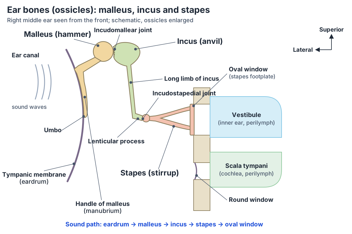
The ear bones sit in the middle ear, which lies within the temporal bone and extends from the tympanic membrane (eardrum) to the lateral wall of the inner ear. Its main space, the tympanic cavity, contains the three auditory ossicles. Above it is a smaller space, the epitympanic recess, which holds the head of the malleus and the body of the incus. (TeachMeAnatomy: The Middle Ear.)
OpenStax describes the middle ear as a space spanned by three small bones. The malleus is attached to the tympanic membrane and articulates with the incus, the incus articulates with the stapes, and the stapes connects to the cochlea of the inner ear through the oval window. (OpenStax Anatomy and Physiology 2e, 14.1: Sensory Perception.)
There are no ossicles inside the inner ear itself. The inner ear is a bony labyrinth, a series of canals embedded within the temporal bone. The three ear bones sit just outside it, in the middle ear, and the stapes connects to the inner ear through the oval window. (OpenStax Anatomy and Physiology 2e, 14.1: Sensory Perception.)
The outer, middle and inner ear together, from the auricle and eardrum to the cochlea and balance organs, in labelled diagrams. Ear anatomy.
How many bones are in the ear?
Each ear has three bones, the malleus, incus and stapes, so the two ears hold six ear bones in total. Cleveland Clinic describes them as three tiny bones in the middle ear that form an interconnected chain and boost the sound signal along the way. (Cleveland Clinic: Ossicles.)
From the outside in, the order is malleus, incus, stapes. The malleus attaches to the eardrum, the incus comes next and joins the last ossicle, and the stapes connects the incus to the oval window of the inner ear. The incus is therefore the middle bone of the chain. (TeachMeAnatomy: The Middle Ear.)
Their names describe their shapes: the malleus is the hammer, the incus the anvil and the stapes the stirrup. Kenhub describes the ossicles as the smallest bones in the body. (Kenhub: Auditory ossicles.)
The auricle and ear canal that funnel sound to the eardrum, with a labelled auricle diagram. External ear.
The three ear bones at a glance
| Bone | Common name | Parts | Joins | Muscle (nerve) |
|---|---|---|---|---|
| Malleus | Hammer | Head, neck, handle (manubrium), anterior and lateral processes | Eardrum (handle and lateral process); incus (head) | Tensor tympani (branch of the mandibular nerve, CN V3) |
| Incus | Anvil | Body, short limb, long limb, lenticular process | Malleus (body); stapes (lenticular process) | None |
| Stapes | Stirrup | Head, neck, anterior and posterior crura, base (footplate) | Incus (head); oval window (footplate, by the annular ligament) | Stapedius (facial nerve, CN VII) |
Parts and joints from Kenhub and Radiopaedia; muscles and nerves from TeachMeAnatomy and Kenhub.
Malleus (hammer): head, neck, handle and processes
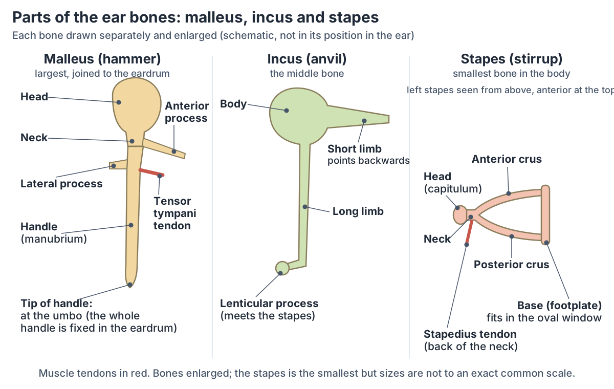
The malleus has a head, a neck and three processes: the manubrium (handle), the anterior process and the lateral process. The handle is connected along its lateral edge to the tympanic membrane, so the malleus moves whenever the eardrum vibrates. (Radiopaedia: Malleus.)
The handle is embedded in the inner surface of the eardrum, and its lower end pulls the membrane inwards to form the umbo, the deepest point of the eardrum. The small, cone-shaped lateral process sits at the root of the handle and is attached to the upper eardrum by the malleolar folds. The longer anterior process is attached to the front wall of the middle ear. (Kenhub: Auditory ossicles.)
The head of the malleus lies above the level of the eardrum, in the epitympanic recess, where it articulates with the incus. TeachMeAnatomy describes the malleus as the largest and most lateral of the ear bones. (TeachMeAnatomy: The Middle Ear.)
Three suspensory ligaments, the anterior, lateral and superior ligaments of the malleus, hold it to the walls of the middle ear. The anterior process runs forwards towards the petrotympanic fissure, and the tendon of tensor tympani inserts on the medial side of the handle near its upper end. (Radiopaedia: Malleus.)
Incus (anvil): the middle ear bone
The incus has four named parts: the body, the short process (short limb), the long process (long limb) and the lenticular (lentiform) process. Its body articulates with the head of the malleus at the incudomallear (incudomalleolar) joint, and its lenticular process articulates with the head of the stapes at the incudostapedial joint. (Radiopaedia: Incus.)
The body sits in the epitympanic recess. The short limb projects backwards and is held by the posterior ligament of the incus. The long limb runs downwards, parallel to the handle of the malleus, and ends in a right-angled bend that forms the lenticular process. (Kenhub: Auditory ossicles.)
The lenticular process is joined to the long process by a short stalk, or pedicle. It was once thought to be a fourth ossicle, but imaging shows that it is part of the incus. (Radiopaedia: Middle ear ossicles.)
The superior ligament of the incus ties its body to the roof of the middle ear. Radiopaedia notes that the long process is the most frequently eroded or resorbed part of the ossicles. (Radiopaedia: Incus.)
The ossicles get their blood from a network of vessels in the mucosa that covers them, fed mainly by the anterior tympanic artery, and from small arteries that run inside the malleus and incus. Usually only one small vessel reaches the end of the long process of the incus. (Manoharan SM et al. Internal vascular channel architecture in human auditory ossicles. J Anat. 2022.)
This sparse blood supply is thought to explain why the long and lenticular processes are the parts most often eroded, for example in chronic otitis media. (Ali KAER et al. Reconstruction of the Long Process of the Incus by Use of Glass Ionomer Cement: Our Experience. Int Arch Otorhinolaryngol. 2022.)
Stapes (stirrup): the smallest bone in the body
The stapes is the smallest bone in the human body and the most medial of the three ossicles. Its concave head (capitulum) meets the lenticular process of the incus. A short neck leads to two limbs, the anterior and posterior crura, which join the oval base, or footplate. The head, neck and crura together are called the superstructure. (Radiopaedia: Stapes.)
The footplate sits in the oval window, held by the annular ligament, which forms the stapediovestibular joint. Through this window the stapes passes vibrations into the cochlea. The stapedius muscle attaches to the back of the stapes neck. (Radiopaedia: Stapes.)
How big is the stapes? Cleveland Clinic says it measures only a few millimetres in width and height. Measurement studies agree that it weighs only a few milligrams: about 2.2 mg on average (range 1.4 to 3.2 mg) in a study of modern human ears by Quam and colleagues, and 3.2 mg in the human reference values reported by Péus and colleagues. (Quam R et al. Early hominin auditory capacities. Sci Adv. 2015.)
Its footplate is just as small. Reported footplate areas are about 2.9 mm² (Quam and colleagues), 3.0 mm² (Péus and colleagues) and about 3.2 mm² (a 2025 review of stapes surgery), and the footplate vibrates by only a few nanometres to move the fluid of the inner ear. (Bruschini L et al. Update on stapes surgery. Acta Otorhinolaryngol Ital. 2025.)
Joints and ligaments of the ear bones
The ossicles articulate with each other through two tiny synovial joints, and the stapes meets the oval window through a third, fibrous joint, which Radiopaedia classes as a syndesmosis. (Radiopaedia: Middle ear ossicles.)
The incudomallear (incudomalleolar) joint lies between the head of the malleus and the body of the incus. The incudostapedial joint lies between the lenticular process of the incus and the head of the stapes. The base of the stapes is bound to the edge of the oval (vestibular) window by the annular ligament, a joint Kenhub calls the tympanostapedial syndesmosis. (Kenhub: Auditory ossicles.)
Ligaments hang the chain inside the middle ear: the anterior, superior and lateral ligaments of the malleus, the superior and posterior ligaments of the incus, and the annular ligament of the stapes. A thin layer of mucosa, the stapedial membrane, spans the stapes. (Kenhub: Auditory ossicles.)
Because the ossicles are linked by true synovial joints, with a capsule, cartilage and synovial fluid, the chain is not a single rigid rod. A 2022 review of middle ear mechanics notes that these joints behave differently at different sound frequencies, one reason the middle ear works in a more complex way than the classic lever model suggests. (Ugarteburu M et al. Mammalian middle ear mechanics: A review. Front Bioeng Biotechnol. 2022.)
Muscles and nerves of the ear bones: tensor tympani and stapedius
Two small muscles act on the chain. Tensor tympani arises from the auditory (Eustachian) tube, attaches to the handle of the malleus and pulls it medially; it is supplied by the tensor tympani nerve, a branch of the mandibular nerve (CN V3). Stapedius attaches to the stapes and is supplied by the facial nerve (CN VII). (TeachMeAnatomy: The Middle Ear.)
Stapedius arises inside the pyramidal eminence on the back (mastoid) wall of the middle ear and inserts on the posterior neck of the stapes; when it contracts it pulls the stapes backwards and limits its movement. Tensor tympani tenses the eardrum, and Kenhub names its nerve as the nerve to medial pterygoid, a branch of the mandibular nerve. (Kenhub: Auditory ossicles.)
In humans, the acoustic reflex works mainly through stapedius. Loud sound makes stapedius contract, which limits the movement of the stapes and reduces the sound transmitted to the inner ear. The human tensor tympani seems not to respond to outside sound; it contracts mainly as part of the startle reaction and with swallowing, speaking or touch to the face. (Rönnblom A et al. Unique fiber phenotype composition and metabolic properties of the stapedius and tensor tympani muscles in the human middle ear. J Anat. 2023.)
The reflex takes about 15 ms to start, so it cannot protect the ear from sudden impulse noise such as a gunshot. (Ugarteburu M et al. Mammalian middle ear mechanics: A review. Front Bioeng Biotechnol. 2022.)
The chorda tympani, a branch of the facial nerve, crosses the middle ear from back to front between the malleus and the incus, at the level of the handle or neck of the malleus. It carries taste from the front two-thirds of the tongue and parasympathetic fibres to the submandibular and sublingual salivary glands. (Radiopaedia: Chorda tympani.)
The nerve to stapedius is the first motor branch of the facial nerve and arises inside the facial canal. TeachMeAnatomy lists hyperacusis, an uncomfortable sensitivity to ordinary sounds in the ear on the same side, among the signs of facial nerve damage inside the skull; Kenhub links it to paralysis of stapedius. (TeachMeAnatomy: The Facial Nerve (CN VII).)
The twelve cranial nerves, including the trigeminal and facial nerves that supply the two muscles of the middle ear. Cranial nerves.
How do the ear bones amplify sound? Area and lever ratios
Sound reaches the ear as vibrations in air, but the inner ear is filled with fluid, and the middle ear is the interface between the two. It is traditionally explained as a transformer: the large eardrum collects the vibration over a wide area and the chain delivers it to the much smaller stapes footplate, which reduces the movement but increases the sound pressure at the oval window. (Ugarteburu M et al. Mammalian middle ear mechanics: A review. Front Bioeng Biotechnol. 2022.)
The difference in area has been measured. In modern human ears the eardrum has an area of about 65 mm² and the stapes footplate about 2.9 mm² (Quam and colleagues). Péus and colleagues use 68.3 mm² for the eardrum and calculate an area ratio of 23.9. Either way, the eardrum is more than 20 times larger than the footplate. (Quam R et al. Early hominin auditory capacities. Sci Adv. 2015.)
The malleus and incus also rotate together like a lever, because the tip of the malleus handle lies a little further from their axis of rotation than the end of the incus does. The human lever ratio is small, 1.25 in the study by Péus and colleagues and 1.26 in the study by Quam and colleagues, so the lever contributes far less to the overall gain than the difference in area does. (Péus D et al. Comparison of sheep and human middle-ear ossicles: anatomy and inertial properties. J Comp Physiol A. 2020.)
Multiplying the two ratios gives the expected pressure gain. Péus and colleagues calculate about 30 times for the human ear, and an OpenStax physics textbook gives pressure waves in the cochlea about 40 times greater than those at the eardrum. A protective muscle reaction to very loud sound reduces this mechanical advantage. (OpenStax College Physics 2e, 17.6: Hearing.)
These ratios are a simplification. The 2022 review of middle ear mechanics concludes that matching impedance to reduce the reflection of sound, rather than a simple gain in pressure, best explains the experimental findings, and that researchers are still learning exactly how the middle ear works. (Ugarteburu M et al. Mammalian middle ear mechanics: A review. Front Bioeng Biotechnol. 2022.)
At the inner end of the chain, the stapes passes the vibrations into the cochlea through the oval window. The scala tympani of the cochlea ends at the round window, which is covered by a membrane. (OpenStax Anatomy and Physiology 2e, 14.1: Sensory Perception.)
Ear bone measurements in adults
| Measure | Value | Sources |
|---|---|---|
| Eardrum (tympanic membrane) area | About 65 to 68 mm² (65.1 ± 5.5 mm²; 68.3 mm²) | Quam et al. 2015; Péus et al. 2020 |
| Stapes footplate area | About 2.9 to 3.2 mm² | Quam et al. 2015; Péus et al. 2020; Bruschini et al. 2025 |
| Area ratio, eardrum to footplate | More than 20 (23.9 in Péus et al.) | Péus et al. 2020; Quam et al. 2015 |
| Malleus–incus lever ratio | 1.25 to 1.26 | Péus et al. 2020; Quam et al. 2015 |
| Expected pressure gain | About 30 to 40 times | Péus et al. 2020 (about 30); OpenStax College Physics (about 40) |
| Mass of the stapes | About 2 to 3 mg (2.2 ± 0.6 mg; 3.2 mg) | Quam et al. 2015; Péus et al. 2020 |
Individual ears vary: Quam and colleagues give ranges of 56.6 to 74.0 mm² for the eardrum and 2.51 to 3.13 mm² for the footplate.
How the ear bones develop
The ear bones come from the pharyngeal (branchial) arches of the embryo. The malleus and incus develop from neural crest cells of the first arch, whose nerve is the mandibular division of the trigeminal nerve. The stapes develops from the second arch, the arch of the facial nerve. (Radiopaedia: Branchial apparatus.)
The two muscles follow the same pattern. Tensor tympani is a first-arch muscle supplied by the trigeminal nerve, while stapedius is a second-arch muscle supplied by the facial nerve, which is why the two muscles of the middle ear have different nerves. (TeachMeAnatomy: The Pharyngeal Arches.)
Having three ear bones is a mammal feature. Reptiles and birds have a single middle-ear bone, the stapes (columella). The malleus and incus are homologous to the articular and quadrate bones, which form the jaw joint in other jawed vertebrates. They could move into the middle ear only after mammals evolved a new jaw joint between the dentary and squamosal bones, the temporomandibular joint in humans. See comparative anatomy for more. (Anthwal N, Joshi L, Tucker AS. Evolution of the mammalian middle ear and jaw: adaptations and novel structures. J Anat. 2013.)
The ossicles are the first bones in the body to ossify fully and are already mature at birth. Radiopaedia adds that they are the only bones that do not grow after birth. (Kenhub: Auditory ossicles.)
Homologous and analogous structures across animals, including how the jaw bones of early vertebrates became the mammalian ear bones. Comparative anatomy.
Ear bone problems: otosclerosis, chain disruption and cholesteatoma
Otosclerosis is a disease of the bony capsule of the inner ear. It usually starts just in front of the oval window, at a small cleft called the fissula ante fenestram, and new bone can grow around the stapes and fix it in place. A fixed stapes cannot pass vibrations on, so the hearing loss is most often conductive, although it can also be sensorineural or mixed. (Radiopaedia: Otosclerosis.)
Radiopaedia notes that otosclerosis typically presents in the fourth and fifth decades of life, is more common in women and is frequently bilateral. (Radiopaedia: Otosclerosis.)
NIDCD explains that otosclerosis is most often caused by the stapes becoming stuck in place, that many cases are thought to be inherited, that white, middle-aged women are most at risk, and that the hearing loss usually starts in one ear and then moves to the other. Mild cases can be helped by a hearing aid; in a stapedectomy, a surgeon inserts a prosthesis into the middle ear to bypass the abnormal bone. (NIDCD (NIH): Otosclerosis.)
Ossicular chain disruption (ossicular discontinuity) means the bones have lost their normal alignment. Causes include trauma, especially a temporal bone fracture, and blast injuries, as well as congenital malformations and erosion by cholesteatoma or chronic infection. The commonest types are separation at the incudomallear joint and at the incudostapedial joint. After an injury it typically causes sudden conductive hearing loss. (Radiopaedia: Ossicular chain disruption.)
Cleveland Clinic lists the main conditions of the ossicles as otosclerosis, ossicular-chain dislocation after a head injury, cholesteatoma (a build-up of skin cells in the middle ear) and, rarely, babies born with an absent, underdeveloped or overdeveloped ossicle. Anyone with new hearing loss, ear pain or discharge should see a clinician; this guide explains anatomy and is not medical advice. (Cleveland Clinic: Ossicles.)
The skull bone that houses the middle and inner ear, including its petrous part and the ear canal. Temporal bone.
Frequently asked questions
What are the 3 bones in the ear called?
The three ear bones are the malleus (hammer), incus (anvil) and stapes (stirrup), together called the auditory ossicles. They sit in the middle ear and form a chain from the eardrum to the oval window of the inner ear, with the malleus on the outside, the incus in the middle and the stapes innermost.
How many bones are in the human ear?
Each middle ear contains three bones, the malleus, incus and stapes, so the two ears hold six ear bones in total. The inner ear has no ossicles of its own: it is a bony labyrinth of canals inside the temporal bone, the skull bone that surrounds the middle and inner ear.
What is the smallest bone in the human body?
The stapes, the innermost of the three ear bones, is the smallest bone in the human body. It is shaped like a stirrup, with a head, two crura and a footplate that sits in the oval window. It weighs only a few milligrams (about 2 to 3 mg in measurement studies), and its footplate has an area of about 3 mm².
What does the stapes do?
The stapes passes sound vibrations from the incus into the inner ear. Its footplate sits in the oval window, so each movement pushes on the fluid of the cochlea. Because the eardrum is more than 20 times larger in area than the footplate, the pressure at the oval window is much higher than at the eardrum. Stapedius can damp it in loud noise.
Which ear bone is attached to the eardrum?
The malleus. Its handle (manubrium) is embedded in the inner surface of the eardrum, and its lower end pulls the membrane inwards to form the umbo, the deepest point of the eardrum. The small lateral process at the top of the handle also attaches to the upper eardrum, so the malleus moves whenever the eardrum vibrates.
What happens if the ear bones are damaged?
If the chain is broken or a bone is fixed, sound cannot pass efficiently to the inner ear, which causes conductive hearing loss. Common causes are otosclerosis, which fixes the stapes, a temporal bone fracture or blast that separates the joints, and erosion by chronic infection or cholesteatoma. Hearing aids or middle ear surgery can help; a clinician assesses each case.
Do the ear bones grow after birth?
No. The ossicles are the first bones in the body to ossify fully and are already mature at birth. Radiopaedia notes that they are the only bones that do not grow after birth, so a newborn's malleus, incus and stapes are essentially the size they will stay for life.
References
- Kenhub. Auditory ossicles.
- TeachMeAnatomy. The Middle Ear.
- TeachMeAnatomy. The Pharyngeal Arches.
- TeachMeAnatomy. The Facial Nerve (CN VII).
- Radiopaedia. Middle ear ossicles.
- Radiopaedia. Malleus.
- Radiopaedia. Incus.
- Radiopaedia. Stapes.
- Radiopaedia. Branchial apparatus.
- Radiopaedia. Chorda tympani.
- Radiopaedia. Otosclerosis.
- Radiopaedia. Ossicular chain disruption.
- OpenStax. Anatomy and Physiology 2e, §14.1 Sensory Perception. Rice University, 2022.
- OpenStax. College Physics 2e, §17.6 Hearing. Rice University, 2022.
- Cleveland Clinic. Ossicles.
- National Institute on Deafness and Other Communication Disorders (NIH). Otosclerosis.
- Péus D, Dobrev I, Pfiffner F, Sim JH. Comparison of sheep and human middle-ear ossicles: anatomy and inertial properties. J Comp Physiol A. 2020;206:683-700.
- Quam R, Martínez I, Rosa M, et al. Early hominin auditory capacities. Sci Adv. 2015;1:e1500355.
- Ugarteburu M, Withnell RH, Cardoso L, Carriero A, Richter CP. Mammalian middle ear mechanics: A review. Front Bioeng Biotechnol. 2022;10:983510.
- Bruschini L, Lazzerini F, De Vito A, Forli F, Berrettini S. Update on stapes surgery. Acta Otorhinolaryngol Ital. 2025;45:S29-S39.
- Rönnblom A, Thornell LE, Shah F, Tano K, Stål P. Unique fiber phenotype composition and metabolic properties of the stapedius and tensor tympani muscles in the human middle ear. J Anat. 2023;243:39-50.
- Manoharan SM, Gray R, Hamilton J, Mason MJ. Internal vascular channel architecture in human auditory ossicles. J Anat. 2022;241:245-258.
- Ali KAER, Ibrahim MA, Hussien MA, Dahy KG. Reconstruction of the Long Process of the Incus by Use of Glass Ionomer Cement: Our Experience. Int Arch Otorhinolaryngol. 2022;26:e697-e700.
- Anthwal N, Joshi L, Tucker AS. Evolution of the mammalian middle ear and jaw: adaptations and novel structures. J Anat. 2013;222:147-160.
Related anatomy
- Ear anatomyEar anatomy with labelled diagrams: auricle, ear canal, normal tympanic membrane, the three ear bones, cochlear duct…
- External earExternal ear anatomy: labelled parts of the auricle (pinna), the outer ear canal, nerves, blood supply, development…
- Temporal boneThe temporal bone (os temporale) explained: petrous part and inner ear, mastoid process, zygomatic and styloid…
- Cranial nervesThe 12 cranial nerves in order with labelled diagrams, plus nerve anatomy of the face: facial nerve branches…
Explore the whole body in the interactive 3D human anatomy model, or browse the A–Z list of human structures.