

Ischium at a glance
- What it is
- The ischium is the lower, posterior part of the hip bone.
- Location
- It lies below and behind the acetabulum. Its body forms part of the hip socket and its ramus joins the inferior ramus of the pubis below the obturator foramen.
- Function
- Its tuberosity bears the body's weight when sitting and gives origin to the hamstring muscles.
- Body system
- Skeletal · paired (left and right) · female body
- FMA ID
- FMA16594 (Foundational Model of Anatomy)
Key facts
- The ischial tuberosity anchors semitendinosus, semimembranosus, the long head of biceps femoris, the hamstring part of adductor magnus and the sacrotuberous ligament.
- The ischial spine anchors the sacrospinous ligament and separates the greater and lesser sciatic notches.
- The distance between the two ischial spines is the narrowest part of the birth canal; the spines are less prominent and further apart in females.
Clinical relevance
The ischial spine is the landmark for a pudendal nerve block, which can be used for pain relief in the second stage of labour.
Where is the ischium?
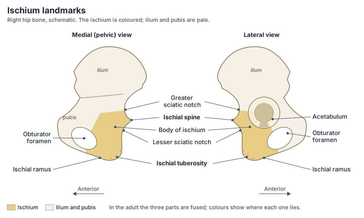
Each hip bone is made of three parts, the ilium, ischium and pubis, which fuse in the late teenage years. The ischium forms the posteroinferior (lower back) region of the hip bone. Its large, roughened lower area, the ischial tuberosity, anchors the posterior thigh muscles and carries the weight of the body when sitting. (OpenStax Anatomy and Physiology 2e, 8.3: The Pelvic Girdle and Pelvis.)
The ischium forms the inferoposterior part of the hip bone and contributes to the lower part of the acetabulum, the socket of the hip joint. It has a body and a ramus. (Radiopaedia: Ischium.)
In the 3D model above, the ischium is highlighted and labelled with the sacrum and coccyx and seen from behind, which shows the tuberosity, the spine and the ramus best; the ilium and pubis are faded with the rest of the skeleton, so the ischium stands out. The ischium is a female-only part: it comes from the Visible Human Female model of the Human Reference Atlas, so the viewer opens the female body. The default male body (BodyParts3D) has a single "Hip bone" mesh and no coccyx, so it cannot show the ischium on its own. The female hip bone is divided along its old fusion lines for teaching, so the edges where the ischium meets the ilium and pubis are cut edges, not real bone surfaces.
The female pelvis in the viewer is fitted to the shared male spine and hip joints, so it sits about 15 to 19 degrees further forward than the anatomical position and its widths cannot be compared with the male body; the pelvis guide linked below explains why.
How the ilium, ischium and pubis make up the hip bone, the pelvic ligaments and the female vs male pelvis. Pelvis and hip bones.
Parts of the ischium: body and ramus
The body of the ischium is its largest part. It projects upwards to join the ilium and the superior pubic ramus and helps form the acetabulum. The ramus runs forwards and inwards from the body and joins the inferior pubic ramus, forming the lower border of the obturator foramen. (Kenhub: Hip bone.)
The ischial ramus is a narrow segment of bone that projects upwards and forwards from the ischial tuberosity. The inferior pubic ramus joins it to form the ischiopubic ramus, and the large opening above, between the ischium and pubis, is the obturator foramen. (OpenStax Anatomy and Physiology 2e, 8.3: The Pelvic Girdle and Pelvis.)
Some sources divide the ischium differently. TeachMeAnatomy describes a body, an inferior ramus and a superior ramus, with the inferior ramus combining with the inferior pubic ramus to form the ischiopubic ramus. (TeachMeAnatomy: The Hip Bone (Ilium, Ischium, Pubis).)
The body has three surfaces. The femoral surface faces down, forwards and outwards; the dorsal surface continues the gluteal surface of the ilium and forms the ischial tuberosity; and the flat pelvic surface faces the pelvic cavity, its lower part forming the side wall of the ischioanal (ischiorectal) fossa. The lower border of the ramus and the inferior pubic ramus bound the subpubic angle and pubic arch. (Radiopaedia: Ischium.)
Landmarks of the ischium
| Landmark | What it is | What attaches or passes |
|---|---|---|
| Body | Largest part; joins the ilium and pubis | Forms about two-fifths of the acetabulum |
| Ischial ramus | Narrow bar running forwards from the tuberosity | Joins the inferior pubic ramus (ischiopubic ramus); adductor part of adductor magnus, part of obturator externus |
| Ischial spine | Projection on the back edge between the sciatic notches | Sacrospinous ligament, superior gemellus, coccygeus, levator ani; pudendal nerve passes just medial to it |
| Lesser sciatic notch | Curved back margin between the spine and the tuberosity | Obturator internus tendon winds over it |
| Ischial tuberosity | Rough lower end of the body; takes your weight when sitting | Hamstrings, hamstring part of adductor magnus, sacrotuberous ligament, inferior gemellus |
| Greater sciatic notch (lower part) | The ischium forms the lower part of the notch below the ilium | Greater sciatic foramen: piriformis, sciatic and pudendal nerves, gluteal vessels and nerves |
| Obturator foramen (back and lower border) | Large opening between the ischium and pubis | Obturator internus and externus arise around it |
Compiled from OpenStax, Radiopaedia, Kenhub, TeachMeAnatomy and Gray's Anatomy (1918); see references.
The ischial spine
The ischial spine is the bony projection on the back edge of the ischium that separates the lesser sciatic notch below from the greater sciatic notch above. (OpenStax Anatomy and Physiology 2e, 8.3: The Pelvic Girdle and Pelvis.)
TeachMeAnatomy describes it as a posteromedial projection near the junction of the body and the superior ramus. The sacrospinous ligament attaches to it and helps form the greater sciatic foramen. (TeachMeAnatomy: The Hip Bone (Ilium, Ischium, Pubis).)
Several pelvic muscles attach to the spine. Coccygeus and levator ani attach to its inner surface, and superior gemellus arises from its outer surface. (Gray H. Anatomy of the Human Body, 20th ed. 1918 (Internet Archive text).)
Iliococcygeus, part of levator ani, arises from the ischial spines and the tendinous arch of the obturator fascia; coccygeus runs from the ischial spines to the lower end of the sacrum and the coccyx. (TeachMeAnatomy: The Pelvic Floor.)
The pudendal nerve, from the ventral rami of S2, S3 and S4, leaves the pelvis through the greater sciatic foramen below piriformis, turns around the sacrospinous ligament just medial to the ischial spine and runs forward through the lesser sciatic foramen into the pudendal canal. (Radiopaedia: Pudendal nerve.)
Levator ani and coccygeus, the muscles that run from the pubis and ischial spines to the coccyx. Pelvic floor.
Lesser sciatic notch, ligaments and the sciatic foramina
The slightly curved back margin of the ischium above the ischial tuberosity is the lesser sciatic notch. (OpenStax Anatomy and Physiology 2e, 8.3: The Pelvic Girdle and Pelvis.)
In life the lesser sciatic notch is smooth and covered with cartilage, with ridges that match the tendon of obturator internus, which winds over it. Through the notch pass that tendon, the nerve to obturator internus and the internal pudendal vessels and nerve. (Gray H. Anatomy of the Human Body, 20th ed. 1918 (Internet Archive text).)
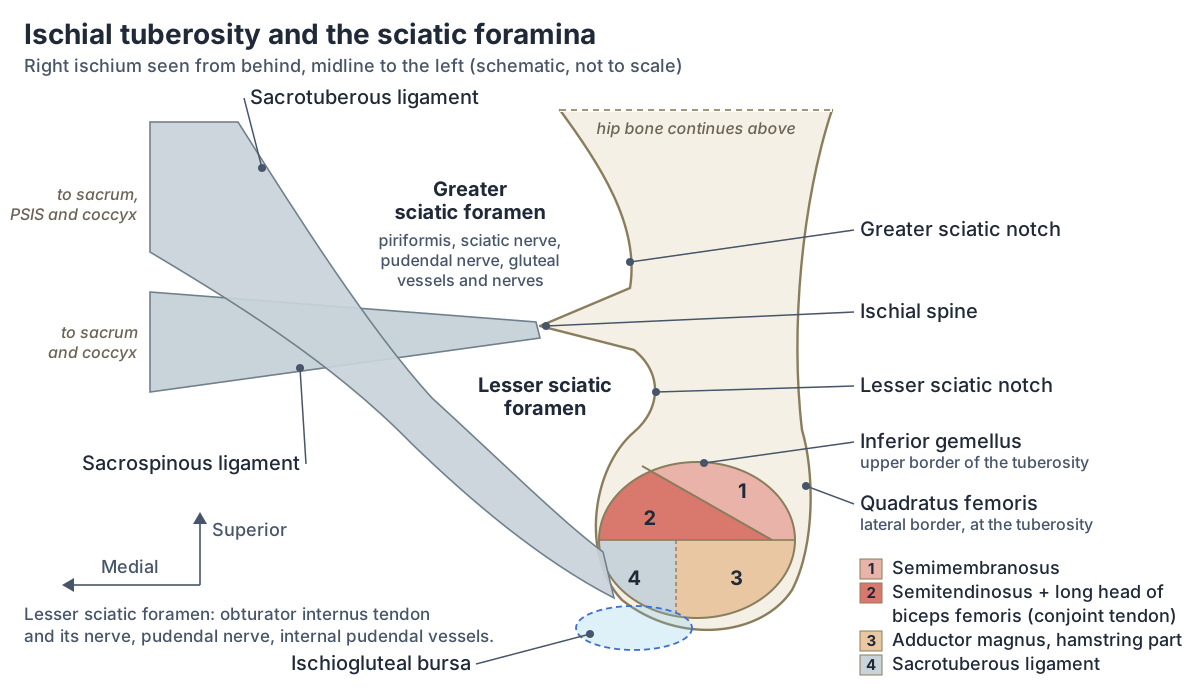
The sacrospinous ligament runs from the sacrum to the ischial spine, and the sacrotuberous ligament from the sacrum to the ischial tuberosity. Together they turn the notches of the hip bone into two openings on the back and side of the pelvis: the greater sciatic foramen, formed by the greater sciatic notch, the sacrum and the sacrospinous ligament, and the smaller, lower lesser sciatic foramen, formed by the lesser sciatic notch. (OpenStax Anatomy and Physiology 2e, 8.3: The Pelvic Girdle and Pelvis.)
The lesser sciatic foramen is bounded above by the sacrospinous ligament and ischial spine, in front by the ischial spine, lesser sciatic notch and ischial tuberosity, and behind by the sacrotuberous ligament. Four structures pass through it: the internal pudendal artery and vein, the pudendal nerve, the tendon of obturator internus and the nerve to obturator internus. (TeachMeAnatomy: The Sciatic Foramina.)
The sacrotuberous ligament arises from the ilium, sacrum and coccyx and attaches below to the ischial tuberosity. (Kenhub: Sacrotuberous ligament.)
The ischial tuberosity (sitting bone) and its attachments
The inferior end of the body forms a rough impression, the ischial tuberosity. The body weight rests on the two ischial tuberosities when you sit. (O'Rahilly R et al. Basic Human Anatomy, Chapter 12: The bones of the lower limb (Dartmouth).)
Gray's Anatomy (1918) maps the tuberosity in two parts. The upper part is smooth and four-sided: semimembranosus arises from its upper outer area, and the long head of biceps femoris and semitendinosus from its lower inner area. The lower part is rough and triangular: adductor magnus attaches to its outer part and the sacrotuberous ligament to its inner part. (Gray H. Anatomy of the Human Body, 20th ed. 1918 (Internet Archive text).)
Ultrasound anatomy agrees. Semitendinosus and the long head of biceps femoris arise by a conjoint tendon from the medial, posterior part of the tuberosity, while semimembranosus attaches more laterally and deeply. (Manske RC et al. Diagnostic musculoskeletal ultrasound in the evaluation of the proximal hamstrings at the ischial tuberosity. Int J Sports Phys Ther. 2026.)
Radiopaedia adds the smaller muscles. Superior gemellus arises from the ischial spine and inferior gemellus from the upper border of the tuberosity; quadratus femoris arises between the origins of inferior gemellus and adductor magnus; and obturator externus and obturator internus take part of their origin from the ramus. (Radiopaedia: Ischium.)
All three hamstrings proper (semitendinosus, semimembranosus and the long head of biceps femoris) are supplied by the tibial part of the sciatic nerve; only the short head of biceps femoris, which arises from the femur, is supplied by the common fibular part. (TeachMeAnatomy: The Hamstrings.)
Adductor magnus has two parts: an adductor part from the ischiopubic ramus, supplied by the obturator nerve, and a hamstring part from the ischial tuberosity, supplied by the sciatic nerve. (Radiopaedia: Adductor magnus muscle.)
The hamstrings and adductors that arise from the ischium, with origin, insertion, nerve and action tables. Thigh muscles.
Muscles and ligaments attached to the ischium
| Structure | Where on the ischium | Nerve (muscles) |
|---|---|---|
| Semimembranosus | Ischial tuberosity, upper outer (lateral) area | Tibial part of the sciatic nerve |
| Semitendinosus and long head of biceps femoris | Ischial tuberosity, upper inner (medial) area, by a conjoint tendon | Tibial part of the sciatic nerve |
| Adductor magnus, hamstring part | Ischial tuberosity, lower outer area | Sciatic nerve (tibial part) |
| Adductor magnus, adductor part | Ischiopubic ramus | Obturator nerve |
| Quadratus femoris | Lateral border of the ischium at the tuberosity | Nerve to quadratus femoris |
| Inferior gemellus | Upper border of the ischial tuberosity | Nerve to quadratus femoris |
| Superior gemellus | Ischial spine (outer surface) | Nerve to obturator internus |
| Obturator internus | Inner surface of the obturator membrane and the bony margin of the obturator foramen | Nerve to obturator internus |
| Coccygeus and levator ani (iliococcygeus) | Ischial spine (inner surface) | Sacral nerves (S4, S5) and nerve to levator ani |
| Sacrospinous ligament | Ischial spine | Ligament |
| Sacrotuberous ligament | Medial (inner) margin of the ischial tuberosity | Ligament |
Attachments from Radiopaedia, Gray's Anatomy (1918), TeachMeAnatomy, Kenhub and the University of Washington musculoskeletal atlas; nerves from TeachMeAnatomy, Kenhub, Radiopaedia and the UW atlas.
The ischium in the hip socket
The ischium forms approximately the posterior two-fifths of the acetabulum; the ilium forms about the superior two-fifths and the pubis about the anterior one-fifth. (O'Rahilly R et al. Basic Human Anatomy, Chapter 12: The bones of the lower limb (Dartmouth).)
Gray's Anatomy (1918) gives the ischium a little more than two-fifths, the largest share of the three. (Gray H. Anatomy of the Human Body, 20th ed. 1918 (Internet Archive text).)
Not every source agrees: Kenhub describes the body of the ischium as forming the posterior one-third of the acetabulum. (Kenhub: Ischium.)
The large upper wing of the hip bone, which forms the roof of the acetabulum. Ilium.
Ossification and growth of the ischium
Gray's Anatomy (1918) places the primary ossification centre of the ischium in its superior ramus at about the third month of fetal life, after the ilium (eighth or ninth week) and before the pubis (fourth to fifth month). (Gray H. Anatomy of the Human Body, 20th ed. 1918 (Internet Archive text).)
Radiopaedia gives an earlier date, the eighth prenatal week, for the centre in the body of the ischium, with bone spreading downwards and forwards to form the ramus. The ischiopubic synchondrosis, the cartilage joint between the ischial and pubic rami, fuses typically in the 7th to 10th year of life, earlier in girls. A single secondary centre forms the ischial tuberosity; it begins to ossify at 10 to 13 years and fuses typically at 16 to 20 years, earlier in females. (Radiopaedia: Ischium.)
Other data give a wider range for the synchondrosis: fusion usually occurs between 4 and 9 years in girls and between 7 and 13 years in boys. The synchondrosis can swell and be painful while it fuses (van Neck-Odelberg disease); in a series of 21 patients the median age at diagnosis was 10 years. (Schneider KN et al. Invasive diagnostic and therapeutic measures are unnecessary in patients with symptomatic van Neck-Odelberg disease. Acta Orthop. 2021.)
Van Neck-Odelberg disease is the enlargement of one of the ischiopubic synchondroses before puberty and is generally considered a normal variant. On X-rays it can mimic a stress fracture, bone infection or a tumour, so the clinical history matters. (Radiopaedia: Van Neck-Odelberg disease.)
A study of ischial tuberosity growth plates on CT and MRI found a five-stage pattern of ossification spanning 13 to 19 years in males and 12 to 17 years in females. (Mitchell BC et al. Classifying ischial tuberosity avulsion fractures by ossification stage and tendon attachment. Orthop J Sports Med. 2021.)
Male vs female ischium
The ischial tuberosities of females are farther apart, which increases the size of the pelvic outlet. (OpenStax Anatomy and Physiology 2e, 8.3: The Pelvic Girdle and Pelvis.)
CT measurements show the size of the difference. In 211 women and 181 men from Latvia, the mean distance between the inner margins of the two tuberosities (the bituberous diameter) was 123.9 mm in women and 103.5 mm in men, and the distance between the two ischial spines (the bispinous diameter) was 112.2 mm in women and 93.6 mm in men. (Kolesova O et al. Age-related trends of lesser pelvic architecture in females and males: a computed tomography pelvimetry study. Anat Cell Biol. 2017.)
The typical female pelvis also has less prominent ischial spines. (TeachMeAnatomy: The Pelvic Girdle.)
Kenhub describes male ischial spines as conical and projecting inwards, and female ones as shorter and blunted. (Kenhub: The pelvis.)
This matters in childbirth because the midpelvis, at the level of the ischial spines, is the plane of smallest pelvic dimensions; its transverse diameter is the distance between the spines. (Gleason RL Jr et al. A safe, low-cost, easy-to-use 3D camera platform to assess risk of obstructed labor due to cephalopelvic disproportion. PLoS One. 2018.)
How to feel your ischial tuberosities
Sit on a firm chair and rock your pelvis from side to side: you can feel the ischial tuberosities, the sitting bones, pressing into the seat. They support the body when sitting. (OpenStax Anatomy and Physiology 2e, 8.3: The Pelvic Girdle and Pelvis.)
Clinical relevance: pudendal block, bursitis, avulsion and impingement
The ischial spine can be felt through the vaginal wall, and the pudendal nerve crosses the sacrospinous ligament close to its attachment to the spine. That makes the spine the landmark for a pudendal nerve block, a form of pain relief occasionally given before vaginal childbirth. (TeachMeAnatomy: The Pudendal Nerve.)
A 2026 expert review of nerve blocks in labour describes the pudendal nerve as lying deep near the ischial spine and discusses the block's use in the second stage of labour, where it found the pain relief limited and inconsistent. (Coviello A et al. Peripheral nerve blocks and fascial plane blocks in labor: expert evidence-based perspectives. Anesth Analg. 2026.)
The ischiogluteal bursa lies deep to the lower part of gluteus maximus, below and behind the ischial tuberosity. Its inflammation, ischiogluteal bursitis or "weaver's bottom", is classically linked with sustained pressure on the tuberosity from prolonged sitting, and because the bursa lies close to the sciatic and posterior femoral cutaneous nerves its pain can mimic nerve-root pain (radiculopathy). (Radiopaedia: Ischiogluteal bursa.)
On MRI in 17 patients, the enlarged bursae lay deep to the gluteus muscles and below and behind the ischial tuberosity, their upper ends touching its inferomedial aspect. (Cho KH et al. Non-infectious ischiogluteal bursitis: MRI findings. Korean J Radiol. 2004.)
In rare cases, mostly in adolescents, a sudden and forceful contraction of the hamstrings pulls a fragment of bone off the ischial tuberosity (an avulsion fracture). A displaced fragment can compress the sciatic nerve. (TeachMeAnatomy: The Hamstrings.)
In a series of 203 adolescent athletes reported by Rossi and Dragoni, the ischial tuberosity was the commonest site of pelvic apophyseal avulsion (54%), ahead of the anterior inferior (22%) and anterior superior (19%) iliac spines. (Radiopaedia: Pelvic apophyseal avulsion fractures (annotated image).)
In ischiofemoral impingement, soft tissues are squeezed between the ischial tuberosity and the lesser trochanter of the femur. It affects females more than males and typically causes long-standing groin or buttock pain without an injury. (Radiopaedia: Ischiofemoral impingement.)
When the space between the lesser trochanter and the ischial tuberosity narrows, quadratus femoris is compressed. The space is sexually dimorphic: in a modelling study of 40,000 virtual pelves built from Belgian imaging data, the mean ischiofemoral distance was 22.2 mm in females and 29.1 mm in males. (Audenaert EA et al. Ischiofemoral impingement: the evolutionary cost of pelvic obstetric adaptation. J Hip Preserv Surg. 2021.)
The short hip rotator that runs from the ischium to the femur and is pinched in ischiofemoral impingement. Quadratus femoris.
Ischium vs ilium vs pubis
The ilium is the fan-like upper region and the largest part of the hip bone, the ischium forms its lower back region, and the pubis forms its front. The three converge to form the acetabulum, and the obturator foramen lies between the ischium and pubis. (OpenStax Anatomy and Physiology 2e, 8.3: The Pelvic Girdle and Pelvis.)
Ischium vs ilium vs pubis
| Feature | Ischium | Ilium | Pubis |
|---|---|---|---|
| Position in the hip bone | Lower back part | Upper, largest part | Front part |
| Share of the acetabulum | About two-fifths (back) | About two-fifths (upper) | About one-fifth (front) |
| Main landmarks | Ischial tuberosity, ischial spine, lesser sciatic notch, ramus | Iliac crest, ASIS, AIIS, PSIS, PIIS, iliac fossa | Pubic body and tubercle, superior and inferior rami |
| Everyday job | Takes your weight when you sit; anchors the hamstrings | Forms the roof of the hip socket; anchors abdominal and gluteal muscles | Joins the other pubis at the pubic symphysis |
Sources: OpenStax 8.3, O'Rahilly's Basic Human Anatomy and Gray's Anatomy (1918). The pelvis guide compares all three in detail.
Structures next to the ischium
The nearest named structures in the 3D model, measured from the centre of each part:
- Hip bone · skeletal
- Obturator internus · muscular
- Femur · skeletal
- Piriformis · muscular
- Gemellus superior · muscular
- Pubis · skeletal
- Gemellus inferior · muscular
- Cardinal ligament of uterus · reproductive
- Broad ligament of uterus · reproductive
- Ilium · skeletal
Frequently asked questions
What is the ischium?
The ischium is the lower, back part of each hip bone, one of the three bones (with the ilium and pubis) that fuse in the teenage years to make it. It has a body, which forms about two-fifths of the hip socket, and a ramus that joins the pubis. Its lower end is the ischial tuberosity, the bone you sit on.
What is the function of the ischium?
The ischium takes your weight when you sit, through the two ischial tuberosities. It forms part of the hip socket and of the pelvic outlet, and anchors many muscles and ligaments: the hamstrings and part of adductor magnus on the tuberosity, the gemelli and quadratus femoris, pelvic floor muscles on the ischial spine, and the sacrotuberous and sacrospinous ligaments.
What is the ischial tuberosity?
The ischial tuberosity is the large, rough lump at the bottom of the ischium, often called the sitting bone. It carries your weight when you sit. Its upper part gives origin to semimembranosus and, by a shared tendon, semitendinosus and the long head of biceps femoris; its lower part takes adductor magnus and the sacrotuberous ligament.
What muscles attach to the ischium?
The hamstrings (semimembranosus, semitendinosus and the long head of biceps femoris) and the hamstring part of adductor magnus attach to the ischial tuberosity. Inferior gemellus and quadratus femoris arise beside it, superior gemellus, coccygeus and levator ani from the ischial spine, and obturator internus and externus and the adductor part of adductor magnus from the ramus and around the obturator foramen.
What passes through the lesser sciatic foramen?
Four structures: the tendon of obturator internus, the nerve to obturator internus, the pudendal nerve and the internal pudendal artery and vein. The foramen is bounded by the lesser sciatic notch of the ischium, the sacrospinous ligament and ischial spine above, and the sacrotuberous ligament behind.
Why can the sitting bones hurt?
Pain over a sitting bone has several possible causes and needs a clinician's assessment. Anatomy explains some of them: the ischiogluteal bursa under gluteus maximus can become inflamed with prolonged sitting, the hamstring tendons attach to the tuberosity, and in teenagers a hard hamstring pull can avulse its growth centre.
Why is the ischial spine important?
It separates the greater and lesser sciatic notches and anchors the sacrospinous ligament and pelvic floor muscles. The pudendal nerve passes just medial to it, so it is the landmark for a pudendal nerve block. The distance between the two spines marks the narrowest part of the birth canal, the midpelvis.
References
- OpenStax. Anatomy and Physiology 2e, §8.3 The Pelvic Girdle and Pelvis. Rice University, 2022.
- Ischium. Radiopaedia reference article.
- Pudendal nerve. Radiopaedia reference article.
- Ischiogluteal bursa. Radiopaedia reference article.
- Pelvic apophyseal avulsion fractures (annotated image). Radiopaedia case.
- Ischiofemoral impingement. Radiopaedia reference article.
- Van Neck-Odelberg disease. Radiopaedia reference article.
- TeachMeAnatomy. The Hip Bone: Ilium, Ischium, Pubis.
- TeachMeAnatomy. The Sciatic Foramina.
- TeachMeAnatomy. The Pudendal Nerve.
- TeachMeAnatomy. The Hamstring Muscles.
- TeachMeAnatomy. Muscles of the Gluteal Region.
- TeachMeAnatomy. The Pelvic Floor.
- TeachMeAnatomy. The Pelvic Girdle.
- Kenhub. Ischium.
- Kenhub. Hip bone.
- Kenhub. The pelvis.
- Kenhub. Sacrotuberous ligament.
- University of Washington Department of Radiology. Musculoskeletal Atlas: Adductor magnus.
- University of Washington Department of Radiology. Musculoskeletal Atlas: Quadratus femoris.
- University of Washington Department of Radiology. Musculoskeletal Atlas: Semimembranosus.
- University of Washington Department of Radiology. Musculoskeletal Atlas: Semitendinosus.
- University of Washington Department of Radiology. Musculoskeletal Atlas: Biceps femoris (long head).
- University of Washington Department of Radiology. Musculoskeletal Atlas: Obturator internus.
- Adductor magnus muscle. Radiopaedia reference article.
- O'Rahilly R, Müller F, Carpenter S, Swenson R. Basic Human Anatomy, Chapter 12: The bones of the lower limb. Dartmouth Medical School.
- Gray H. Anatomy of the Human Body. 20th ed. Philadelphia: Lea & Febiger, 1918 (Internet Archive full text).
- Manske RC, Wolfe C, Page P, Voight M. Diagnostic musculoskeletal ultrasound in the evaluation of the proximal hamstrings at the ischial tuberosity. Int J Sports Phys Ther. 2026;21(4):477-482.
- Mitchell BC, Bomar JD, Wenger D, Pennock AT. Classifying ischial tuberosity avulsion fractures by ossification stage and tendon attachment. Orthop J Sports Med. 2021;9(7 suppl3):2325967121S00077.
- Schneider KN, Lampe LP, Gosheger G, et al. Invasive diagnostic and therapeutic measures are unnecessary in patients with symptomatic van Neck-Odelberg disease (ischiopubic synchondrosis). Acta Orthop. 2021;92(3):347-351.
- Kolesova O, Kolesovs A, Vetra J. Age-related trends of lesser pelvic architecture in females and males: a computed tomography pelvimetry study. Anat Cell Biol. 2017;50(4):265-274.
- Gleason RL Jr, Yigeremu M, Debebe T, et al. A safe, low-cost, easy-to-use 3D camera platform to assess risk of obstructed labor due to cephalopelvic disproportion. PLoS One. 2018;13(9):e0203865.
- Vitale MR, Shtirmer D, Ashley BP, Brown AC, Verrier SP, Lobo S. Modern utility of pelvimetry: a relevant tool or an outdated concept? Cureus. 2025;17(10):e95522.
- Coviello A, Frigo MG, Cirillo D, et al. Peripheral nerve blocks and fascial plane blocks in labor: expert evidence-based perspectives. Anesth Analg. 2026;143(1):59-67.
- Kale A, Usta T, Basol G, et al. Comparison of ultrasound-guided transgluteal and finger-guided transvaginal pudendal nerve block techniques: which one is more effective? Int Neurourol J. 2019;23(4):310-320.
- Cho KH, Lee SM, Lee YH, et al. Non-infectious ischiogluteal bursitis: MRI findings. Korean J Radiol. 2004;5(4):280-286.
- Audenaert EA, Duquesne K, De Roeck J, et al. Ischiofemoral impingement: the evolutionary cost of pelvic obstetric adaptation. J Hip Preserv Surg. 2021;7(4):677-687.
- Foundational Model of Anatomy: Ischium (FMA16594). EMBL-EBI Ontology Lookup Service.
- Mitsuhashi N. et al. (2009). BodyParts3D: 3D structure database for anatomical concepts. Nucleic Acids Research 37, D782–D785. Data © The Database Center for Life Science, CC BY 4.0.
- Browne K., Schlehlein H. 3D Reference Organ Set for United, Female (Visible Human Female). Human Reference Atlas 3D Reference Object Library, HuBMAP / NIH. Data from the NLM Visible Human Project. CC BY 4.0.
Related anatomy
- PelvisPelvis and hip bone anatomy in 3D: ilium, ischium and pubis, ASIS and PSIS, ligaments, female pelvic anatomy and a…
- IliumIlium anatomy with a labelled diagram and 3D model: iliac crest, ASIS, AIIS, PSIS, PIIS, iliac fossa, gluteal lines…
- PubisPubis: The pubis is the anterior part of the hip bone. See where it is in an interactive 3D human body, what it does…
- Hip musclesMuscles of the hip explained: gluteus maximus, medius and minimus, TFL, piriformis, obturator internus and the other…
- Thigh musclesThigh muscles explained: quadriceps and sartorius at the front, adductor muscles of the inner thigh, hamstrings at the…
- Pelvic floor and perineumPerineum and pelvic floor anatomy with labelled diagrams and a 3D model: levator ani, coccygeus, perineal body, both…
Explore the whole body in the interactive 3D human anatomy model, or browse the A–Z list of human structures.