

Ilium at a glance
- What it is
- The ilium is the large, fan-shaped upper part of the hip bone.
- Location
- It forms the upper part of each hip bone. Its crest runs from the anterior superior iliac spine (ASIS) to the posterior superior iliac spine, and its auricular surface meets the sacrum at the sacroiliac joint.
- Function
- It carries body weight from the sacrum to the hip joint, forms the upper part of the acetabulum and gives attachment to the abdominal, back, gluteal and iliacus muscles.
- Body system
- Skeletal · paired (left and right) · female body
- FMA ID
- FMA16591 (Foundational Model of Anatomy)
Key facts
- It forms about two-fifths of the acetabulum; the ischium forms two-fifths and the pubis one-fifth.
- The arcuate line on its inner surface is part of the pelvic brim (linea terminalis), the border between the greater and lesser pelvis.
- The ASIS anchors the inguinal ligament and sartorius; the anterior inferior iliac spine anchors the straight head of rectus femoris.
- The greater sciatic notch below the posterior ilium is wider in females than in males.
Clinical relevance
The posterior iliac crest is the usual site for bone marrow aspiration and biopsy, and the iliac crest is a common donor site for bone grafts.
Where is the ilium?
The pelvis consists of four bones: the right and left hip bones, the sacrum and the coccyx. Each adult hip bone is formed by three bones that fuse in the late teenage years, the ilium, ischium and pubis, and the ilium is the fan-like upper region that forms the largest part of it. The hip bone is the attachment point for each lower limb, and the pelvic girdle passes the weight of the body from the vertebral column through the hip joints into the legs. (OpenStax Anatomy and Physiology 2e, 8.3: The Pelvic Girdle and Pelvis.)
The ilium has a smaller lower part, its body, which helps form the acetabulum, and an expanded, fan-shaped upper part, its wing (ala), which supports the lower abdomen, the region also called the false pelvis. (Kenhub: Ilium.)
In the adult the ilium, ischium and pubis are fused into one hip bone, mainly at the acetabulum; in the immature skeleton they are separate. The ilium is not to be confused with the ileum, which only sounds the same (see the comparison below). (Radiopaedia: Ilium.)
In the 3D model above, the ilium is highlighted and labelled with the sacrum, seen from the left side so that its outer gluteal surface faces you; the ischium and pubis are faded with the rest of the skeleton, so the ilium stands out. The ilium is a female-only part: it comes from the Visible Human Female model of the Human Reference Atlas, so the viewer opens the female body. The default male body (BodyParts3D) has a single "Hip bone" mesh and no coccyx, so it cannot show the ilium on its own. In the female model the adult hip bone is divided along its old fusion lines for teaching, so the edges where the ilium meets the ischium and pubis are cut edges, not real bone surfaces.
The female pelvis in the viewer is fitted to the shared male spine and hip joints, so it sits about 15 to 19 degrees further forward than the anatomical position and its widths cannot be compared with the male body; the pelvis guide linked below explains why. Compare shapes in the model, not distances.
The whole bony pelvis, how the ilium, ischium and pubis fit together, and how the female and male pelvis differ. Pelvis and hip bones.
Parts of the ilium: body, wing, borders and surfaces
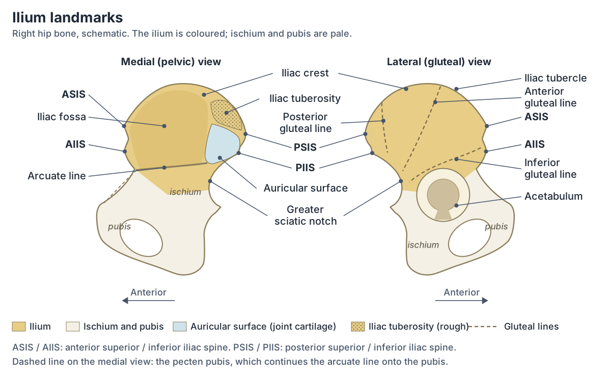
The body of the ilium is the smaller lower part that helps form the acetabulum. The ala (wing) is the expanded upper part, and it has four borders and three surfaces. (Kenhub: Hip bone.)
The superior border is the iliac crest, which runs from the anterior superior iliac spine (ASIS) to the posterior superior iliac spine (PSIS). The anterior border descends to the acetabulum and carries the anterior inferior iliac spine (AIIS). The posterior border carries the posterior inferior iliac spine (PIIS), with the greater sciatic notch below it. The medial border, on the inside of the pelvis, forms part of the iliopectineal line. (Radiopaedia: Ilium.)
The three surfaces are the gluteal surface, which faces backwards and outwards and is marked by the gluteal lines; the iliac fossa on the inner side, which gives attachment to iliacus; and the sacropelvic surface behind the fossa, which carries the iliac tuberosity and the auricular surface for the sacrum. The pelvic part of this surface forms part of the side wall of the true pelvis. (Radiopaedia: Ilium.)
The iliac crest and the level of the top of the hips
The iliac crest is the prominent upper border of the ilium and a site of attachment for many muscles. It has an inner and an outer lip separated by an intermediate zone, and the outer lip bears a prominence near the front of the crest, the iliac tubercle. (Kenhub: Hip bone.)
Gray's Anatomy (1918) gives the classic map of the crest: tensor fasciae latae, external oblique and latissimus dorsi attach to the outer lip, internal oblique to the intermediate line, and transversus abdominis and quadratus lumborum to the inner lip. (Gray H. Anatomy of the Human Body, 20th ed. 1918 (Internet Archive text).)
The highest point of the iliac crest is slightly behind its midpoint. The supracristal plane, a horizontal plane joining the highest points of the two crests, lies at the level of the fourth lumbar vertebra (L4). (O'Rahilly R et al. Basic Human Anatomy, Chapter 12: The bones of the lower limb (Dartmouth).)
On an X-ray the same line is called the intercristal line, or Tuffier's or Jacoby's line. Its level varies between people, anywhere from the lower end plate of L4 to the upper end plate of L5. It is used to number the lumbar vertebrae and as a landmark to avoid operating or injecting at the wrong spinal level; Jacoby and Tuffier both described it as a guide for lumbar injections. (Radiopaedia: Intercristal line.)
Feeling for the level by hand is less precise than it sounds. Anaesthetists use the intercristal line, which is said to cross the L4 vertebra or the L4 to L5 disc, to choose the space for spinal anaesthesia; in a 2016 study of 105 adults, the interspace chosen by palpation matched the level found by ultrasound in only 39 (37%). (Parate LH et al. Inaccurate level of intervertebral space estimated by palpation: the ultrasonic revelation. Saudi J Anaesth. 2016.)
ASIS, AIIS, PSIS and PIIS
The anterior superior iliac spine (ASIS) is a prominent bony projection at the front end of the iliac crest. Sartorius, tensor fasciae latae and the lateral end of the inguinal ligament attach to it. Because it is easy to feel, clinicians use it to find the inguinal ligament, the common femoral artery and the base of the appendix, and as the top point when measuring true leg length. (Kenhub: Anterior superior iliac spine.)
Below the ASIS, the anterior inferior iliac spine (AIIS) gives origin to the straight head of rectus femoris and attachment to the iliofemoral ligament of the hip joint. The reflected head of rectus femoris arises from the bone just above the acetabulum. (Radiopaedia: Ilium.)
At the back, the crest ends at the posterior superior iliac spine (PSIS). It is not as easy to feel as the ASIS, but it is often marked by a dimple above the inner buttock. Multifidus, the posterior sacroiliac ligaments and the sacrotuberous ligament attach to it. (Kenhub: Posterior superior iliac spine.)
Lower down is the posterior inferior iliac spine (PIIS), at the lower end of the rough auricular surface, and below it the large, inverted U-shaped greater sciatic notch on the back edge of the lower ilium. (OpenStax Anatomy and Physiology 2e, 8.3: The Pelvic Girdle and Pelvis.)
The greater sciatic notch is closed off by the sacrospinous and sacrotuberous ligaments to form the greater sciatic foramen, the main passage from the pelvis to the buttock. Above piriformis it carries the superior gluteal vessels and nerve. Below piriformis it carries the sciatic nerve, the pudendal nerve, the inferior gluteal vessels and nerve, the posterior femoral cutaneous nerve and the nerves to obturator internus and quadratus femoris. (TeachMeAnatomy: The Sciatic Foramina.)
Landmarks of the ilium
| Landmark | What it is | What attaches or passes |
|---|---|---|
| Iliac crest | Curved upper border from the ASIS to the PSIS, with inner and outer lips | External and internal oblique, transversus abdominis, quadratus lumborum, latissimus dorsi, tensor fasciae latae |
| Iliac tubercle | Prominence on the outer lip near the front of the crest | The lateralmost part of the crest; a surface landmark |
| ASIS | Front end of the iliac crest | Sartorius, tensor fasciae latae, lateral end of the inguinal ligament |
| AIIS | Rounded projection below the ASIS, just above the acetabulum | Straight head of rectus femoris, iliofemoral ligament |
| PSIS | Back end of the iliac crest, under a skin dimple | Multifidus, posterior sacroiliac ligaments, sacrotuberous ligament |
| PIIS | Below the PSIS, at the lower end of the auricular surface | Sacrotuberous ligament; upper edge of the greater sciatic notch |
| Gluteal lines | Posterior, anterior and inferior ridges on the outer surface | Borders of the gluteus maximus, medius and minimus origins |
| Iliac fossa | Smooth, concave inner surface of the wing | Iliacus |
| Arcuate line | Ridge at the lower edge of the iliac fossa | Part of the pelvic brim, between the greater and lesser pelvis |
| Auricular surface | Ear-shaped, cartilage-covered surface behind the iliac fossa | Sacroiliac joint with the sacrum |
| Iliac tuberosity | Rough, raised area behind the auricular surface | Posterior sacroiliac ligaments |
| Greater sciatic notch | Large notch on the back edge below the PIIS | With the sacrospinous and sacrotuberous ligaments forms the greater sciatic foramen: piriformis, sciatic nerve, gluteal vessels and nerves, pudendal nerve |
Compiled from Radiopaedia, Kenhub, OpenStax, TeachMeAnatomy, O'Rahilly's Basic Human Anatomy and Gray's Anatomy (1918); see references.
Gluteal surface and the gluteal lines
The outer (gluteal) surface of the wing faces backwards and outwards and shows three long, prominent ridges: the posterior, anterior and inferior gluteal lines. (Radiopaedia: Ilium.)
Gray's Anatomy (1918) describes their course. The posterior gluteal line is the shortest: it begins at the crest and runs down to the upper part of the greater sciatic notch. The anterior gluteal line curves downwards and backwards from the crest to the upper part of the notch. The inferior gluteal line curves backwards and downwards above the acetabulum and ends near the middle of the notch. (Gray H. Anatomy of the Human Body, 20th ed. 1918 (Internet Archive text).)
Gluteus maximus arises from the iliac surface behind the posterior gluteal line, gluteus medius from the area between the posterior and anterior lines, and gluteus minimus from the area between the anterior and inferior lines. (Radiopaedia: Gluteal muscles.)
Between the inferior gluteal line and the rim of the acetabulum is a shallow supra-acetabular groove, the attachment of the reflected head of rectus femoris. (Kenhub: Hip bone.)
The hip abductor that arises between the anterior and posterior gluteal lines of the ilium. Gluteus medius.
Muscles attached to the ilium
| Muscle | Where it attaches on the ilium | Origin or insertion |
|---|---|---|
| Iliacus | Iliac fossa | Origin |
| Gluteus maximus | Gluteal surface behind the posterior gluteal line | Origin |
| Gluteus medius | Gluteal surface between the anterior and posterior gluteal lines | Origin |
| Gluteus minimus | Gluteal surface between the anterior and inferior gluteal lines | Origin |
| Tensor fasciae latae | ASIS and the front of the iliac crest | Origin |
| Sartorius | ASIS | Origin |
| Rectus femoris | Straight head from the AIIS; reflected head from the groove above the acetabulum | Origin |
| External oblique | Iliac crest (outer lip) | Insertion |
| Internal oblique | Iliac crest (intermediate line) | Origin |
| Transversus abdominis | Iliac crest (inner lip) | Origin |
| Quadratus lumborum | Iliac crest (inner lip) and iliolumbar ligament | Origin |
| Latissimus dorsi | Iliac crest (outer lip) | Origin |
| Multifidus | PSIS | Attachment |
Origins and insertions from Radiopaedia, the University of Washington musculoskeletal atlas, Kenhub, TeachMeAnatomy and Gray's Anatomy (1918).
Inner surface: iliac fossa, arcuate line and auricular surface
The iliac fossa is the large, concave front-and-inner surface of the wing. It forms the smooth back and side wall of the greater pelvis. (Kenhub: Hip bone.)
The lower edge of the iliac fossa is the arcuate line of the ilium. The pelvic brim, the upper edge of the lesser (true) pelvis, runs through the pubic symphysis in front and, further back, along the pectineal line of the pubis, the arcuate line of the ilium and the sacral promontory. (OpenStax Anatomy and Physiology 2e, 8.3: The Pelvic Girdle and Pelvis.)
Behind the iliac fossa is a rough area in two parts. The front part is the auricular surface, which is covered with cartilage and joins the sacrum. The back part is the raised, rough iliac tuberosity, which anchors the posterior sacroiliac ligaments. Below the fossa, the arcuate line runs downwards, forwards and inwards. (Gray H. Anatomy of the Human Body, 20th ed. 1918 (Internet Archive text).)
The auricular surface is ear-shaped and forms the sacroiliac joint with the sacrum; the iliac tuberosity is a roughened, raised area for ligaments. The arcuate line runs forwards and downwards from the auricular surface and marks the transition between the body and the wing of the ilium. (Kenhub: Hip bone.)
The other half of the sacroiliac joint, with its auricular surface, ligaments and joint movement. Sacrum.
Joints of the ilium and its share of the acetabulum
All three parts of the hip bone form the acetabulum. The ilium forms about the superior two-fifths, the ischium about the posterior two-fifths and the pubis about the anterior one-fifth. (O'Rahilly R et al. Basic Human Anatomy, Chapter 12: The bones of the lower limb (Dartmouth).)
Gray's Anatomy (1918) is a little more precise: a little less than two-fifths comes from the ilium, a little more than two-fifths from the ischium and the remaining fifth from the pubis. (Gray H. Anatomy of the Human Body, 20th ed. 1918 (Internet Archive text).)
The body of the ilium forms the roof of the acetabulum. The head of the femur sits in the acetabulum to form the hip joint. (TeachMeAnatomy: The Hip Bone (Ilium, Ischium, Pubis).)
At the back, the irregular, rough auricular surface of the ilium articulates with the sacrum at the sacroiliac joint. So the ilium takes part in two joints, the sacroiliac joint and the hip joint, while its junctions with the ischium and pubis become bone in the adult. (Radiopaedia: Ilium.)
The ball-and-socket joint between the acetabulum and the head of the femur. Hip joint.
Blood supply of the ilium
A branch of the iliolumbar artery enters a nutrient foramen on the inner (medial) side of the ilium. (Radiopaedia: Ilium.)
An MRI study of 245 people confirmed that the nutrient arteries of the ilium arise mainly from the iliolumbar artery and enter the inner surface of the bone towards its upper side, in front of the sacroiliac joint. A nutrient channel with a clear entry point was seen on 243 of 245 right sides and on every left side. (Albanese L et al. Prevalence and origin of prominent nutrient channels of the ilium bone on MR-imaging. Skeletal Radiol. 2025.)
Ossification: when does the ilium fuse?
Each hip bone ossifies from three primary centres, one each for the ilium, ischium and pubis. The centre for the ilium appears about the eighth or ninth week of fetal life. (Gray H. Anatomy of the Human Body, 20th ed. 1918 (Internet Archive text).)
A study of 42 fetuses describes ossification of the ilium beginning at week 9 of gestation, just above the greater sciatic notch at the upper edge of the future acetabulum, and spreading upwards towards the iliac crest. (Baumgart M et al. Quantitative anatomy of the ilium's primary ossification center in the human fetus. Surg Radiol Anat. 2018.)
In the growing skeleton the ilium, ischium and pubis meet at a Y-shaped growth plate in the acetabulum, the triradiate cartilage. It closes typically by 13 to 14 years in girls and 15 to 16 years in boys, beginning at the acetabulum and finishing at the greater sciatic notch between the ilium and ischium. (Radiopaedia: Triradiate cartilage.)
TeachMeAnatomy gives a slightly later timetable, with fusion beginning only at the age of 15 to 17. (TeachMeAnatomy: The Hip Bone (Ilium, Ischium, Pubis).)
The iliac crest grows from its own cap of cartilage. Secondary ossification centres for the crest appear at puberty and join the rest of the ilium between about 15 and 25 years of age. (Radiopaedia: Ilium.)
A CT study of 531 people aged 10 to 29 in Ahvaz, Iran, saw no complete fusion of the crest before 17 years in males or 18 years in females (20 years on the right side), and some people were still not fully fused at 29. (Norouzi M et al. Computed tomography-based age estimation of iliac crests calcification in 10-29 year-old individuals. J Family Med Prim Care. 2019.)
Because the crest cap ossifies in stages, radiologists use it to judge skeletal maturity. The Risser grade runs from no ossification of the iliac crest apophysis (grade 0) to complete ossification and fusion (grade 5) and is used mainly when planning surgery for scoliosis. (Radiopaedia: Risser classification.)
Male vs female ilium
The clearest sex difference in the ilium is the greater sciatic notch: in the male hip bone it is narrower and deeper than the broader notch of females. (OpenStax Anatomy and Physiology 2e, 8.3: The Pelvic Girdle and Pelvis.)
The notch is used to estimate sex from a skeleton. In 567 adult pelves from Australia, a five-point notch score sorted 87.5% correctly (94.6% of females and 81.6% of males). Older women had higher scores on average, meaning that the notch tends to become narrower with age. (DesMarais A et al. The influence of age on greater sciatic notch morphology: testing the Walker method in an Australian population. Int J Legal Med. 2024.)
How to feel the ilium on yourself
Place your hands on your waist: the arching ridge you feel along your waistline is the iliac crest, the upper margin of the ilium. Behind, the crest curves down to end at the posterior superior iliac spine (PSIS); muscles and ligaments surround the PSIS but do not cover it, so it often shows as a dimple on the lower back. (OpenStax Anatomy and Physiology 2e, 8.3: The Pelvic Girdle and Pelvis.)
Follow the crest forwards to the anterior superior iliac spine, which can be felt and sometimes seen at the front of the hip. The tubercle of the crest is its lateralmost part. The dimple over the posterior superior iliac spine lies in the skin just lateral to the sacrum. (O'Rahilly R et al. Basic Human Anatomy, Chapter 12: The bones of the lower limb (Dartmouth).)
Clinical relevance: bone marrow, bone grafts and fractures
Because the iliac crest lies close to the surface and is easy to feel, it is often used for bone marrow biopsy. A cutting needle is passed through the crest under local anaesthetic to take a sample of marrow, which helps diagnose and stage conditions, especially blood diseases. (Kenhub: Hip bone.)
For safety and comfort, the posterior iliac crest is the preferred site for the biopsy. (Alsayegh H et al. Endovascular management of iatrogenic arterial injury post bone marrow biopsy: a report of 3 cases. Radiol Case Rep. 2023.)
The iliac crest is also the gold standard donor site for autologous bone graft, bone taken from a patient's own body. Both tricortical grafts and purely cancellous grafts can be harvested: grafts from the front of the crest are used in procedures such as anterior cervical fusion, and grafts from the back of the crest in posterior spinal fusion. Donor-site pain is the commonest complication. (Pokharel RK et al. Iliac crest bone graft harvesting: modified technique for reduction of complications. JNMA J Nepal Med Assoc. 2022.)
Donor-site problems can last. In a survey of 170 patients an average of about 3.6 years after posterior iliac crest graft harvest, about one in five still had substantial pain and disability. (Schwartz CE et al. Prospective evaluation of chronic pain associated with posterior autologous iliac crest bone graft harvest. Health Qual Life Outcomes. 2009.)
In young athletes the growth centres (apophyses) where muscles attach have not yet fused, so a sudden, forceful contraction of the muscle can pull off a fragment of bone, an apophyseal avulsion fracture. (Radiopaedia: Pelvic apophyseal avulsion fractures (annotated image).)
AIIS avulsion, where the straight head of rectus femoris pulls off the spine, is one of the main types. It occurs mostly in adolescents aged 14 to 17, most often in sports that involve kicking, and almost all cases are managed without surgery. (Radiopaedia: Anterior inferior iliac spine avulsion injury.)
A review of eight studies with 314 patients (mean age 14.5 years, 77% male) found the AIIS the commonest site (46%), ahead of the ASIS, pulled by sartorius (about 31%), the ischial tuberosity (12%) and the iliac crest (11%). (Calderazzi F et al. Apophyseal avulsion fractures of the pelvis. A review. Acta Biomed. 2018.)
Other series rank the sites differently. In 203 adolescent athletes reported by Rossi and Dragoni, the ischial tuberosity was the commonest site (54%), followed by the AIIS (22%) and the ASIS (19%), with soccer and gymnastics the commonest sports. (Radiopaedia: Pelvic apophyseal avulsion fractures (annotated image).)
A direct blow to the ilium can break the iliac wing on its own, a Duverney fracture. It is regarded as a stable injury, but it may be operated on if the bone is badly shattered. (Radiopaedia: Duverney fracture.)
Ilium vs ischium vs pubis (and ilium vs ileum)
The ilium is the fan-like upper region and the largest part of the hip bone, the ischium forms its lower back (posteroinferior) region, and the pubis forms its front (anterior) portion. The three converge to form the acetabulum. The ischial tuberosity carries the weight of the body when sitting, and the two pubic bodies meet at the pubic symphysis. (OpenStax Anatomy and Physiology 2e, 8.3: The Pelvic Girdle and Pelvis.)
The ileum, spelt with an e, is part of the gut, not the skeleton: it is the final and longest segment of the small intestine, with no clear boundary from the jejunum above it, and it joins the caecum at the ileocaecal valve. (OpenStax Anatomy and Physiology 2e, 23.5: The Small and Large Intestines.)
The lower back part of the hip bone: ischial tuberosity, ischial spine, ramus and the hamstring attachments. Ischium.
Ilium vs ischium vs pubis
| Feature | Ilium | Ischium | Pubis |
|---|---|---|---|
| Position in the hip bone | Upper, largest part | Lower back part | Front part |
| Share of the acetabulum | About two-fifths (upper) | About two-fifths (back) | About one-fifth (front) |
| Main landmarks | Iliac crest, ASIS, AIIS, PSIS, PIIS, iliac fossa, auricular surface | Ischial tuberosity, ischial spine, lesser sciatic notch, ischial ramus | Pubic body and tubercle, superior and inferior pubic rami |
| Joins | Sacrum (sacroiliac joint) | Pubis below the obturator foramen (ischiopubic ramus) | Other pubis (pubic symphysis) |
| Everyday job | Forms the roof of the hip socket; anchors abdominal, back and gluteal muscles | Takes your weight when you sit; anchors the hamstrings | Joins the other pubis at the pubic symphysis |
Sources: OpenStax 8.3, O'Rahilly's Basic Human Anatomy and Gray's Anatomy (1918). The full comparison is in the pelvis guide.
Structures next to the ilium
The nearest named structures in the 3D model, measured from the centre of each part:
- Hip bone · skeletal
- Iliacus · muscular
- Sacrum · skeletal
- Descending colon · digestive
- Femur · skeletal
- Gluteus maximus · muscular
- Ascending colon · digestive
- Taenia omentalis · digestive
- Sartorius · muscular
- Cecum · digestive
Frequently asked questions
What is the ilium?
The ilium is the largest and uppermost of the three bones that make up each hip bone; the other two are the ischium and the pubis. It has a body, which forms the upper part of the hip socket, and a broad, fan-shaped wing. Its upper edge is the iliac crest, which you can feel at your waist.
Where is the ilium located?
The ilium forms the upper part of each side of the pelvis. It runs from the iliac crest at your waistline down to the hip socket (acetabulum). At the back it joins the sacrum at the sacroiliac joint, and below it is fused with the ischium and pubis to make one hip bone.
What is the function of the ilium?
The ilium helps pass the weight of the trunk from the sacrum to the hip joint and the leg, forms the roof of the hip socket and anchors many muscles: iliacus on its inner surface, the gluteal muscles on its outer surface, and the abdominal wall muscles, quadratus lumborum and latissimus dorsi on the iliac crest.
What is the difference between the ilium and the ileum?
They sound the same but are unrelated. The ilium, with an i, is the large upper bone of the hip. The ileum, with an e, is the last and longest part of the small intestine; it follows the jejunum without a clear boundary and empties into the caecum, the first part of the large intestine, at the ileocaecal valve.
At what vertebral level is the top of the iliac crest?
A line joining the highest points of the two iliac crests usually lies at the level of the fourth lumbar vertebra (L4) or the L4 to L5 disc. It varies between people, from the lower end plate of L4 to the upper end plate of L5, so it is only an approximate guide to spinal level.
When do the ilium, ischium and pubis fuse?
They meet at a Y-shaped triradiate cartilage in the hip socket, which closes in the teenage years: about 13 to 14 years in girls and 15 to 16 in boys according to Radiopaedia, with other sources giving 15 to 17 or the late teens. The cap on the iliac crest fuses later, between about 15 and 25 years.
Why is bone marrow taken from the iliac crest?
The iliac crest lies just under the skin and is easy to feel, so a needle can reach the marrow inside it safely under local anaesthetic. The posterior iliac crest is the preferred site for safety and comfort. The same crest is also the classic donor site for bone grafts.
References
- OpenStax. Anatomy and Physiology 2e, §8.3 The Pelvic Girdle and Pelvis. Rice University, 2022.
- OpenStax. Anatomy and Physiology 2e, §23.5 The Small and Large Intestines. Rice University, 2022.
- Ilium. Radiopaedia reference article.
- Anterior superior iliac spine. Radiopaedia reference article.
- Intercristal line. Radiopaedia reference article.
- Gluteal muscles. Radiopaedia reference article.
- Triradiate cartilage. Radiopaedia reference article.
- Risser classification. Radiopaedia reference article.
- Anterior inferior iliac spine avulsion injury. Radiopaedia reference article.
- Pelvic apophyseal avulsion fractures (annotated image). Radiopaedia case.
- Duverney fracture. Radiopaedia reference article.
- TeachMeAnatomy. The Hip Bone: Ilium, Ischium, Pubis.
- TeachMeAnatomy. The Sciatic Foramina.
- Kenhub. Ilium.
- Kenhub. Hip bone.
- Kenhub. Anterior superior iliac spine.
- Kenhub. Posterior superior iliac spine.
- O'Rahilly R, Müller F, Carpenter S, Swenson R. Basic Human Anatomy, Chapter 12: The bones of the lower limb. Dartmouth Medical School.
- Gray H. Anatomy of the Human Body. 20th ed. Philadelphia: Lea & Febiger, 1918 (Internet Archive full text).
- Parate LH, Manjunath B, Tejesh CA, Pujari V. Inaccurate level of intervertebral space estimated by palpation: the ultrasonic revelation. Saudi J Anaesth. 2016;10(3):270-275.
- Albanese L, Germann C, Pauli C, Gascho D, Sutter R. Prevalence and origin of prominent nutrient channels of the ilium bone on MR-imaging. Skeletal Radiol. 2025;54(10):2127-2135.
- Baumgart M, Wiśniewski M, Grzonkowska M, et al. Quantitative anatomy of the ilium's primary ossification center in the human fetus. Surg Radiol Anat. 2018;40(9):1047-1054.
- Norouzi M, Hanafi MQ, Gharibvand MM. Computed tomography-based age estimation of iliac crests calcification in 10-29 year-old individuals. J Family Med Prim Care. 2019;8(6):1947-1952.
- DesMarais A, Obertova Z, Franklin D. The influence of age on greater sciatic notch morphology: testing the Walker method in an Australian population. Int J Legal Med. 2024;138(1):239-247.
- Alsayegh H, Apostolidis J, Dhahi T, et al. Endovascular management of iatrogenic arterial injury post bone marrow biopsy: a report of 3 cases. Radiol Case Rep. 2023;18(3):948-956.
- Pokharel RK, Paudel S, Lakhey RB. Iliac crest bone graft harvesting: modified technique for reduction of complications. JNMA J Nepal Med Assoc. 2022;60(247):325-328.
- Schwartz CE, Martha JF, Kowalski P, et al. Prospective evaluation of chronic pain associated with posterior autologous iliac crest bone graft harvest and its effect on postoperative outcome. Health Qual Life Outcomes. 2009;7:49.
- Calderazzi F, Nosenzo A, Galavotti C, et al. Apophyseal avulsion fractures of the pelvis. A review. Acta Biomed. 2018;89(4):470-476.
- Foundational Model of Anatomy: Ilium (FMA16591). EMBL-EBI Ontology Lookup Service.
- Mitsuhashi N. et al. (2009). BodyParts3D: 3D structure database for anatomical concepts. Nucleic Acids Research 37, D782–D785. Data © The Database Center for Life Science, CC BY 4.0.
- Browne K., Schlehlein H. 3D Reference Organ Set for United, Female (Visible Human Female). Human Reference Atlas 3D Reference Object Library, HuBMAP / NIH. Data from the NLM Visible Human Project. CC BY 4.0.
Related anatomy
- PelvisPelvis and hip bone anatomy in 3D: ilium, ischium and pubis, ASIS and PSIS, ligaments, female pelvic anatomy and a…
- IschiumIschium anatomy with labelled diagrams and a 3D model: body, ramus, ischial spine, ischial tuberosity, hamstring…
- PubisPubis: The pubis is the anterior part of the hip bone. See where it is in an interactive 3D human body, what it does…
- SacrumSacrum anatomy explained: promontory, ala, sacral foramina, crests and hiatus, joints and ligaments, sacrum vs coccyx…
- Hip jointHip anatomy explained: the ball-and-socket joint, acetabulum and labrum, iliofemoral and other ligaments, movements…
- Hip musclesMuscles of the hip explained: gluteus maximus, medius and minimus, TFL, piriformis, obturator internus and the other…
Explore the whole body in the interactive 3D human anatomy model, or browse the A–Z list of human structures.