

What is the perineum?
Anatomically, the perineum is the region of the pelvis between the thighs. It is the lowest part of the pelvic outlet and is separated from the pelvic cavity above by the pelvic floor. It supports the urinary, reproductive and digestive organs and has a part in urination, defecation, sexual intercourse and childbirth. (TeachMeAnatomy: The Perineum.)
Clinicians often use the word for the area between the external genitalia and the anus only, but in anatomy the perineum is the whole diamond-shaped region. (TeachMeAnatomy: The Perineum.)
On the surface, the perineum lies between the vaginal opening and the anus in females and between the scrotum and the anus in males. Cleveland Clinic gives its average length as about 1.5 inches (about 4 cm) in females and about 2 inches (about 5 cm) or more in males. Under the skin are layers of connective tissue, fat, muscles and membranes, with the perineal body, the anal sphincter, part of the urethra and branches of the pudendal nerve. The skin carries many nerve endings, and Cleveland Clinic describes the area as an erogenous zone. (Cleveland Clinic: Perineum.)
Borders of the perineum: the urogenital and anal triangles
The perineum is bounded in front by the pubic symphysis, behind by the tip of the coccyx, and at the sides by the inferior pubic and ischial rami and the sacrotuberous ligaments. Its roof is the pelvic floor and its base is the skin and fascia. Seen with the thighs apart, the surface borders are the mons pubis or the base of the penis in front, the inner thighs at the sides and the top of the cleft between the buttocks behind. (TeachMeAnatomy: The Perineum.)
OpenStax describes the same diamond more simply, with the pubic symphysis in front, the coccyx behind and the ischial tuberosities at the sides. (OpenStax Anatomy and Physiology 2e, 11.4: Axial Muscles of the Abdominal Wall and Thorax (muscles of the pelvic floor and perineum).)
A transverse line between the two ischial tuberosities, the interischial line, divides the diamond into two triangles. The urogenital triangle in front, between the pubic symphysis and the ischiopubic rami, holds the external genitalia and the openings of the urethra (and vagina). The anal triangle behind, between the coccyx and the sacrotuberous ligaments, holds the anal canal, its sphincters and the fat-filled ischioanal fossae. (Kenhub: Perineal region.)
The urogenital triangle has an extra layer of strong deep fascia, the perineal membrane, which anchors the muscles of the external genitalia and is pierced by the urethra (and the vagina in females). The anal triangle has no such membrane. (TeachMeAnatomy: The Perineum.)
The pubic symphysis, ischiopubic rami and ischial tuberosities that frame the perineum, with male vs female pelvis. Pelvis bones.
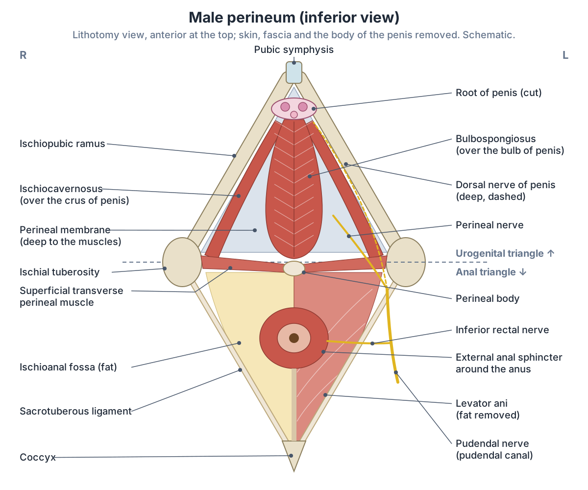
The two triangles of the perineum
| Triangle | Boundaries | Main contents | Nerve |
|---|---|---|---|
| Urogenital (anterior) | Pubic symphysis, ischiopubic rami, interischial line | External genitalia, urethra (and vagina), perineal membrane, deep and superficial perineal pouches | Perineal nerve and dorsal nerve of the penis or clitoris (pudendal) |
| Anal (posterior) | Interischial line, sacrotuberous ligaments, coccyx | Anal canal, external anal sphincter, ischioanal fossae, anococcygeal body | Inferior rectal nerve (pudendal) |
Sources: TeachMeAnatomy (perineum, pudendal nerve) and Kenhub (perineal region).
Pelvic floor muscles: levator ani and coccygeus
The pelvic floor, or pelvic diaphragm, is a funnel-shaped sheet that attaches to the walls of the lesser pelvis and separates the pelvic cavity from the perineum below. It has three components: levator ani, the largest; coccygeus; and the fascia covering both muscles. It supports the bladder, rectum, genital organs and the end of the urethra. (TeachMeAnatomy: The Pelvic Floor.)
Levator ani is usually described as three paired muscles: puborectalis, pubococcygeus and iliococcygeus. Coccygeus (also called ischiococcygeus) completes the pelvic diaphragm behind them but is not part of levator ani. (Kenhub: Muscles of the pelvic floor.)
Sources divide levator ani differently. OpenStax names only two parts, pubococcygeus and iliococcygeus, while TeachMeAnatomy, Kenhub and Radiopaedia describe three. (OpenStax Anatomy and Physiology 2e, 11.4: Axial Muscles of the Abdominal Wall and Thorax (muscles of the pelvic floor and perineum).)
The two halves of levator ani leave a gap in the midline, the levator hiatus. Fibres passing in front of the rectum divide it into a urogenital hiatus in front, which transmits the urethra and, in females, the vagina, and an anal hiatus behind for the rectum. The levator hiatus is a potential weak spot, especially in women. (Kenhub: Levator ani muscle.)
Puborectalis arises from the back of the pubis and forms a U-shaped sling around the anal canal that joins the muscle of the other side. Its constant tone bends the anal canal forwards, creating the anorectal angle that helps hold in faeces; it relaxes during defecation. Fibres in front of the rectum flank the urethra and vagina and help prevent leaks when pressure rises suddenly, as in a sneeze. (TeachMeAnatomy: The Pelvic Floor.)
Kenhub gives the anorectal angle made by a contracted puborectalis as about 90 degrees. Pubococcygeus arises from the back of the pubis lateral to puborectalis and inserts into the anococcygeal ligament, the coccyx, the perineal body and the muscle of the prostate or vagina. Iliococcygeus arises from the tendinous arch of the obturator internus fascia and the ischial spine and inserts into the anococcygeal ligament and coccyx. (Kenhub: Muscles of the pelvic floor.)
Iliococcygeus is the thinnest of the three and forms the back and side of the funnel; TeachMeAnatomy also lists the perineal body among its insertions. Coccygeus is a small triangular muscle behind levator ani that runs from the ischial spine to the lower sacrum and coccyx; it supports the pelvic organs and flexes the coccyx. (TeachMeAnatomy: The Pelvic Floor.)
The iliococcygeus fibres of the two sides meet in the midline at the anococcygeal raphe, which runs from the anal verge to the tip of the coccyx. Coccygeus is partly fused with the sacrospinous ligament. Thinning or absence of part of levator ani on one or both sides is common on MRI, at about 50% in one study of women who had not given birth. (Radiopaedia: Levator ani (pelvic floor).)
Newer anatomical work questions the classic three-part scheme. A study of 31 female cadavers found that the levator ani bundles twist between origin and insertion, and its authors describe the bundles by where they insert rather than as puborectalis, pubococcygeus and iliococcygeus. (Muro S, Moue S, Akita K. Twisted orientation of the muscle bundles in the levator ani functional parts in women. J Anat. 2024.)
The bones at the back of the pelvic floor, where coccygeus and levator ani insert. Sacrum and coccyx.
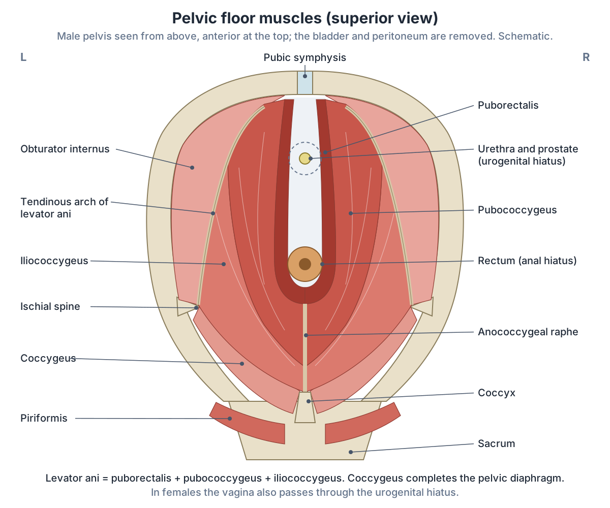
Pelvic floor muscles: origin, insertion, nerve and action
| Muscle | Origin | Insertion | Nerve | Main action |
|---|---|---|---|---|
| Puborectalis (levator ani) | Back of the body of the pubis | Sling behind the anorectal junction, joining the other side | Nerve to levator ani and pudendal nerve | Holds the anorectal angle; relaxes for defecation |
| Pubococcygeus (levator ani) | Back of the pubis, lateral to puborectalis; front of the tendinous arch | Anococcygeal ligament, coccyx, perineal body, prostate or vagina | Nerve to levator ani and pudendal branches | Supports the pelvic organs; forms the bulk of levator ani |
| Iliococcygeus (levator ani) | Tendinous arch of the obturator internus fascia, ischial spine | Anococcygeal ligament (raphe) and coccyx | Nerve to levator ani (S4, or S3 to S4) | Raises the pelvic floor and anorectal canal |
| Coccygeus (ischiococcygeus) | Ischial spine | Lower sacrum and coccyx | Anterior rami of S4 to S5 (Texas Tech: S3 to S4) | Supports the pelvic organs; flexes the coccyx |
Sources: TeachMeAnatomy, Kenhub, Radiopaedia and Texas Tech anatomy tables. Root values differ between sources; both are shown.
The perineal body (central tendon of the perineum)
The perineal body is an irregular fibromuscular mass in the midline, just under the skin where the urogenital and anal triangles meet. It contains skeletal muscle, smooth muscle and collagen and elastic fibres, and it anchors levator ani, bulbospongiosus, the superficial and deep transverse perineal muscles, the external anal sphincter and fibres of the external urethral sphincter. In women it acts as a tear-resistant body between the vagina and the external anal sphincter and supports the back wall of the vagina against prolapse. (TeachMeAnatomy: The Perineum.)
It lies between the anal canal and the vagina in females, or the bulb of the penis in males. It has three layers: superficial fibres of the external anal sphincter that continue forwards into bulbospongiosus, the superficial transverse perineal muscle in the middle layer, and the deep transverse perineal muscle in the deepest layer. Whether puborectalis truly joins it is disputed. In young women it is about twice the size of the male perineal body. (Radiopaedia: Perineal body.)
Above, the perineal body continues as the rectovaginal septum in females or the rectoprostatic septum in males. Damage to it from childbirth, trauma or surgery may contribute to pelvic organ prolapse, urinary or faecal incontinence and sexual dysfunction. (Kenhub: Perineal region.)
Layers of the perineum: perineal membrane and the deep and superficial pouches
The perineal membrane is a thin, triangular, horizontal layer of dense fascia in the urogenital triangle. Its sides attach to the lower edges of the ischiopubic rami, and its thickened back edge forms the transverse perineal ligament, which holds the perineal body in the midline. In males the membranous urethra and the ducts of the bulbourethral glands pass through it; in females the urethra and vagina do. (Radiopaedia: Perineal membrane.)
Above the membrane is the deep perineal pouch, a potential space below the fascia of the pelvic floor. It contains part of the urethra and the external urethral sphincter, the vagina in females, and the bulbourethral glands and deep transverse perineal muscles in males. Below the membrane is the superficial perineal pouch, bounded by the superficial perineal fascia and closed behind at the perineal body. It holds the erectile tissue of the penis or clitoris, the ischiocavernosus, bulbospongiosus and superficial transverse perineal muscles, and in females the greater vestibular (Bartholin) glands. (TeachMeAnatomy: The Perineum.)
Both pouches also carry branches of the pudendal nerve and internal pudendal vessels. The deep layer of superficial fascia, Colles' fascia, continues from Scarpa's fascia of the abdominal wall, fuses with the ischiopubic rami and the perineal membrane, and in males continues into the scrotum around the dartos muscle. (Kenhub: Perineal region.)
Older textbooks describe a urogenital diaphragm made simply of the deep transverse perineal muscles. A 2014 review of cadaver histology argues that this region is made of several fibrous structures rather than a simple muscle sheet, though it does not reject the concept completely. (Hinata N, Murakami G. The urethral rhabdosphincter, levator ani muscle, and perineal membrane: a review. Biomed Res Int. 2014.)
The root of the penis, bulbospongiosus and ischiocavernosus in the superficial pouch. Male reproductive system.
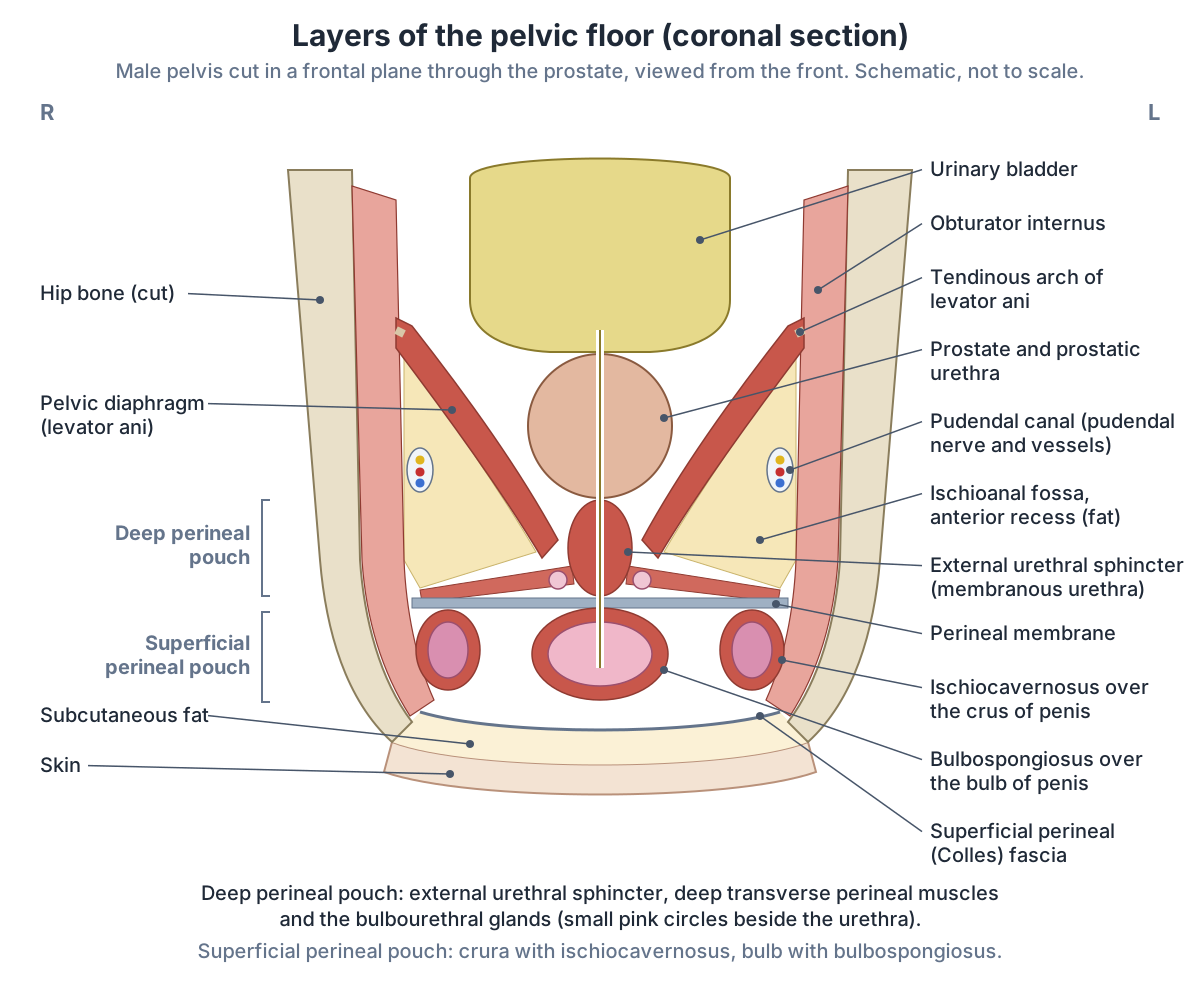
Contents of the perineal pouches in males and females
| Layer | Males | Females |
|---|---|---|
| Pelvic diaphragm (above) | Levator ani and coccygeus around the urethra and anal canal | Levator ani and coccygeus around the urethra, vagina and anal canal |
| Deep perineal pouch | Membranous urethra, external urethral sphincter, deep transverse perineal muscles, bulbourethral glands | Urethra, external urethral sphincter (with compressor urethrae and urethrovaginal sphincter), part of the vagina |
| Perineal membrane | Pierced by the membranous urethra and bulbourethral ducts | Pierced by the urethra and vagina |
| Superficial perineal pouch | Crura and bulb of the penis, ischiocavernosus, bulbospongiosus, superficial transverse perineal muscles | Crura of the clitoris, bulbs of the vestibule, greater vestibular glands, ischiocavernosus, bulbospongiosus, superficial transverse perineal muscles |
| Superficial fascia | Colles' fascia, continuing into the scrotum | Colles' fascia |
Sources: TeachMeAnatomy (perineum), Kenhub (perineal region), OpenStax 11.4 and Radiopaedia (perineal membrane).
Perineal muscles: bulbospongiosus, ischiocavernosus and the transverse perineal muscles
Below the pelvic diaphragm, the perineum has superficial and deep muscle layers. Bulbospongiosus compresses the urethra at the end of urination in both sexes and during ejaculation in males, and helps erection; ischiocavernosus compresses veins to maintain erection of the penis or clitoris; the superficial transverse perineal muscle supports the perineal body; the external urethral sphincter compresses the urethra voluntarily; and the external anal sphincter closes the anus. OpenStax lists the compressor urethrae and sphincter urethrovaginalis as female muscles and the deep transverse perineal muscle as a male muscle. (OpenStax Anatomy and Physiology 2e, 11.4: Axial Muscles of the Abdominal Wall and Thorax (muscles of the pelvic floor and perineum).)
Ischiocavernosus arises from the ischial tuberosity and ischiopubic ramus and covers the crus of the penis or clitoris. Bulbospongiosus arises from the perineal body (and the midline raphe of the penile bulb in males) and inserts into the perineal membrane and the erectile tissue. The superficial and deep transverse perineal muscles run from the ischial ramus to the perineal body, separated by the perineal membrane, and both steady the perineal body. (Texas Tech University HSC El Paso, Anatomy Tables: Muscles of the Pelvis and Perineum.)
The perineal nerve supplies the transverse perineal muscles, bulbospongiosus and ischiocavernosus, and the inferior rectal nerve supplies the external anal sphincter. Both are branches of the pudendal nerve. (Kenhub: Perineal region.)
Perineal muscles at a glance
| Muscle | Attachments | Nerve | Main action |
|---|---|---|---|
| Bulbospongiosus | Perineal body and midline raphe to the perineal membrane and erectile tissue | Perineal nerve (pudendal) | Empties the urethra; helps erection; narrows the vaginal orifice in females |
| Ischiocavernosus | Ischial tuberosity and ischiopubic ramus to the crus of the penis or clitoris | Perineal nerve (pudendal) | Compresses the crus to maintain erection |
| Superficial transverse perineal | Ischial ramus to the perineal body | Perineal nerve (pudendal) | Steadies the perineal body |
| Deep transverse perineal | Ischial ramus to the perineal body, above the perineal membrane | Perineal nerve (pudendal) | Steadies the perineal body |
| External urethral sphincter | Around the membranous urethra (males) or urethra and vagina (females) | Perineal branches of the pudendal nerve | Voluntary closure of the urethra |
| External anal sphincter | Perineal body to the anococcygeal ligament, around the anal canal | Inferior rectal nerve (pudendal) | Voluntary closure of the anus |
Sources: Texas Tech anatomy tables, OpenStax 11.4, Kenhub (perineal region) and TeachMeAnatomy.
Anal triangle: anal canal, sphincters and ischioanal fossa
The anal canal is about 4 cm long and sits in the anal triangle between the right and left ischioanal fossae. The internal anal sphincter, a thickening of the circular smooth muscle of the gut wall, surrounds the upper two-thirds and is involuntary. The external anal sphincter, voluntary skeletal muscle, surrounds the lower two-thirds and blends above with puborectalis. Together with puborectalis they form the anorectal ring, which can be felt on rectal examination. (TeachMeAnatomy: The Anal Canal.)
Each ischioanal fossa is a fat-filled space beside the anal canal that lets the canal expand during defecation. It extends from the skin up to the pelvic diaphragm. (TeachMeAnatomy: The Perineum.)
The medial wall of the fossa is the external anal sphincter and the lower part of levator ani, the lateral wall is obturator internus with its fascia, and the base is the skin. In females the anal triangle is generally wider because the pelvic outlet is broader. (Kenhub: Perineal region.)
Below the pectinate line, the anal canal is supplied by the inferior rectal nerves, branches of the pudendal nerve, and is sensitive to pain, temperature, touch and pressure; its lymph drains to the superficial inguinal nodes in the groin. (TeachMeAnatomy: The Anal Canal.)
The last part of the large intestine, which becomes the anal canal at the pelvic floor. Rectum.
Nerve and blood supply: the pudendal nerve
The pudendal nerve arises from the ventral rami of S2, S3 and S4. It leaves the pelvis through the greater sciatic foramen, crosses the sacrospinous ligament close to the ischial spine, re-enters through the lesser sciatic foramen and runs forwards with the internal pudendal artery and vein in the pudendal (Alcock's) canal, a tunnel in the obturator internus fascia. In the canal it gives off the inferior rectal nerve and the perineal nerve and continues as the dorsal nerve of the penis or clitoris. (TeachMeAnatomy: The Pudendal Nerve.)
It supplies bulbospongiosus, ischiocavernosus, the external urethral and anal sphincters and part of levator ani, and carries sensation from the external genitalia and the skin of the perineum and anus; the mnemonic is 'S2, S3, S4 keeps the poo off the floor'. A pudendal nerve block injects local anaesthetic around the ischial spine before some vaginal births; the needle is aspirated first because the internal pudendal artery runs beside the nerve. (TeachMeAnatomy: The Pudendal Nerve.)
Sources disagree on the nerve supply of levator ani. Kenhub gives the main supply as the nerve to levator ani from S4, with a small contribution from the pudendal nerve (S2 to S4). (Kenhub: Levator ani muscle.)
Radiopaedia gives branches of S3 and S4 to the upper (pelvic) surface of levator ani and pudendal branches to its lower (perineal) surface. (Radiopaedia: Levator ani (pelvic floor).)
Texas Tech lists ventral rami of S3 and S4 for both levator ani and coccygeus, whereas TeachMeAnatomy and Kenhub give anterior rami of S4 and S5 for coccygeus. (Texas Tech University HSC El Paso, Anatomy Tables: Muscles of the Pelvis and Perineum.)
The internal pudendal artery, a branch of the internal iliac artery, enters the perineum through the pudendal canal. It gives the perineal artery to the superficial structures and the inferior rectal artery to the anal canal and external anal sphincter, and ends as the dorsal and deep (cavernosal) arteries of the penis or clitoris. The internal pudendal vein drains to the internal iliac vein. (Kenhub: Perineal region.)
What does the pelvic floor do?
The pelvic floor does three things. Its constant tone supports the abdominal and pelvic organs; it resists rises in pressure inside the abdomen and pelvis, such as in coughing or heavy lifting; and its fibres act as sphincters on the rectum and urethra, then relax to allow urination and defecation. (TeachMeAnatomy: The Pelvic Floor.)
Most pelvic floor fibres are type I (slow-twitch). The muscles are contracted unconsciously at rest and tighten further when you cough, sneeze or lift, and during childbirth they support the presenting part of the baby. (Kenhub: Muscles of the pelvic floor.)
Levator ani resists the pressure created when the abdominal muscles contract, which directs that pressure towards the colon during defecation and towards the uterus during childbirth. (OpenStax Anatomy and Physiology 2e, 11.4: Axial Muscles of the Abdominal Wall and Thorax (muscles of the pelvic floor and perineum).)
Cleveland Clinic adds sexual function (vaginal contractions, erection and ejaculation) and describes the pelvic floor as the base of the core, working with the abdominal muscles, back muscles and diaphragm. It counts 14 pelvic floor muscles in all, with levator ani forming the bulk. (Cleveland Clinic: Pelvic Floor Muscles.)
The organ the pelvic floor supports and helps keep closed. Urinary bladder.
Male vs female pelvic floor and perineum
The pelvic floor has two openings in males (urethra and anal canal) and three in females, which adds the vagina. The medial fibres of pubococcygeus are called puboprostaticus in males, where they surround the prostate, and pubovaginalis in females, where they run beside the back wall of the vagina. Pelvic floor disorders are more common in females. (Kenhub: Muscles of the pelvic floor.)
In females the pelvic floor also supports the vagina and uterus; in males it holds the prostate in place. (Cleveland Clinic: Pelvic Floor Muscles.)
In the urogenital triangle the same muscles are arranged around different structures. Males have the crura and bulb of the penis and the bulbourethral glands; females have the crura of the clitoris, the bulbs of the vestibule around the vaginal orifice and the greater vestibular glands. Colles' fascia continues into the scrotum only in males. (Kenhub: Perineal region.)
The external urethral sphincter differs too. In males it is omega-shaped and sits between the two slings of levator ani, joined to them by fascia rich in elastic fibres, and the perineal membrane is thin but solid. In females it is semicircular, the vagina lies between it and levator ani, and the perineal membrane is thick and multilayered. (Hinata N, Murakami G. The urethral rhabdosphincter, levator ani muscle, and perineal membrane: a review. Biomed Res Int. 2014.)
The vagina, uterus and vulva that the female pelvic floor supports. Female reproductive system.
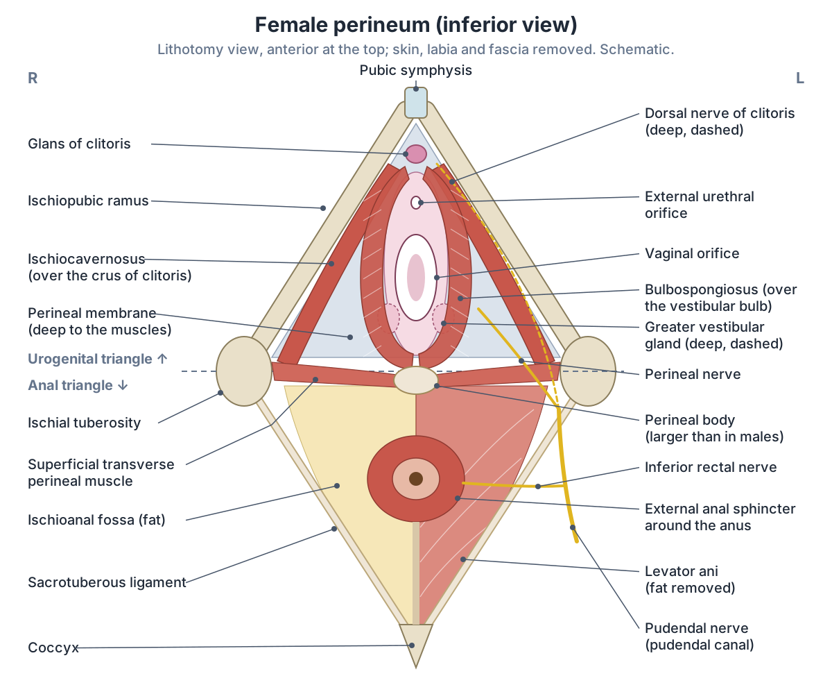
Pelvic floor problems: incontinence, prolapse and childbirth tears
In a national US survey of 1,961 women, 23.7% had at least one symptomatic pelvic floor disorder: 15.7% urinary incontinence, 9.0% faecal incontinence and 2.9% pelvic organ prolapse. The share rose from 9.7% at ages 20 to 39 to 49.7% at 80 and over, and from 12.8% in women who had not given birth to 32.4% in those with three or more deliveries. (Nygaard I et al. Prevalence of symptomatic pelvic floor disorders in US women. JAMA. 2008.)
Weak pelvic floor muscles remove support from the urethra, vagina and anal canal and can lead to incontinence, prolapse, pelvic pain and sexual dysfunction. Causes include obstetric trauma, ageing, obesity and chronic straining. (TeachMeAnatomy: The Pelvic Floor.)
Pelvic organ prolapse is when the womb, bladder or bowel bulges into the vagina. It is common in women over 50, and risk rises with age (especially after the menopause), childbirth, excess weight, long-term constipation, persistent coughing and pelvic surgery such as hysterectomy. (NHS: Pelvic organ prolapse.)
Childbirth is the most common cause of perineal injury: Cleveland Clinic states that about 85% of people who give birth vaginally have a perineal tear. Deeper tears can damage the pelvic floor muscles or the anal sphincter. (Cleveland Clinic: Perineum.)
The NHS gives a figure for first births: up to 9 in 10 first-time mothers who have a vaginal birth have some tear, graze or episiotomy. An episiotomy is a cut in the perineum; when possible it is made as a small diagonal cut from the back of the vagina, directed down and out to one side. (NHS: Episiotomy and perineal tears.)
The mediolateral direction keeps the cut away from the external anal sphincter. In males, a tear of the urethra below the perineal membrane (for example from a straddle injury) lets urine leak into the superficial perineal pouch; Colles' fascia limits its spread sideways and backwards but lets it track into the scrotum, penis and lower abdominal wall. (Kenhub: Perineal region.)
Raised pressure from pregnancy, obesity or a chronic cough can weaken the pelvic floor over time. About 6% of people have intermittent painful spasms of levator ani (levator ani syndrome), whose cause is unclear. (Kenhub: Levator ani muscle.)
Kenhub describes the classic pelvic floor (Kegel) exercise as squeezing the muscles for about 5 seconds and relaxing for 5, repeated 4 to 5 times up to three times a day, and advises against regularly stopping the urine stream as an exercise. This is educational information, not a treatment plan. (Kenhub: Muscles of the pelvic floor.)
Pelvic floor and perineum in the 3D model
- Coccygeus
- A small muscle of the posterior pelvic floor.
- External anal sphincter
- A ring of voluntary skeletal muscle surrounding the anal canal.
- Iliococcygeus
- The posterior part of levator ani in the pelvic floor.
- Obturator internus
- A lateral rotator of the hip that arises inside the pelvis and leaves through the lesser sciatic foramen.
- Prostate
- The prostate is a walnut-sized gland surrounding the first part of the male urethra.
- Pubococcygeus
- A part of levator ani forming the main muscular floor of the pelvis.
- Puborectalis
- The most medial part of levator ani, forming a sling around the anorectal junction.
- Rectum
- The rectum is the final straight portion of the large intestine before the anal canal.
- Tendinous arch of levator ani
- A fibrous thickening of the obturator fascia from which part of levator ani arises.
- Urethra
- The urethra is the tube carrying urine out of the bladder; in males it also carries semen.
Frequently asked questions
What is the medical name for the taint?
The medical name is the perineum. In everyday speech it means the strip of skin between the genitals and the anus; in anatomy the perineum is the whole diamond-shaped region below the pelvic floor, from the pubic symphysis to the coccyx and out to the ischial tuberosities. It contains muscles, the perineal body and branches of the pudendal nerve.
What muscles make up the pelvic floor?
The pelvic floor (pelvic diaphragm) is formed by levator ani and coccygeus, with their fascia. Levator ani has three parts on each side: puborectalis, a sling around the anorectal junction; pubococcygeus, the bulk of the muscle; and iliococcygeus, the thin back part. Coccygeus runs from the ischial spine to the sacrum and coccyx. Smaller perineal muscles lie below them.
Do men have a pelvic floor?
Yes. Males have the same levator ani and coccygeus muscles as females, but the pelvic floor has two openings in males (urethra and anal canal) instead of three. In males its medial fibres surround the prostate and it helps with urinary control, erection and ejaculation. Weakness can follow prostate surgery.
What are the two triangles of the perineum?
A line between the two ischial tuberosities divides the perineum into the urogenital triangle in front and the anal triangle behind. The urogenital triangle holds the external genitalia, the urethral (and vaginal) openings and the perineal membrane. The anal triangle holds the anal canal, the external anal sphincter and the fat-filled ischioanal fossae.
What is the perineal body?
The perineal body, or central tendon of the perineum, is a fibromuscular knot in the midline between the anal canal and the vagina or the bulb of the penis. Levator ani, bulbospongiosus, the transverse perineal muscles and the external anal sphincter attach to it. It is larger in women and can be torn in childbirth, which weakens support for the vagina.
Which nerve supplies the perineum?
The pudendal nerve (S2 to S4) supplies most of the perineum. It runs in the pudendal canal on the side wall of the ischioanal fossa and divides into the inferior rectal nerve (external anal sphincter and skin around the anus), the perineal nerve (perineal muscles and skin) and the dorsal nerve of the penis or clitoris. Levator ani also receives direct sacral branches.
What is the difference between the pelvic floor and the perineum?
The pelvic floor is the muscular sheet (levator ani and coccygeus) that closes the bottom of the pelvic cavity. The perineum is the region below it, between the thighs, which contains the perineal membrane, the perineal pouches with the external genital muscles, the perineal body, the anal canal and the ischioanal fossae. The pelvic floor forms the roof of the perineum.
References
- OpenStax. Anatomy and Physiology 2e, §11.4 Axial Muscles of the Abdominal Wall and Thorax (Muscles of the Pelvic Floor and Perineum). Rice University, 2022.
- TeachMeAnatomy. The Pelvic Floor.
- TeachMeAnatomy. The Perineum.
- TeachMeAnatomy. The Pudendal Nerve.
- TeachMeAnatomy. The Anal Canal.
- Kenhub. Muscles of the pelvic floor.
- Kenhub. Levator ani muscle.
- Kenhub. Perineal region.
- Radiopaedia. Levator ani (pelvic floor).
- Radiopaedia. Perineal body.
- Radiopaedia. Perineal membrane.
- Texas Tech University Health Sciences Center El Paso. Anatomy Tables: Muscles of the Pelvis and Perineum.
- Cleveland Clinic. Pelvic Floor Muscles.
- Cleveland Clinic. Perineum.
- NHS. Pelvic organ prolapse.
- NHS. Episiotomy and perineal tears.
- Nygaard I, Barber MD, Burgio KL, et al. Prevalence of symptomatic pelvic floor disorders in US women. JAMA. 2008;300(11):1311-1316.
- Hinata N, Murakami G. The urethral rhabdosphincter, levator ani muscle, and perineal membrane: a review. Biomed Res Int. 2014;2014:906921.
- Muro S, Moue S, Akita K. Twisted orientation of the muscle bundles in the levator ani functional parts in women: implications for pelvic floor support mechanism. J Anat. 2024;244(3):486-496.
- Mitsuhashi N. et al. (2009). BodyParts3D: 3D structure database for anatomical concepts. Nucleic Acids Research 37, D782–D785. Data © The Database Center for Life Science, CC BY 4.0.
Related anatomy
- PelvisPelvis and hip bone anatomy in 3D: ilium, ischium and pubis, ASIS and PSIS, ligaments, female pelvic anatomy and a…
- SacrumSacrum anatomy explained: promontory, ala, sacral foramina, crests and hiatus, joints and ligaments, sacrum vs coccyx…
- Male reproductive systemMale reproductive anatomy with labelled diagrams: scrotum, testes, epididymis, ductus deferens, seminal gland…
- Female reproductive systemLabelled female reproductive system diagrams: uterus anatomy and layers, uterine tube parts, ovaries, ligaments…
- Urinary bladderBladder anatomy with a labelled diagram and 3D model: apex, body, fundus, neck and trigone, detrusor and sphincters…
- GroinGroin anatomy with labelled diagrams and a 3D model: inguinal ligament and canal, femoral triangle, groin muscles…
Explore the whole body in the interactive 3D human anatomy model, or browse the A–Z list of human structures.