

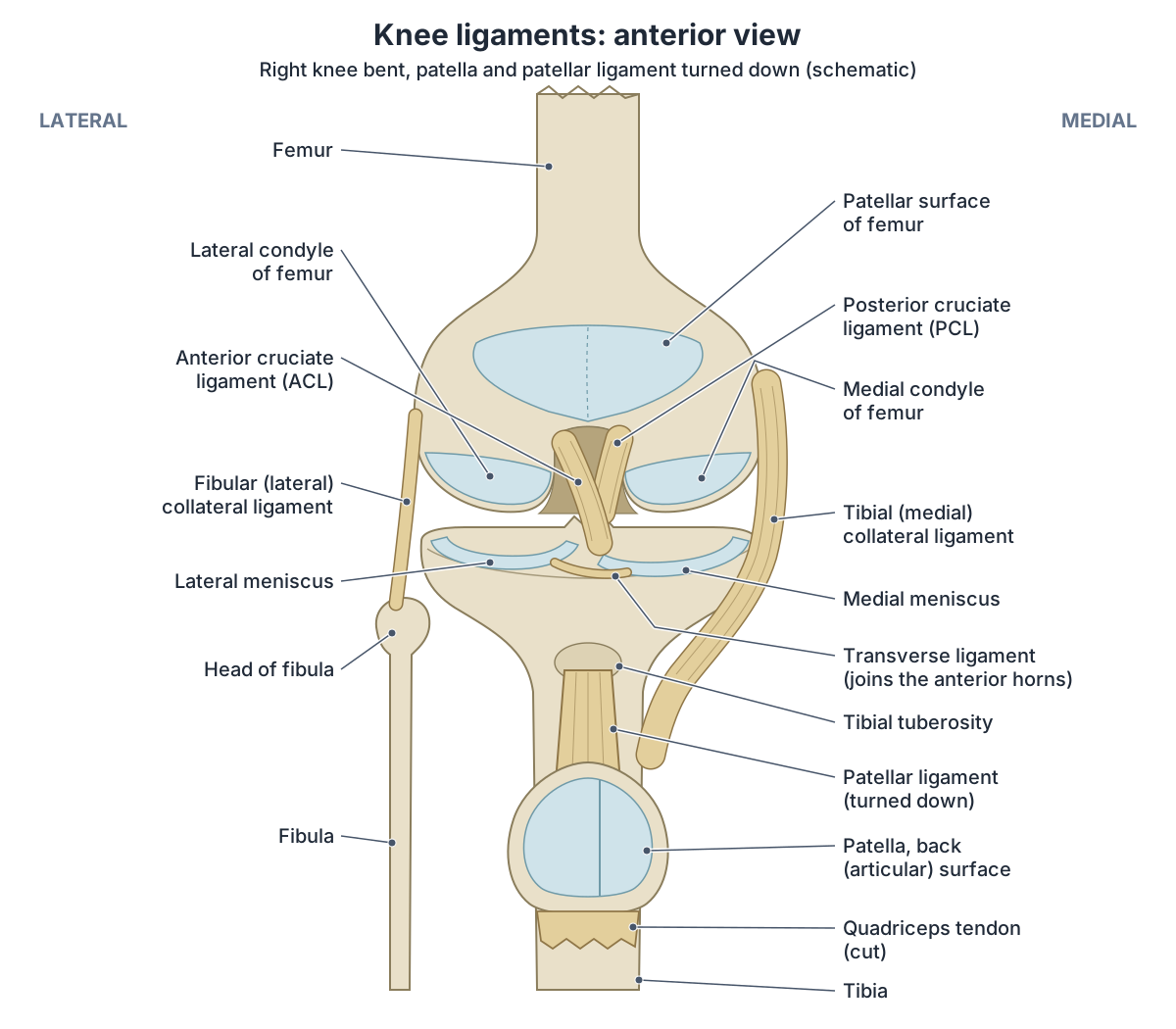
How many ligaments are in the knee?
The usual answer is four main ligaments. Cleveland Clinic describes two collateral ligaments on the sides of the knee and two cruciate ligaments inside the joint, where the PCL crosses behind the ACL to form an X. It describes knee ligaments as bands of strong connective tissue containing collagen and some slightly stretchy elastic fibres. (Cleveland Clinic: Knee Ligaments.)
OpenStax groups the four by position. The fibular (lateral) and tibial (medial) collateral ligaments lie outside the fibrous joint capsule, so they are extrinsic or extracapsular. The anterior and posterior cruciate ligaments lie inside it, so they are intracapsular. (OpenStax Anatomy and Physiology 2e, 9.6: Anatomy of Selected Synovial Joints.)
Anatomy references list many more. Kenhub's extracapsular list has the patellar ligament, the medial and lateral patellar retinacula, the two collateral ligaments, the oblique and arcuate popliteal ligaments and the anterolateral ligament. Inside the capsule it puts the two cruciate ligaments together with the menisci, and it adds the transverse ligament, the meniscofemoral and meniscotibial (coronary) ligaments, the medial and lateral patellomeniscal ligaments, the ligamentum mucosum and the popliteofibular and fabellofibular ligaments. (Kenhub: Knee joint.)
Totals differ partly because some 'ligaments' are thickenings of the joint capsule rather than separate cords. The deep part of the medial collateral ligament is a condensation of the capsule that ties the middle of the medial meniscus to the femur, and the coronary ligament is the part of the capsule that ties the edge of each meniscus to the tibia. (Fox AJS, Bedi A, Rodeo SA. The Basic Science of Human Knee Menisci. Sports Health. 2012.)
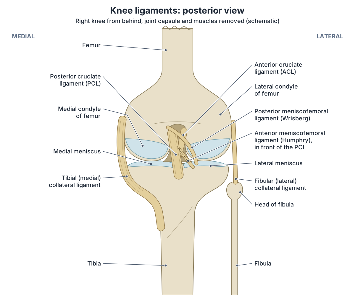
Other ligaments are not present in everyone. The meniscofemoral ligament splits around the PCL into an anterior band (of Humphry) and a posterior band (of Wrisberg); Radiopaedia gives about 80% of people at least one band, with the posterior band the more common. (Radiopaedia: Meniscofemoral ligament.)
A 2021 review puts the share of knees with at least one meniscofemoral ligament higher, at more than 90%, and agrees that the posterior band is the more common. Either way, a count that includes both bands will not fit every knee. (Winkler PW, et al. Evolving evidence in the treatment of posterior cruciate ligament injuries, part 1: anatomy, biomechanics and diagnostics. KSSTA. 2021.)
The patellar ligament is a special case: it runs from the patella to the tibia, but it is the continuation of the quadriceps tendon below the kneecap, so some lists count it as a tendon rather than a ligament. (Radiopaedia: Patellar tendon.)
No source gives one agreed total; counts run from about 10 to more than 15 depending on the list. For example, adding these bands from Kenhub's list to the four main ligaments gives 11: the patellar ligament, the oblique and arcuate popliteal ligaments, the transverse ligament, the two meniscofemoral ligaments and the coronary ligaments, counting the coronary ligament of each meniscus together as one. Counting the medial and lateral coronary ligaments separately gives 12, and adding the popliteofibular, fabellofibular, anterolateral and patellomeniscal ligaments and the patellar retinacula takes the total past 15. (Kenhub: Knee joint.)
The whole knee joint: bones, menisci, muscles, bursae and the back of the knee. Knee anatomy.
Ligaments of the knee: the full list
| Ligament | Where it is | What kind of ligament | In the 3D model? |
|---|---|---|---|
| Anterior cruciate ligament (ACL) | Centre of the joint, in the notch between the femoral condyles | Main ligament, inside the capsule | Yes |
| Posterior cruciate ligament (PCL) | Centre of the joint, behind the ACL | Main ligament, inside the capsule | Yes |
| Tibial (medial) collateral ligament (MCL) | Inner side | Main ligament, superficial and deep parts | Yes |
| Fibular (lateral) collateral ligament (LCL) | Outer side, to the head of the fibula | Main ligament, outside the capsule | Yes |
| Patellar ligament (patellar tendon) | Front, below the kneecap | Continuation of the quadriceps tendon | Yes |
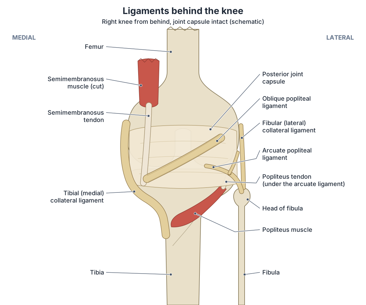
| Oblique popliteal ligament | Back, across the capsule | Expansion of the semimembranosus tendon | No (diagram) |
| Arcuate popliteal ligament | Back, outer corner | Thickening of the posterolateral capsule | No (diagram) |
| Transverse (intermeniscal) ligament | Front, between the menisci | Meniscal ligament | No (diagram) |
| Coronary (meniscotibial) ligaments | Rim of each meniscus to the tibia | Part of the capsule | No (diagram) |
| Anterior meniscofemoral ligament (Humphry) | Centre, in front of the PCL | Meniscal ligament, often absent | No (diagram) |
| Posterior meniscofemoral ligament (Wrisberg) | Centre, behind the PCL | Meniscal ligament, sometimes absent | No (diagram) |
| Posterior oblique ligament | Back of the inner side | Part of the MCL complex | No |
| Medial patellofemoral ligament | Inner side of the kneecap | Main medial stabiliser of the patella | No |
| Popliteofibular ligament | Back of the outer side | Posterolateral corner | No |
| Anterolateral ligament | Front of the outer side | Debated; deep to the iliotibial band | No (diagram) |
Compiled from Kenhub, Radiopaedia, Fox et al. (2012), Meyer et al. (2022), Aframian et al. (2017) and the ALL Expert Group (2017). Kenhub also lists the patellar retinacula, the medial and lateral patellomeniscal ligaments, the ligamentum mucosum and the fabellofibular ligament.
Knee ligaments in the 3D model
The 3D model above highlights, on both knees, the anterior and posterior cruciate ligaments, the tibial (medial) and fibular (lateral) collateral ligaments, the medial and lateral menisci and the patellar ligament, on a ghosted skeleton. These parts come from the Human Reference Atlas Visible Human Male model, fitted to the BodyParts3D skeleton. The smaller ligaments (the oblique and arcuate popliteal, meniscofemoral, transverse, coronary and anterolateral ligaments) are not in the model, so they are shown in the diagrams on this page. Click any highlighted part to open its own page.
Turn the model to look into the notch between the two femoral condyles. The ACL runs from the front of the tibia to the posterior part of the inner (medial) surface of the lateral femoral condyle, and the PCL runs from the back of the tibia to the front of the inner (lateral) surface of the medial condyle, which is why the two ligaments cross. (Kenhub: Knee joint.)
Use the model for position and relations rather than exact sizes. The BodyParts3D patella sits low, so the model's patellar ligament is shorter than in a real knee, where it is about 5 cm long, roughly the height of the patella. (Radiopaedia: Patellar tendon.)
The patellar ligament (patellar tendon) on its own page, from the apex of the patella to the tibial tuberosity. Patellar ligament.
The main knee ligaments: attachments, parts and job
| Ligament | Upper attachment | Lower attachment | Parts | What it resists |
|---|---|---|---|---|
| ACL | Posteromedial part of the lateral femoral condyle (inner wall of the notch) | Anteromedial intercondylar area of the tibia | Anteromedial and posterolateral bundles | Tibia sliding forward on the femur; hyperextension; rotational instability |
| PCL | Anterolateral surface of the medial femoral condyle (inner wall of the notch) | Posterior intercondylar area of the tibia, just below the joint line (the PCL facet) | Larger anterolateral and smaller posteromedial bundles | Tibia being pushed backward on the femur, especially with the knee bent |
| MCL (tibial collateral) | Medial femoral epicondyle | Upper medial tibia behind the pes anserinus; deep part to the medial meniscus and tibial rim | Superficial MCL, deep MCL; with the posterior oblique ligament forms the MCL complex | Valgus force (knee pushed inward); rotation |
| LCL (fibular collateral) | Just behind and above the lateral femoral epicondyle | Lateral side of the head of the fibula | Single cord; not attached to the lateral meniscus | Varus force (knee bowed outward) |
| Patellar ligament | Apex of the patella | Tibial tuberosity | Continuation of the quadriceps tendon | Carries the pull of quadriceps that straightens the knee |
From Radiopaedia, Kenhub, OpenStax, TeachMeAnatomy, Banovetz et al. (2023), Winkler et al. (2021) and Meyer et al. (2022).
ACL and PCL: the cruciate ligaments inside the knee
The anterior cruciate ligament (ACL) arises from the anteromedial part of the intercondylar area of the tibia and runs upward and backward to the posteromedial part of the lateral femoral condyle. Radiopaedia gives it a length of 31 to 38 mm. It lies inside the joint capsule but outside the synovial membrane that lines the joint (intracapsular but extrasynovial), and its tibial footprint overlaps the attachment of the front of the lateral meniscus. (Radiopaedia: Anterior cruciate ligament.)
Measured ACL lengths depend on the person: a 2023 review gives a range of 26 to 38 mm across studies. (Runer A, et al. Current trends in graft choice for anterior cruciate ligament reconstruction, part I: anatomy, biomechanics, graft incorporation and fixation. J Exp Orthop. 2023.)
There is wide agreement that the ACL has two bundles, named after where they attach on the tibia: the anteromedial (AM) and posterolateral (PL) bundles. On its way from femur to tibia the ACL passes in front of the PCL, and its main blood supply is the middle genicular artery, a branch of the popliteal artery. (Banovetz MT, et al. Anatomy of the anterior cruciate ligament and the common autograft specimens for ACL reconstruction. Ann Jt. 2023.)
The ACL stops the tibia sliding forward out from under the femur and also gives the knee rotational stability. It runs diagonally through the middle of the knee. (OrthoInfo (AAOS): Anterior Cruciate Ligament (ACL) Injuries.)
OpenStax adds that the ACL tightens as the knee straightens and so resists hyperextension. The posterior cruciate ligament (PCL) is the stronger of the two. It supports the knee when it is bent and carrying weight, as when walking downhill. (OpenStax Anatomy and Physiology 2e, 9.6: Anatomy of Selected Synovial Joints.)
The PCL arises from the anterolateral surface of the medial femoral condyle inside the notch and runs down and back to the posterior intercondylar area of the tibia, just below the joint line. Radiopaedia gives an average length of 38 mm and width of 13 mm, and describes a larger anterolateral bundle and a smaller posteromedial bundle. The posterior meniscofemoral ligament lies directly behind it. (Radiopaedia: Posterior cruciate ligament.)
A 2021 review places the centre of the PCL's tibial attachment slightly below the joint surface, in a shallow groove called the PCL facet between the two tibial condyles. It describes the anterolateral bundle as larger and stronger and the posteromedial bundle as weaker and more variable. The two bundles were long thought to work in turn, each taking over at a different angle of bending; newer studies find they share the load (codominance), and both matter more when the knee is bent than when it is straight. The PCL's main job is to stop the tibia being pushed backward on the femur. (Winkler PW, et al. Evolving evidence in the treatment of posterior cruciate ligament injuries, part 1: anatomy, biomechanics and diagnostics. KSSTA. 2021.)
The ACL on its own page, highlighted in the 3D model with its attachments and nearby structures. Anterior cruciate ligament (ACL).
What ligament is on the inside of your knee? The MCL and its neighbours
The ligament on the inside of the knee is the medial collateral ligament (MCL), also called the tibial collateral ligament. It is a wide, flat band that runs from the medial epicondyle of the femur to the medial condyle of the tibia. (TeachMeAnatomy: The Knee Joint.)
Radiopaedia describes the MCL as a flat, triangular band with superficial and deep parts. The superficial MCL starts on the medial femoral epicondyle and ends at two places on the upper medial tibia, the second just behind the attachment of the pes anserinus tendons. The deep MCL, also called the mid-third capsular ligament, is a thickening of the joint capsule with meniscofemoral and meniscotibial parts. The MCL resists valgus force, the force that pushes the knee inward. (Radiopaedia: Medial collateral ligament of the knee.)
The tibial collateral ligament is firmly attached on its deep side to the joint capsule and to the medial meniscus. Both collateral ligaments are taut when the knee is fully straight. (OpenStax Anatomy and Physiology 2e, 9.6: Anatomy of Selected Synovial Joints.)
A 2022 review describes an MCL complex with three parts: the superficial MCL, the deep MCL and the posterior oblique ligament. Together they steady the knee against valgus and rotational forces. (Meyer P, et al. Imaging of the medial collateral ligament of the knee: a systematic review. Arch Orthop Trauma Surg. 2022.)
The tendons of sartorius, gracilis and semitendinosus cross the inner knee on top of the superficial MCL and join to form the pes anserinus ('goose foot'). It inserts on the tibia above and in front of the MCL's tibial attachment, with sartorius uppermost, then gracilis, then semitendinosus. (Banovetz MT, et al. Anatomy of the anterior cruciate ligament and the common autograft specimens for ACL reconstruction. Ann Jt. 2023.)
Higher up, on the inner side of the kneecap, the medial patellofemoral ligament (MPFL) runs from an area between the adductor tubercle, the medial femoral epicondyle and the gastrocnemius tubercle to the upper inner edge of the patella. It is the major soft-tissue stabiliser on the inner side of the patella. (Aframian A, et al. Origin and insertion of the medial patellofemoral ligament: a systematic review of anatomy. KSSTA. 2017.)
The medial collateral ligament on its own page, with its attachments and the 3D model. Tibial collateral ligament (MCL).
Ligament on the outside of the knee: the LCL and the posterolateral corner
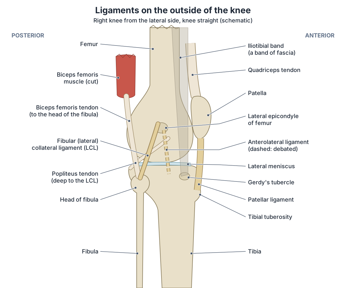
The main ligament on the outside of the knee is the lateral collateral ligament (LCL), also called the fibular collateral ligament. Radiopaedia places its upper end in a small hollow just behind and above the lateral femoral epicondyle and its lower end on the front and outer part of the head of the fibula. It is usually cord-like rather than flat, it is not attached to the joint capsule or to the lateral meniscus, and it is a major stabiliser against varus force, the force that bows the knee outward. (Radiopaedia: Lateral collateral ligament of the knee.)
The popliteus tendon runs deep to the LCL and separates it from the lateral meniscus, and at the fibular head the LCL splits the tendon of biceps femoris. (Kenhub: Knee joint.)
The LCL belongs to the posterolateral corner of the knee. A review of this region names the LCL, the popliteus tendon and the popliteofibular ligament as the three main stabilisers of the outer knee. The LCL resists varus force at every angle of bending and carries most load at about 30 degrees of flexion, when the other structures help least. (Crespo B, James EW, Metsavaht L, LaPrade RF. Injuries to posterolateral corner of the knee: a comprehensive review from anatomy to surgical treatment. Rev Bras Ortop. 2015.)
Authors differ on what else belongs to the posterolateral corner. A 2025 review lists the fibular collateral, fabellofibular, anterolateral, arcuate popliteal, popliteofibular, meniscofibular, meniscotibial and meniscofemoral ligaments and the PCL. Together these structures resist varus force, outward (external) rotation of the tibia and backward movement of the tibia. (Angelats X, et al. Anatomy of the ligaments of the posterolateral corner of the knee: a narrative literature review. J Exp Orthop. 2025.)
The lateral collateral ligament on its own page, from the femoral epicondyle to the head of the fibula. Fibular collateral ligament (LCL).
Ligaments behind the knee: PCL, oblique popliteal and arcuate popliteal ligaments
The deepest ligament behind the knee is the PCL, attached to the back of the tibia between the condyles. On the back of the joint capsule lie two flatter ligaments. The oblique popliteal ligament is an expansion of the semimembranosus tendon: it starts at the back of the medial tibial condyle and runs upward and laterally toward the lateral femoral condyle. The arcuate popliteal ligament arises from the back of the head of the fibula and arches upward and medially into the back of the capsule, reinforcing its outer corner. (Kenhub: Knee joint.)
Radiopaedia describes the oblique popliteal ligament as a broad, flat band lying on the back of the capsule and running obliquely upward and laterally. Medially it comes from the semimembranosus tendon, the back of the medial tibia and the capsule; laterally it blends with the capsule, the fabella when one is present, and the origins of the lateral head of gastrocnemius and plantaris. Its main job is to stop the knee overstraightening (hyperextension), and it may also limit outward rotation. (Radiopaedia: Oblique popliteal ligament.)
The arcuate popliteal ligament is a condensation of the posterolateral capsule of variable thickness, often Y-shaped. Its lower end attaches to the apex of the fibular head just behind the LCL. Some authors use 'arcuate ligament complex' for a whole group of posterolateral structures, which is a common source of confusion. (Angelats X, et al. Anatomy of the ligaments of the posterolateral corner of the knee: a narrative literature review. J Exp Orthop. 2025.)
Radiopaedia adds that the arcuate popliteal ligament arches over the popliteus tendon, and that the popliteofibular ligament runs from the popliteus tendon, near the junction of muscle and tendon, to the fibula. (Radiopaedia: Knee joint.)
The PCL on its own page, with the 3D model showing it behind the ACL in the femoral notch. Posterior cruciate ligament (PCL).
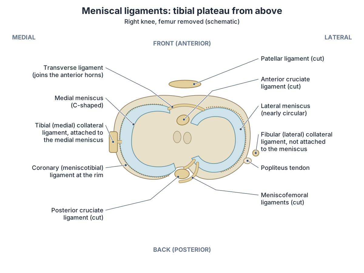
Meniscal ligaments: transverse, coronary and meniscofemoral ligaments
Each meniscus is anchored by its two ends (horns) to the tibia. Around its outer edge, the coronary ligament is the part of the joint capsule that attaches the meniscus to the tibia. The transverse or intermeniscal ligament joins the anterior horn of the medial meniscus to the anterior horn of the lateral meniscus. The lateral meniscus is not attached to the LCL. (Fox AJS, Bedi A, Rodeo SA. The Basic Science of Human Knee Menisci. Sports Health. 2012.)
Kenhub describes medial and lateral meniscotibial (coronary) ligaments running from the rim of each meniscus to the edge of the tibial condyles. It notes that the exact role of the transverse ligament is uncertain, although it may steady the menisci during movement. (Kenhub: Knee joint.)
The coronary ligaments matter in injury: in acute trauma they can tear, so that the meniscus lifts off the tibial plateau. Radiologists call this a floating meniscus. (Radiopaedia: Floating meniscus.)
The meniscofemoral ligaments run from the posterior horn of the lateral meniscus to the inner (lateral) side of the medial femoral condyle. The anterior meniscofemoral ligament (of Humphry) passes in front of the PCL and the posterior one (of Wrisberg) passes behind it. Radiopaedia gives the posterior ligament in 50 to 70% of people and the anterior one in 10 to 25%. (Radiopaedia: Meniscofemoral ligament.)
A 2021 meta-analysis of 41 studies (4,220 limbs) found the anterior meniscofemoral ligament in 55.5% of knees overall, in 60.0% of knees in cadaver studies but only 35.8% in MRI studies, so the method used changes the answer. (Pękala PA, et al. Clinical Anatomy of the Anterior Meniscofemoral Ligament of Humphrey: MRI Study, Meta-analysis, and Systematic Review. Orthop J Sports Med. 2021.)
A companion meta-analysis of 47 studies (4,940 limbs) found the posterior meniscofemoral ligament in 70.4% of knees, with a mean length of 27.7 mm. As in Radiopaedia's figures, the posterior band is the more common of the two. (Pękala PA, et al. Clinical Anatomy of the Posterior Meniscofemoral Ligament of Wrisberg: MRI Study, Meta-analysis, and Systematic Review. Orthop J Sports Med. 2021.)
The lateral meniscus also has a muscle attached to it: popliteus arises partly from the lateral meniscus as well as from the lateral femoral condyle. (TeachMeAnatomy: Muscles in the Posterior Compartment of the Leg.)
The C-shaped medial meniscus on its own page, with the 3D model and its attachments. Medial meniscus.
The anterolateral ligament of the knee: a debated ligament
The anterolateral ligament (ALL) lies on the outer front of the knee, in front of the LCL. The French surgeon Paul Segond reported a band here in 1879, and in 2013 Claes and colleagues described it in detail, finding a well-defined ligament in 40 of 41 cadaver knees (97%). A 2019 review (Ahn et al.) notes that other dissection studies using similar methods have found it in anywhere from 45.5% to 100% of knees, probably because there is no clear agreed way to tell it apart from the capsulo-osseous layer of the iliotibial band. (Ahn JH, Patel NA, Lin CC, Lee TQ. The anterolateral ligament of the knee joint: a review of the anatomy, biomechanics, and anterolateral ligament surgery. Knee Surg Relat Res. 2019.)
A 2025 review of the posterolateral corner gives an even wider range: anatomical studies have reported the ALL in anywhere from 0% to 100% of knees. (Angelats X, et al. Anatomy of the ligaments of the posterolateral corner of the knee: a narrative literature review. J Exp Orthop. 2025.)
In 2017 an expert group agreed that the ALL is a distinct ligament lying deep to the iliotibial band. It attaches on the femur just behind and above the lateral epicondyle, on the tibia between Gerdy's tubercle and the head of the fibula (about 4 to 10 mm below the joint line), and always to the lateral meniscus. It helps resist inward (internal) rotation of the tibia, and its injury is associated with the Segond fracture and often occurs together with an acute ACL injury. (Sonnery-Cottet B, et al. Anterolateral Ligament Expert Group consensus paper. J Orthop Traumatol. 2017.)
Not every study agrees on the femoral end. Radiopaedia, following the 2013 description, places the origin on the prominence of the lateral femoral epicondyle, slightly in front of the LCL, while agreeing on the tibial insertion between Gerdy's tubercle and the fibular head and the firm attachment to the lateral meniscus. (Radiopaedia: Anterolateral ligament of the knee.)
A 2013 dissection of 20 knees likewise found the ALL in every knee, with its femoral attachment a few millimetres below and in front of the LCL's, and two lower attachments: one to the lateral meniscus and one to the tibia between Gerdy's tubercle and the fibular head. (Helito CP, et al. Anatomy and Histology of the Knee Anterolateral Ligament. Orthop J Sports Med. 2013.)
Patellar ligament vs patellar tendon: is it a ligament or a tendon?
Both names describe the same strong band at the front of the knee. It begins at the apex (lowest point) of the patella and ends on the tibial tuberosity. Radiopaedia calls it the patellar tendon and notes that 'patellar ligament' is less correct, because the patella is a sesamoid bone embedded in the quadriceps tendon, so the band below it is really the lower part of that tendon. It is about 5 cm long and 20 to 30 mm wide, has no tendon sheath, and has the infrapatellar (Hoffa) fat pad behind it. (Radiopaedia: Patellar tendon.)
A 2023 anatomy review gives a similar length, 48.6 mm on average, and reports a mean of 4.8 cm (range 3 to 6 cm) in a study of 50 cadavers. Most of its fibres continue from the central fibres of rectus femoris. (Banovetz MT, et al. Anatomy of the anterior cruciate ligament and the common autograft specimens for ACL reconstruction. Ann Jt. 2023.)
OpenStax keeps the name patellar ligament and describes the band as continuing from the patella to the front of the tibia just below the knee, so it joins one bone to another, as ligaments do. (OpenStax Anatomy and Physiology 2e, 9.6: Anatomy of Selected Synovial Joints.)
The largest sesamoid bone: its surfaces, the tendons around it and how it tracks. Patella (kneecap).
Tendons and ligaments of the knee: quadriceps, hamstring and popliteus tendons and the IT band
Ligaments are strong bands of fibrous connective tissue that join bones and limit how far a joint can move; a tendon is the dense connective tissue that attaches a muscle to bone. Several tendons cross the knee and work alongside its ligaments. (OpenStax Anatomy and Physiology 2e, 9.4: Synovial Joints.)
At the front, the quadriceps tendon is made from all four parts of quadriceps femoris and classically has three layers: rectus femoris on the surface, vastus medialis and vastus lateralis in the middle, and vastus intermedius deepest. Below the patella it continues as the patellar tendon. (Radiopaedia: Quadriceps tendon.)
At the back and sides are the hamstring tendons. Biceps femoris, the outer hamstring, inserts into the head of the fibula; semitendinosus attaches to the medial surface of the tibia and semimembranosus to the medial tibial condyle. All three bend the knee, and they also rotate the bent knee: biceps femoris laterally, the other two medially. (TeachMeAnatomy: The Hamstrings.)
Popliteus is a small muscle at the back of the knee whose tendon starts on the lateral femoral condyle and lateral meniscus. Its body ends on the back of the upper tibia, above the origin of soleus. By rotating the femur laterally on the tibia it 'unlocks' the straightened knee so that bending can begin. The two heads of gastrocnemius also cross the knee, arising from the medial and lateral femoral condyles. (TeachMeAnatomy: Muscles in the Posterior Compartment of the Leg.)
The iliotibial band (IT band) runs down the outside of the thigh. It is a thick band of fascia, a thickening of the fascia lata, rather than a ligament. Its superficial layer acts as its tendon and inserts on Gerdy's tubercle on the front and outer side of the tibia, and its deep layer attaches to the lower femur. (Radiopaedia: Iliotibial band.)
At the knee, the iliotibial band lies superficial to the LCL, while the popliteus tendon passes deep to it. (Radiopaedia: Lateral collateral ligament of the knee.)
Quadriceps, hamstrings and adductors with origin, insertion, nerve and action tables. Thigh muscles.
Tendons around the knee
| Tendon or band | Muscle | Where it attaches at the knee | Side of the knee |
|---|---|---|---|
| Quadriceps tendon | Rectus femoris and the three vasti | Patella | Front, above the kneecap |
| Patellar tendon (patellar ligament) | Quadriceps, continued below the patella | Tibial tuberosity | Front, below the kneecap |
| Biceps femoris tendon | Biceps femoris (outer hamstring) | Head of the fibula, around the LCL | Back and outer side |
| Semimembranosus tendon | Semimembranosus | Medial tibial condyle; gives off the oblique popliteal ligament | Back and inner side |
| Pes anserinus | Sartorius, gracilis, semitendinosus | Upper medial tibia, in front of the MCL attachment | Inner side |
| Popliteus tendon | Popliteus | Lateral femoral condyle and lateral meniscus, deep to the LCL | Back and outer side |
| Gastrocnemius heads | Gastrocnemius | Medial and lateral femoral condyles | Back |
| Iliotibial band | Tensor fasciae latae and gluteus maximus, via the fascia lata | Gerdy's tubercle on the tibia | Outer side |
From Radiopaedia (quadriceps tendon, patellar tendon, iliotibial band, oblique popliteal ligament, LCL), TeachMeAnatomy (hamstrings, posterior leg), Kenhub and Banovetz et al. (2023).
Torn ligament in the knee: where it hurts and how it usually happens
An overstretched knee ligament is called a sprain and a torn one a rupture. Cleveland Clinic lists the usual ways it happens: a force on the back of the partly bent knee, a force on the front of the bent knee (often called a dashboard injury), a force on the side of the knee with the foot on the ground, the knee forced into hyperextension, or the knee twisting in an unnatural way. It names the ACL as the most commonly injured knee ligament. (Cleveland Clinic: Knee Ligaments.)
Sources rank the commonest injury differently. TeachMeAnatomy calls collateral ligament injury the most common problem of the knee joint, caused by a force to the side of the knee with the foot planted. (TeachMeAnatomy: The Knee Joint.)
A 2022 systematic review of MCL imaging goes further and calls the MCL complex the most commonly injured structure of the knee. (Meyer P, et al. Imaging of the medial collateral ligament of the knee: a systematic review. Arch Orthop Trauma Surg. 2022.)
ACL. Most ACL injuries happen without a blow to the knee: changing direction quickly, stopping suddenly, slowing down while running or landing badly from a jump. People often hear a pop and feel the knee give way. The ACL lies deep in the middle of the knee, and its injury is felt as pain with swelling, which develops within 24 hours, and tenderness along the joint line; the menisci and MCL are often injured at the same time. (OrthoInfo (AAOS): Anterior Cruciate Ligament (ACL) Injuries.)
A 2018 clinical review describes the usual pattern for each cruciate ligament. An ACL tear typically follows a sudden pivot, often with an audible pop and a feeling of instability, and the knee swells within one to two hours. The PCL lies at the back of the joint, and its injury, usually from a blow to the front of the shin or a sudden overbending or overstraightening of the knee, is felt as pain at the back of the knee, often worse when kneeling. (Bunt CW, Jonas CE, Chang JG. Knee Pain in Adults and Adolescents: The Initial Evaluation. Am Fam Physician. 2018.)
The swelling comes on quickly because a torn ACL bleeds into the joint (haemarthrosis), giving a large, tense swelling within about two hours. A meniscus injury or a ligament sprain usually causes a milder swelling that builds up over a day or more. (Calmbach WL, Hutchens M. Evaluation of Patients Presenting with Knee Pain: Part I. Am Fam Physician. 2003.)
MCL and LCL. The MCL is usually sprained by a valgus force, a blow or misstep that pushes the knee inward, and the tenderness is at the medial joint line on the inside of the knee. The LCL is sprained by a varus force and is tender at the lateral joint line on the outside. Pes anserine bursitis, a few centimetres below the joint line on the front of the inner tibia, where the sartorius, gracilis and semitendinosus tendons insert, is easily mistaken for an MCL sprain. (Calmbach WL, Hutchens M. Evaluation of Patients Presenting with Knee Pain: Part II. Differential Diagnosis. Am Fam Physician. 2003.)
OrthoInfo describes the same pattern: pain on the inside of the knee with an MCL injury and on the outside with an LCL injury, swelling over the injured ligament, sometimes a pop, and a feeling that the knee is giving out. A blow to the inner side of the knee is what usually tears the LCL. (OrthoInfo (AAOS): Collateral Ligament Injuries.)
A powerful blow to the outer side of the knee can injure three structures in sequence: the tibial collateral ligament, then the medial meniscus, then the ACL. This is the 'terrible' or 'unhappy' triad. (OpenStax Anatomy and Physiology 2e, 9.6: Anatomy of Selected Synovial Joints.)
Injuries to the posterolateral corner usually come from a blow to the front and inner side of the tibia, from hyperextension, or from the tibia twisting outward on a planted foot. They cause pain over the joint line, bruising and swelling, and most happen together with an ACL or PCL tear. (Crespo B, James EW, Metsavaht L, LaPrade RF. Injuries to posterolateral corner of the knee: a comprehensive review from anatomy to surgical treatment. Rev Bras Ortop. 2015.)
Cleveland Clinic advises seeing a healthcare provider after a knee injury with severe pain or swelling, a knee that is unstable or cannot be used normally, a numb foot, or a leg or foot that changes colour or feels cold. (Cleveland Clinic: PCL (Posterior Cruciate Ligament) Tears.)
This section explains anatomy, not how to diagnose a knee. The NHS advises getting urgent advice (from NHS 111 in the UK) if a knee is very painful, cannot move or take weight, is badly swollen or has changed shape, or locks or gives way. (NHS: Knee pain.)
Which knee structures lie at the front, sides and back, and the problems linked with each area. Knee anatomy and pain by location.
Where knee ligament injuries are usually felt
| Structure | Usual mechanism | Where it is felt | What is usually noticed |
|---|---|---|---|
| ACL | Sudden pivot, stop or change of direction; bad landing; usually no contact | Inside the knee: swelling of the joint and tenderness along the joint line | Often a pop and a feeling of giving way; the torn ligament bleeds into the joint, so the knee swells within an hour or two |
| PCL | Blow to the front of the shin with the knee bent (dashboard injury); fall on a bent knee; overbending or overstraightening | Back of the knee | Pain on kneeling; often torn together with other ligaments |
| MCL | Valgus force: blow to the outer side or a misstep pushing the knee inward | Inner side, at the medial joint line | Swelling over the ligament; because it is attached to the medial meniscus, the meniscus may be torn as well |
| LCL and posterolateral corner | Varus force or a blow to the inner side; hyperextension; twisting outward | Outer side, at the lateral joint line | Bruising and swelling on the outer side; often torn together with the ACL or PCL |
| Patellar tendon | Repeated running or jumping (jumper's knee) | Front, between the kneecap and the shin | Pain when quadriceps pulls on the tendon, as in straightening the knee against resistance |
From OrthoInfo, Cleveland Clinic, the NHS, TeachMeAnatomy, Crespo et al. (2015), Bunt et al. (2018) and Calmbach and Hutchens (2003). For learning only: a knee injury needs to be examined by a clinician.
Knee ligaments in the 3D model
- Anterior cruciate ligament
- The anterior cruciate ligament (ACL) is one of the two cruciate ligaments in the middle of the knee. It stops the tibia from sliding forward on the femur.
- Fibular collateral ligament
- The fibular collateral ligament, usually called the lateral collateral ligament (LCL), is the cord-like ligament on the outer side of the knee.
- Lateral meniscus
- The lateral meniscus is the smaller of the two knee menisci: a ring of fibrocartilage between the lateral condyles of the femur and tibia, closed off almost into a circle.
- Medial meniscus
- The medial meniscus is the larger, C-shaped (almost semicircular) ring of fibrocartilage between the medial condyles of the femur and tibia.
- Patellar ligament
- The patellar ligament (patellar tendon) is the strong band below the kneecap that carries the pull of the quadriceps muscle to the tibia.
- Posterior cruciate ligament
- The posterior cruciate ligament (PCL) is one of the two cruciate ligaments in the middle of the knee. It stops the tibia from being pushed backward on the femur.
- Tibial collateral ligament
- The tibial collateral ligament, usually called the medial collateral ligament (MCL), is the wide, flat ligament on the inner side of the knee.
Frequently asked questions
How many ligaments are in the knee?
The knee has four main ligaments: the anterior and posterior cruciate ligaments inside the joint and the medial and lateral collateral ligaments at its sides. Adding the smaller named bands gives totals from about 10 to more than 15, depending on the list. For example, the patellar ligament, the two popliteal ligaments, the transverse ligament, the two meniscofemoral ligaments and the coronary ligaments (counted as one) bring the total to 11.
What are the 4 main ligaments of the knee?
The four main knee ligaments are the anterior cruciate ligament (ACL), the posterior cruciate ligament (PCL), the medial (tibial) collateral ligament (MCL) and the lateral (fibular) collateral ligament (LCL). The ACL and PCL cross inside the joint and stop the tibia sliding forward or backward on the femur. The MCL and LCL run down the inner and outer sides and stop the knee bending sideways.
What ligament is on the inside of your knee?
The medial collateral ligament (MCL), also called the tibial collateral ligament, is the wide, flat ligament on the inside of the knee. It runs from the medial epicondyle of the femur to the upper medial tibia and is attached on its deep side to the medial meniscus. It resists forces that push the knee inward. The posterior oblique ligament and the medial patellofemoral ligament are smaller neighbours.
What ligament is on the outside of the knee?
The lateral collateral ligament (LCL), also called the fibular collateral ligament, is the cord-like ligament on the outside of the knee. It runs from just behind the lateral femoral epicondyle to the head of the fibula, is not attached to the lateral meniscus, and resists forces that bow the knee outward. The anterolateral ligament, the popliteofibular ligament and the iliotibial band lie nearby.
What ligament is behind the knee?
The posterior cruciate ligament (PCL) is the main ligament at the back of the knee, attached to the back of the tibia between its condyles. On the back of the joint capsule lie the oblique popliteal ligament, an expansion of the semimembranosus tendon, and the arcuate popliteal ligament from the head of the fibula. The meniscofemoral ligaments run beside the PCL.
Is the patellar tendon a ligament or a tendon?
It is both, depending on the point of view. The band runs from the patella to the tibial tuberosity, joining bone to bone like a ligament, so textbooks call it the patellar ligament. But the patella sits inside the quadriceps tendon, so the band is really the lower part of that tendon, and Radiopaedia prefers the name patellar tendon. It is about 5 cm long.
Which knee ligament is torn most often?
Sources disagree. Cleveland Clinic names the ACL as the most commonly injured knee ligament, while TeachMeAnatomy calls collateral ligament injury the most common knee joint problem and a 2022 review calls the MCL complex the most commonly injured structure of the knee. The PCL is stronger and is injured much less often than the ACL, usually together with other ligaments.
What is the anterolateral ligament of the knee?
The anterolateral ligament is a band on the outer front of the knee, deep to the iliotibial band, running from near the lateral femoral epicondyle to the tibia between Gerdy's tubercle and the head of the fibula, with an attachment to the lateral meniscus. It helps resist inward rotation of the tibia. Studies disagree on how often it is present and exactly where it starts on the femur.
References
- OpenStax. Anatomy and Physiology 2e, §9.6 Anatomy of Selected Synovial Joints. Rice University, 2022.
- OpenStax. Anatomy and Physiology 2e, §9.4 Synovial Joints. Rice University, 2022.
- TeachMeAnatomy. The Knee Joint.
- TeachMeAnatomy. The Hamstrings.
- TeachMeAnatomy. Muscles in the Posterior Compartment of the Leg.
- Kenhub. Knee joint.
- Kenhub. Anterior cruciate ligament.
- Kenhub. Posterior cruciate ligament.
- Radiopaedia. Anterior cruciate ligament.
- Radiopaedia. Posterior cruciate ligament.
- Radiopaedia. Medial collateral ligament of the knee.
- Radiopaedia. Lateral collateral ligament of the knee.
- Radiopaedia. Patellar tendon.
- Radiopaedia. Oblique popliteal ligament.
- Radiopaedia. Meniscofemoral ligament.
- Radiopaedia. Anterolateral ligament of the knee.
- Radiopaedia. Knee joint.
- Radiopaedia. Floating meniscus.
- Radiopaedia. Iliotibial band.
- Radiopaedia. Quadriceps tendon.
- Radiopaedia. Medial patellofemoral ligament.
- Cleveland Clinic. Knee Ligaments: What They Are, Anatomy and Function.
- Cleveland Clinic. PCL (Posterior Cruciate Ligament) Tears.
- American Academy of Orthopaedic Surgeons, OrthoInfo. Collateral Ligament Injuries.
- American Academy of Orthopaedic Surgeons, OrthoInfo. Anterior Cruciate Ligament (ACL) Injuries.
- Calmbach WL, Hutchens M. Evaluation of Patients Presenting with Knee Pain: Part I. History, Physical Examination, Radiographs, and Laboratory Tests. Am Fam Physician. 2003;68(5):907-912.
- Calmbach WL, Hutchens M. Evaluation of Patients Presenting with Knee Pain: Part II. Differential Diagnosis. Am Fam Physician. 2003;68(5):917-922.
- Bunt CW, Jonas CE, Chang JG. Knee Pain in Adults and Adolescents: The Initial Evaluation. Am Fam Physician. 2018;98(9):576-585.
- NHS. Knee pain.
- Fox AJS, Bedi A, Rodeo SA. The Basic Science of Human Knee Menisci: Structure, Composition, and Function. Sports Health. 2012;4(4):340-351. doi:10.1177/1941738111429419
- Winkler PW, Zsidai B, Wagala NN, et al. Evolving evidence in the treatment of primary and recurrent posterior cruciate ligament injuries, part 1: anatomy, biomechanics and diagnostics. Knee Surg Sports Traumatol Arthrosc. 2021;29(3):672-681. doi:10.1007/s00167-020-06357-y
- Pękala PA, Rosa MA, Łazarz DP, et al. Clinical Anatomy of the Anterior Meniscofemoral Ligament of Humphrey: An Original MRI Study, Meta-analysis, and Systematic Review. Orthop J Sports Med. 2021;9(2):2325967120973192. doi:10.1177/2325967120973192
- Pękala PA, Łazarz DP, Rosa MA, et al. Clinical Anatomy of the Posterior Meniscofemoral Ligament of Wrisberg: An Original MRI Study, Meta-analysis, and Systematic Review. Orthop J Sports Med. 2021;9(2):2325967120973195. doi:10.1177/2325967120973195
- Banovetz MT, Familiari F, Kennedy NI, et al. Anatomy of the anterior cruciate ligament and the common autograft specimens for anterior cruciate ligament reconstruction. Ann Jt. 2023;8:28. doi:10.21037/aoj-22-49
- Runer A, Keeling L, Wagala N, et al. Current trends in graft choice for anterior cruciate ligament reconstruction, part I: anatomy, biomechanics, graft incorporation and fixation. J Exp Orthop. 2023;10(1):37. doi:10.1186/s40634-023-00600-4
- Meyer P, Reiter A, Akoto R, et al. Imaging of the medial collateral ligament of the knee: a systematic review. Arch Orthop Trauma Surg. 2022;142(12):3721-3736. doi:10.1007/s00402-021-04200-8
- Crespo B, James EW, Metsavaht L, LaPrade RF. Injuries to posterolateral corner of the knee: a comprehensive review from anatomy to surgical treatment. Rev Bras Ortop. 2015;50(4):363-370. doi:10.1016/j.rboe.2014.12.008
- Angelats X, Carrera A, Tubbs RS, et al. Anatomy of the ligaments of the posterolateral corner of the knee: A narrative literature review. J Exp Orthop. 2025;12(4):e70562. doi:10.1002/jeo2.70562
- Ahn JH, Patel NA, Lin CC, Lee TQ. The anterolateral ligament of the knee joint: a review of the anatomy, biomechanics, and anterolateral ligament surgery. Knee Surg Relat Res. 2019;31(1):12. doi:10.1186/s43019-019-0012-4
- Helito CP, Demange MK, Bonadio MB, et al. Anatomy and Histology of the Knee Anterolateral Ligament. Orthop J Sports Med. 2013;1(7):2325967113513546. doi:10.1177/2325967113513546
- Sonnery-Cottet B, Daggett M, Fayard JM, et al. Anterolateral Ligament Expert Group consensus paper on the management of internal rotation and instability of the anterior cruciate ligament-deficient knee. J Orthop Traumatol. 2017;18(2):91-106. doi:10.1007/s10195-017-0449-8
- Aframian A, Smith TO, Tennent TD, Cobb JP, Hing CB. Origin and insertion of the medial patellofemoral ligament: a systematic review of anatomy. Knee Surg Sports Traumatol Arthrosc. 2017;25(12):3755-3772. doi:10.1007/s00167-016-4272-1
- Mitsuhashi N. et al. (2009). BodyParts3D: 3D structure database for anatomical concepts. Nucleic Acids Research 37, D782–D785. Data © The Database Center for Life Science, CC BY 4.0.
- Browne K., Schlehlein H. (2026). 3D Reference Organ Set for Male, v1.10 (Visible Human Male). Human Reference Atlas 3D Reference Object Library, HuBMAP / NIH. CC BY 4.0.
About this page
Drafted with AI assistance from the sources listed above, checked against them and reviewed by Dr Shaista Andleeb; last reviewed on 3 November 2026. The editorial policy explains how sources are chosen and how errors are corrected. To report a mistake, use the contact page.
Related anatomy
- Knee jointKnee anatomy with labelled diagrams and a 3D model: femur, tibia and patella, the ACL, PCL, MCL and LCL, menisci…
- Anterior cruciate ligamentAnterior cruciate ligament: The anterior cruciate ligament (ACL) is one of the two cruciate ligaments in the middle of…
- Posterior cruciate ligamentPosterior cruciate ligament: The posterior cruciate ligament (PCL) is one of the two cruciate ligaments in the middle…
- Tibial collateral ligamentTibial collateral ligament: The tibial collateral ligament, usually called the medial collateral ligament (MCL), is…
- Fibular collateral ligamentFibular collateral ligament: The fibular collateral ligament, usually called the lateral collateral ligament (LCL), is…
- Thigh musclesThigh muscles explained: quadriceps and sartorius at the front, adductor muscles of the inner thigh, hamstrings at the…
Explore the whole body in the interactive 3D human anatomy model, or browse the A–Z list of human structures.