

What are nails? Nails in anatomy
A nail is an accessory structure of the skin. The nail bed is a specialised region of the epidermis at the tips of the fingers and toes, and on it forms the nail body, made of densely packed dead keratinocytes. The nail protects the fingertips and toes, the parts of the limbs under the most mechanical stress, and gives a firm backing that helps with picking up small objects. (OpenStax Anatomy and Physiology 2e, 5.2: Accessory Structures of the Skin.)
Like hair, nails are skin appendages: they are made from skin cells. InformedHealth.org describes the nail plate as mostly keratin, about half a millimetre thick and slightly curved, and adds that nails are used as tools, for example for scratching, and contribute to the sense of touch. (InformedHealth.org (IQWiG): Structure of the nails.)
Nails form before birth. In OpenStax's timeline of fetal development the fingers and toes begin to develop nails in weeks 9 to 12, and in the last weeks of pregnancy, from about week 31, the nails grow to the tips of the fingers and toes. (OpenStax Anatomy and Physiology 2e, 28.3: Fetal Development.)
The epidermis, dermis and hypodermis in 3D, the layers the nail grows from. Layers of the skin.
Parts of the nail (the nail unit)
The nail unit is made of the nail plate, the nail matrix, the nail bed and the soft tissues around the nail: the eponychium (cuticle), the proximal and lateral nail folds and the hyponychium. The plate extends from the eponychium at its base, passes over the lunula and meets the onychodermal band near the hyponychium at the tip. (Radiopaedia: Nail unit (anatomy).)
The Merck Manual puts the parts in plain terms. The nail plate is the hard visible part, made of the protein keratin; the nail bed is the tissue under the nail that holds it to the finger; the matrix is the area at the base where new nail growth starts; the cuticle is the tissue that joins the top of the plate to the skin behind it; the lunula is the pale half-moon at the base; and the nail folds are the bands of hard skin along the base and sides of the plate. (Merck Manual Consumer Version: Overview of Nail Disorders.)
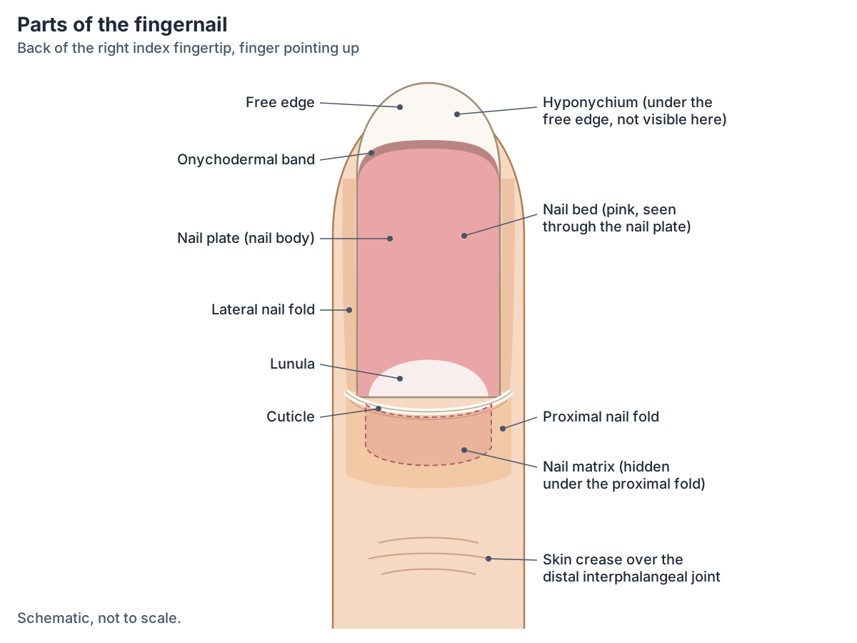
Parts of the nail at a glance
| Part | Where it is | What it does |
|---|---|---|
| Nail plate (nail body) | The hard, visible nail, from the proximal nail fold to the fingertip | Protects the tip; made of densely packed dead keratinocytes |
| Free edge | The part of the plate that projects beyond the nail bed | Overhangs the hyponychium |
| Nail root | The base of the plate, hidden under the proximal nail fold | Where new plate is added |
| Nail matrix | Under the nail root and proximal nail fold | Dividing cells that make the nail plate |
| Lunula | Pale half-moon at the base of the plate | The front edge of the matrix, seen through the plate |
| Nail bed | Under the nail plate, on the distal phalanx | Holds the plate; its blood vessels make the nail look pink |
| Onychodermal band | Narrow pink-grey line at the far end of the nail bed | Seals the nail bed where the plate lifts off it |
| Hyponychium | Skin under the free edge | Thickened stratum corneum that seals the tip |
| Proximal nail fold | Fold of skin over the base of the nail | Covers and protects the matrix |
| Cuticle and eponychium | At the edge of the proximal nail fold | Seal the fold to the plate (terms used differently by different sources) |
| Lateral nail folds | Skin along each side of the plate | Overlap and anchor the sides of the nail |
Compiled from OpenStax, Radiopaedia, FPnotebook, the Merck Manual and a nail-care anatomy text (Shelverton 2024).
The nail plate: what are nails made of?
Nails are made of keratin, an intracellular fibrous protein that gives hair, nails and skin their hardness and water-resistant properties. (OpenStax Anatomy and Physiology 2e, 5.1: Layers of the Skin.)
The nail body is a plate of densely packed dead keratinocytes. It forms at the nail root, which contains a matrix of dividing cells, and this allows the nail to grow continuously. (OpenStax Anatomy and Physiology 2e, 5.2: Accessory Structures of the Skin.)
Radiopaedia describes the nail plate as a trilaminar (three-layered) keratinised structure anchored to the nail bed. On ultrasound it shows bright dorsal and ventral plates with a darker layer between them, and its thickness, measured from the ventral to the dorsal plate, should not exceed 0.65 mm. That fits the figure of about half a millimetre given by InformedHealth.org. (Radiopaedia: Nail unit (anatomy).)
A nail-care anatomy text adds that the hardness of the plate comes from keratin in two forms: one fills the cells and makes them hard, and the other forms thin fibrils that interlock with neighbouring cells and bond the layers together. (Shelverton TA. The Nail Unit: Essential Anatomy and Physiology for the Beauty Industry. NailKnowledge, 2024.)
Nail matrix and lunula: where does a nail grow from?
A nail grows from its matrix, the layer of dividing cells under the nail root. The matrix lies under the skin of the proximal nail fold, and its convex front edge continues into the nail bed and may be visible through the plate as the lunula. The matrix makes the nail plate, and its proximal part is far more proliferative than its distal part. (Radiopaedia: Nail unit (anatomy).)
OpenStax describes the lunula as the crescent-shaped area at the base of the nail, where a thick layer of epithelium covers the nail matrix. (OpenStax Anatomy and Physiology 2e, 5.2: Accessory Structures of the Skin.)
Different parts of the matrix make different layers of the plate. FPnotebook explains that the proximal matrix forms the superficial (upper) part of the plate and the distal matrix the deeper part, so damage to the proximal matrix may cause superficial pitting and damage to the distal matrix deep nail ridges. (FPnotebook: Nail Anatomy.)
Why is the lunula white? A nail-care anatomy text explains that the plate cells formed there are still soft and not yet fully keratinised, so they are opaque and hide the pink nail bed beneath. The same text notes that the size and shape of the matrix set the thickness and curve of the nail. (Shelverton TA. The Nail Unit: Essential Anatomy and Physiology for the Beauty Industry. NailKnowledge, 2024.)
Everyone has lunulae, but they may be hidden under the cuticle. They are usually largest on the thumb and the big toe, are more noticeable in childhood and tend to shrink with age. (Cleveland Clinic Health Essentials: What your half-moon nails (lunulae) can tell you.)
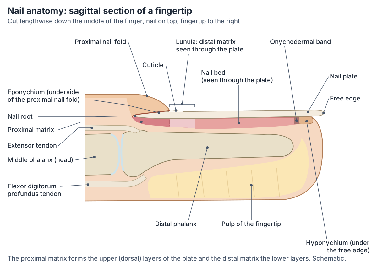
Cuticle vs eponychium: are they the same?
Sources use these two words differently. OpenStax treats them as one structure: where the proximal nail fold meets the nail body it forms the nail cuticle, also called the eponychium. (OpenStax Anatomy and Physiology 2e, 5.2: Accessory Structures of the Skin.)
FPnotebook separates them: the cuticle is the point where the nail plate meets the overlying skin at the base of the nail, and the eponychium is the skin just proximal to the cuticle, a fold that binds the nail to the underlying skin. (FPnotebook: Nail Anatomy.)
Nail-care anatomy texts draw the line most sharply. The eponychium is living skin on the underside of the proximal nail fold, and it sheds a thin, colourless layer of dead cells, the cuticle, which sticks to the emerging nail plate and seals the fold to the plate against water, chemicals and microbes. (Shelverton TA. The Nail Unit: Essential Anatomy and Physiology for the Beauty Industry. NailKnowledge, 2024.)
In the diagrams on this page the eponychium is the underside of the proximal nail fold and the cuticle is the thin layer it leaves on the nail. Where a book uses cuticle and eponychium as synonyms, both words refer to this seal at the base of the nail.
Nail bed, hyponychium and onychodermal band
The nail bed is the specialised epidermis on which the nail plate rests. It is rich in blood vessels, which is why nails look pink. The hyponychium is the area under the free edge of the nail, farthest from the cuticle, and consists of a thickened stratum corneum, the tough outer layer of the epidermis. (OpenStax Anatomy and Physiology 2e, 5.2: Accessory Structures of the Skin.)
FPnotebook calls the nail bed the sterile matrix. It attaches to the nail through squamous epithelium, makes the plate thicker and stronger, and contains blood vessels and nerves. The hyponychium is where the nail bed meets the skin at the tip of the digit. (FPnotebook: Nail Anatomy.)
The onychodermal band is the narrow, slightly greyish line seen through the plate at the far end of the nail bed, just before the free edge. It marks the tight seal where the nail bed epithelium meets the hyponychium and protects the nail bed against allergens and microbes. (Shelverton TA. The Nail Unit: Essential Anatomy and Physiology for the Beauty Industry. NailKnowledge, 2024.)
At the fingertip or toe tip the nail and the nail bed separate, and the nail projects beyond the tip as the free edge. (InformedHealth.org (IQWiG): Structure of the nails.)
Proximal and lateral nail folds
Folds of skin frame the nail on three sides. The lateral nail folds overlap the sides of the nail and help to anchor it, and the proximal nail fold at the base forms the cuticle where it meets the nail body. (OpenStax Anatomy and Physiology 2e, 5.2: Accessory Structures of the Skin.)
FPnotebook calls the lateral borders of the nail the perionychium or paronychial edge. This is where hangnails form and where paronychia, an infection of the nail fold, occurs. (FPnotebook: Nail Anatomy.)
The proximal nail fold covers and protects the matrix. Together with the cuticle, the two lateral folds and the hyponychium, it seals the nail unit so that infection cannot easily get under the plate to the matrix and the bone. (Shelverton TA. The Nail Unit: Essential Anatomy and Physiology for the Beauty Industry. NailKnowledge, 2024.)
Blood supply and nerves of the nail
The nail is supplied by the digital arteries, which in the hand come from the radial and ulnar arteries through the superficial and deep palmar arches and the dorsal metacarpal arch. A dorsal arcade just beyond the insertion of flexor digitorum profundus supplies the proximal nail fold, the extensor tendon insertion and part of the matrix. Its connections with the palmar arch form matrix arches and a capillary network that perfuses the nail bed, matrix, folds and hyponychium. Digital nerves supply the proximal nail fold, nail bed and matrix. (Radiopaedia: Nail unit (anatomy).)
The nail unit covers much of the back (dorsal surface) of the distal phalanx, with the nail bed lying directly on the bone. The extensor tendon inserts on the distal phalanx close to the matrix, and Radiopaedia notes that this closeness may let disease spread between the two. (Radiopaedia: Nail unit (anatomy).)
The fingertip, distal phalanx and the nerves that supply each finger. Hand anatomy.
Fingernail vs toenail anatomy and growth
Toenails have the same parts as fingernails but grow more slowly. In a study of 22 healthy American young adults, Yaemsiri and colleagues (2010) measured average growth of 3.47 mm a month for fingernails and 1.62 mm a month for toenails, so fingernails grew more than twice as fast. The little fingernail grew more slowly than the other fingernails and the big toenail faster than the other toenails, while right and left did not differ. Younger age and male sex went with faster growth, but those differences were not statistically significant. (Yaemsiri S, Hou N, Slining MM, He K. Growth rate of human fingernails and toenails in healthy American young adults. J Eur Acad Dermatol Venereol 2010.)
InformedHealth.org gives a rounder figure of about 3 mm a month for fingernails and says that toenails grow somewhat more slowly. As the matrix keeps producing keratin, the nail is pushed forward. (InformedHealth.org (IQWiG): Structure of the nails.)
A whole fingernail takes about 6 months to regrow, while a toenail takes 12 to 18 months, two to three times as long. Any systemic illness can stop nail growth for a while and leave deep transverse grooves called Beau's lines. (FPnotebook: Nail Anatomy.)
With age, nails grow more slowly and may become dull, brittle, yellowed or cloudy. Toenails may thicken, ingrown toenails become more common, and fingernail tips may break more easily. (MedlinePlus Medical Encyclopedia: Aging changes in hair and nails.)
The Merck Manual notes that toenails need extra care in older adults and in people with diabetes or peripheral vascular disease, because reduced feeling in the feet raises the risk of injury when trimming them. (Merck Manual Consumer Version: Overview of Nail Disorders.)
The toes and big toe, with a labelled toenail and big toe section. Foot anatomy.
Fingernail vs toenail
| Feature | Fingernail | Toenail |
|---|---|---|
| Average growth in a 2010 study of young adults | 3.47 mm per month | 1.62 mm per month |
| Typical figure in patient information | About 3 mm per month | Somewhat slower |
| Time to regrow a whole nail | About 6 months | 12 to 18 months |
| Largest lunula | Thumb | Big toe |
| Fastest and slowest in the 2010 study | Little fingernail slowest | Big toenail fastest |
| Changes with age | Grows more slowly; may become brittle and ridged | Grows more slowly; may thicken; ingrown nails more common |
Growth: Yaemsiri and colleagues 2010, InformedHealth.org and FPnotebook. Lunula: Cleveland Clinic. Ageing: MedlinePlus.
What nails can show: clubbing, spoon nails and other signs
Clubbing is a change in the tissue under and around the fingernails or toenails. The nail bed softens so the nail seems to float, the nail forms a sharper angle with the cuticle, the fingertip looks enlarged and the nail curves downward like the bowl of an upside-down spoon. Lung cancer is the most common cause; others include chronic lung infections, congenital heart defects, infective endocarditis and some liver and bowel diseases. (MedlinePlus Medical Encyclopedia: Clubbing of the fingers or toes.)
Koilonychia, or spoon nails, is a nail that is thin and curved inward with raised ridges, and is linked to iron deficiency anaemia. Beau lines are horizontal depressions across the nail after illness, injury or chemotherapy; pitting, small depressions in the nail surface, is seen in psoriasis and alopecia areata; and leukonychia means white streaks or spots on the nail. (MedlinePlus Medical Encyclopedia: Nail abnormalities.)
Changes in the colour or size of the lunulae are occasionally linked with diseases of the kidney, liver, heart or lungs, but a Cleveland Clinic physician stresses that no disease is diagnosed from the lunulae alone. (Cleveland Clinic Health Essentials: What your half-moon nails (lunulae) can tell you.)
These signs are listed to show how nail anatomy (matrix, plate and bed) is affected; they are not a guide to diagnosis. New changes in the nails are assessed by a clinician.
Nails and the 3D model
This page has no 3D view because the BodyParts3D model, built from MRI of an adult male, contains no nails. The bone under each nail, the distal phalanx, is in the model and can be explored on the hand and foot pages. The two diagrams above show the nail unit itself. (Mitsuhashi N et al. BodyParts3D: 3D structure database for anatomical concepts. Nucleic Acids Res 2009.)
The fingertip bone under the nail, in 3D. Distal phalanx of the index finger.
Frequently asked questions
What are the parts of a fingernail?
A fingernail has a hard nail plate with a free edge, a nail root under the skin at its base, and a nail matrix that makes the plate. The pale lunula is the visible front of the matrix. The plate rests on the nail bed and is framed by the proximal and lateral nail folds, the cuticle and, under the free edge, the hyponychium.
What is the white half-moon on your nails?
It is the lunula, the front edge of the nail matrix seen through the nail plate. It looks pale because a thick layer of epithelium covers the matrix there and the new plate cells are not yet fully keratinised. Lunulae are usually largest on the thumbs and big toes and may be hidden under the cuticle on other nails.
Is the cuticle the same as the eponychium?
Some textbooks, such as OpenStax, use the words as synonyms. Others separate them: the eponychium is the living skin of the proximal nail fold, especially its underside, and the cuticle is the thin layer of dead cells it leaves on the nail plate. Both describe the seal that protects the nail matrix at the base of the nail.
What are nails made of?
Nails are made of keratin, the same tough protein found in hair and the outer skin. The nail plate is built from densely packed dead keratinocytes produced by the nail matrix. It has three layers and is about half a millimetre thick, normally no more than about 0.65 mm.
How fast do fingernails and toenails grow?
Fingernails grow about 3 to 3.5 mm a month and toenails about 1.6 mm a month, so fingernails grow more than twice as fast. A lost fingernail takes about 6 months to grow back completely and a toenail 12 to 18 months. Nail growth slows with age and can pause during illness.
What is the skin under the tip of the nail called?
The skin under the free edge of the nail is the hyponychium. It is made of thickened stratum corneum and seals the space under the nail at the fingertip. Just in front of it, the onychodermal band is the narrow pink-grey line where the nail plate leaves the nail bed.
Where does a nail grow from?
A nail grows from the nail matrix, a layer of dividing cells under the nail root, hidden beneath the proximal nail fold. The proximal part of the matrix forms the upper layers of the plate and the distal part, seen as the lunula, forms the lower layers. New cells push the plate forward over the nail bed.
References
- Betts JG, et al. Anatomy and Physiology 2e, §5.2 Accessory Structures of the Skin. OpenStax, Rice University, 2022.
- Betts JG, et al. Anatomy and Physiology 2e, §5.1 Layers of the Skin. OpenStax, Rice University, 2022.
- Betts JG, et al. Anatomy and Physiology 2e, §28.3 Fetal Development. OpenStax, Rice University, 2022.
- Radiopaedia. Nail unit (anatomy). Accessed 2026-10-24.
- InformedHealth.org. Structure of the nails. Institute for Quality and Efficiency in Health Care (IQWiG), updated 2024.
- FPnotebook. Nail Anatomy. Accessed 2026-10-24.
- Yaemsiri S, Hou N, Slining MM, He K. Growth rate of human fingernails and toenails in healthy American young adults. J Eur Acad Dermatol Venereol. 2010;24(4):420-423. doi:10.1111/j.1468-3083.2009.03426.x
- Lipner S. Overview of Nail Disorders. Merck Manual Consumer Version, reviewed October 2025.
- MedlinePlus Medical Encyclopedia. Clubbing of the fingers or toes. US National Library of Medicine.
- MedlinePlus Medical Encyclopedia. Nail abnormalities. US National Library of Medicine.
- MedlinePlus Medical Encyclopedia. Aging changes in hair and nails. US National Library of Medicine.
- Cleveland Clinic Health Essentials. What your half-moon nails (lunulae) can tell you. Accessed 2026-10-24.
- Shelverton TA. The Nail Unit: Essential Anatomy and Physiology for the Beauty Industry. NailKnowledge, 2024.
- Mitsuhashi N, et al. BodyParts3D: 3D structure database for anatomical concepts. Nucleic Acids Res. 2009;37:D782-D785.
Related anatomy
- Hand anatomyHand anatomy with labelled diagrams and a 3D model: 27 bones, finger and thumb joints, flexor and extensor tendons…
- Foot anatomyFoot anatomy with labelled diagrams and a 3D model: the 26 bones, joints, arches, muscles, tendons, nerves, vessels…
- Hand bonesHow many bones are in the human hand? Learn the 8 carpal bones, 5 metacarpals and 14 phalanges by name, plus the thumb…
- Hair anatomyHair anatomy with labelled diagrams: shaft layers, follicle root sheaths, bulb and dermal papilla, arrector pili, hair…
- Distal phalanx of index fingerDistal phalanx of index finger: The smallest, end bone of the index finger (second digit), supporting the fingertip…
Explore the whole body in the interactive 3D human anatomy model, or browse the A–Z list of human structures.