

Parts of the hand: wrist, palm, fingers and thumb
The upper limb has three regions: the arm between shoulder and elbow, the forearm between elbow and wrist, and the hand, which lies distal to the wrist. The digits are numbered from the thumb: the thumb (pollex) is digit 1, the index finger digit 2, then the middle and ring fingers, and the little finger is digit 5. (OpenStax Anatomy and Physiology 2e, 8.2: Bones of the Upper Limb.)
Two fleshy mounds shape the palm. The thenar eminence is the rounded contour at the base of the thumb, formed by the thenar muscles, and the hypothenar eminence is the rounded contour at the base of the little finger. (OpenStax Anatomy and Physiology 2e, 11.5: Muscles of the Pectoral Girdle and Upper Limbs.)
The skin of the palm and the palmar surface of the digits is thick skin. Thick skin, found only on the palms and the soles, has a fifth epidermal layer, the stratum lucidum, and its surface ridges form fingerprints, which are unique to each person. (OpenStax Anatomy and Physiology 2e, 5.1: Layers of the Skin.)
The carpal bones, wrist joints and ligaments, carpal tunnel and anatomical snuffbox. Wrist anatomy.
Bones of the hand: carpals, metacarpals and phalanges
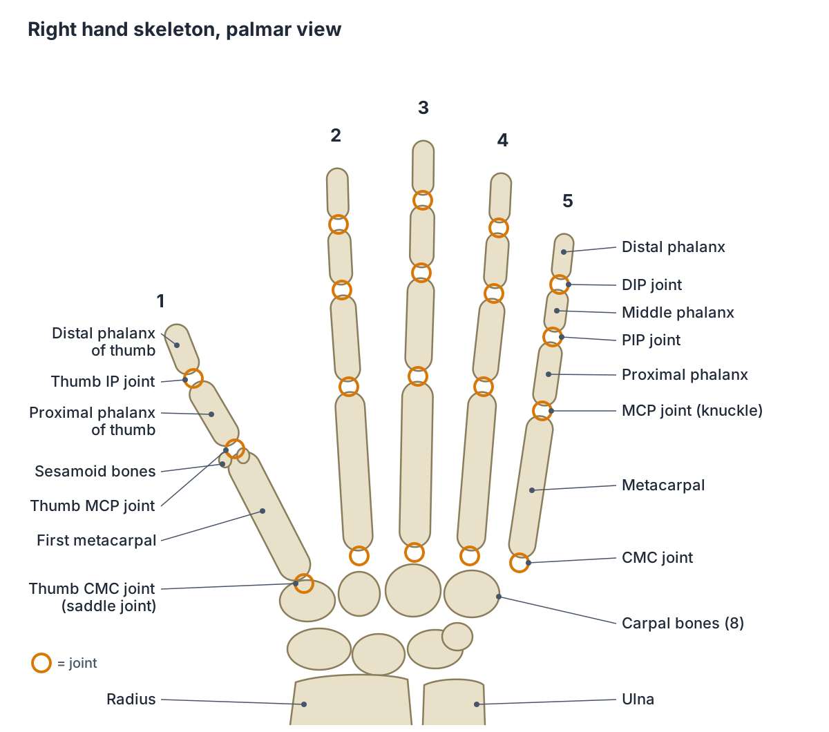
Each upper limb has 30 bones, and 27 of them are in the wrist and hand: 8 carpal bones in the wrist, 5 metacarpal bones in the palm and 14 phalanges in the digits. The metacarpals are numbered 1 to 5 starting at the thumb. The first metacarpal is separated from the others, which lets the thumb move independently. (OpenStax Anatomy and Physiology 2e, 8.2: Bones of the Upper Limb.)
Each metacarpal has a base, a shaft and a head. The metacarpals articulate proximally with the carpal bones and distally with the proximal phalanges. The pisiform, one of the carpal bones, is a sesamoid bone that forms within the tendon of flexor carpi ulnaris. (TeachMeAnatomy: Bones of the Hand.)
The thumb also has two small sesamoid bones embedded in the palmar plate of its metacarpophalangeal joint. They are not counted among the 27. (Kenhub: Metacarpophalangeal (MCP) joints.)
Every carpal, metacarpal and phalanx named, with the sesamoid bones and common fractures. Hand bones.
Finger anatomy: phalanges and finger joints
Each hand has 14 phalanges: five proximal, four middle and five distal, because the thumb has no middle phalanx. Every phalanx has a proximal base, a shaft and a distal head, and the distal tuberosity at the tip of each distal phalanx anchors the pulp of the fingertip. (Kenhub: Phalanges of the hand.)
Each finger has three joints: the metacarpophalangeal (MCP) joint between the metacarpal and the proximal phalanx, the proximal interphalangeal (PIP) joint between the proximal and middle phalanges, and the distal interphalangeal (DIP) joint between the middle and distal phalanges. (Kenhub: Phalanges of the hand.)
The PIP and DIP joints are uniaxial synovial hinge joints. Each has two collateral ligaments at its sides and a thick fibrocartilage palmar (volar) plate on the palm side. The palmar plate is shaped like an inverted U, and its two legs act as check-rein ligaments that limit hyperextension. (Kenhub: Interphalangeal joints of the hand.)
Kenhub gives typical ranges of about 100 to 110 degrees of flexion at the PIP joints and 70 to 80 degrees at the DIP joints, with almost no active extension beyond straight. Flexor digitorum profundus is the only muscle that flexes the DIP joint. (Kenhub: Interphalangeal joints of the hand.)
Wrist position changes grip strength: extending the wrist lengthens the finger flexors and strengthens the grip, while flexing it slackens them and weakens the grip. (Kenhub: Interphalangeal joints of the hand.)
One finger bone in detail: its joints, attachments and fractures. Proximal phalanx of the index finger.
Knuckles: the metacarpophalangeal (MCP) joints
The knuckles are the metacarpophalangeal joints, where the metacarpals meet the proximal phalanges. eOrthopod describes the volar plate on the palm side of the PIP joint as its strongest ligament, which keeps the joint from bending back too far (hyperextending). (eOrthopod: Hand Anatomy.)
Each MCP joint is a synovial condyloid joint between the rounded head of a metacarpal and the shallow base of a proximal phalanx. Its collateral ligaments are the main stabilisers and chiefly limit flexion, the palmar ligament (palmar plate) prevents hyperextension, and the deep transverse metacarpal ligaments link MCP joints 2 to 5 and steady them during grip. (Kenhub: Metacarpophalangeal (MCP) joints.)
The finger MCP joints flex to about 90 degrees, extend from about 10 degrees at the index finger to 30 degrees at the little finger, and allow about 25 to 30 degrees of side-to-side movement. Spreading and closing the fingers is not possible when the MCP joints are fully flexed, largely because of the way the collateral ligaments are arranged. (Kenhub: Metacarpophalangeal (MCP) joints.)
Thumb anatomy: why the thumb is different
The thumb (pollex) has only two phalanges, a proximal and a distal phalanx. Its first metacarpal is separated from the other metacarpals, which gives it freedom of motion independent of them. By contrast, the second and third metacarpals are firmly anchored and immobile, and the fourth and fifth have limited forward and backward movement that helps the palm close around an object in a grip. (OpenStax Anatomy and Physiology 2e, 8.2: Bones of the Upper Limb.)
The key joint is the carpometacarpal (CMC) joint at the base of the thumb, also called the trapeziometacarpal or basal joint. It is a synovial saddle joint: the concave surface of the trapezium meets the convex base of the first metacarpal. Kenhub gives it three degrees of freedom, with about 40 to 50 degrees of flexion-extension, 80 degrees of abduction-adduction and 70 to 110 degrees of axial rotation, held by the radial carpometacarpal ligament and the anterior and posterior oblique ligaments. (Kenhub: Trapeziometacarpal joint.)
Opposition is the movement that brings the tip of the thumb into contact with the tip of a finger; returning the thumb to lie beside the index finger is reposition. (OpenStax Anatomy and Physiology 2e, 9.5: Types of Body Movements.)
The thumb MCP joint moves about 60 degrees, almost all of it flexion, and its palmar plate contains two sesamoid bones. A tear of its ulnar collateral ligament, often from a fall with the thumb pulled outwards or from a ski pole, is called gamekeeper's or skier's thumb. The thumb's single interphalangeal joint flexes up to about 90 degrees. (Kenhub: Metacarpophalangeal (MCP) joints.)
The thumb CMC joint is highly vulnerable to arthritis with age. Women are 2 to 3 times more likely than men to develop it, and typical symptoms are pain at the base of the thumb with gripping or pinching, weakness, swelling and a bony bump. (OrthoInfo (AAOS): Arthritis of the Thumb.)
The thumb metacarpal, its saddle joint with the trapezium and the Bennett fracture. First metacarpal bone.
Joints of the hand, fingers and thumb
| Joint | Bones | Type | Main movements | Typical range |
|---|---|---|---|---|
| Thumb CMC (trapeziometacarpal) | Trapezium and first metacarpal | Saddle | Flexion, extension, abduction, adduction, rotation (opposition) | 40 to 50° flexion-extension; 80° abduction-adduction |
| Finger CMC (2 to 5) | Distal carpal row and metacarpals 2 to 5 | Synovial | 2nd and 3rd immobile; 4th and 5th move slightly forwards and back | Small, greatest at the 5th |
| MCP (knuckles) | Metacarpal head and proximal phalanx | Condyloid | Flexion, extension, abduction, adduction | About 90° flexion; 25 to 30° side to side |
| Thumb MCP | First metacarpal and proximal phalanx of thumb | Condyloid (moves mainly as a hinge) | Mainly flexion | About 60° |
| PIP | Proximal and middle phalanges | Hinge | Flexion and extension | About 100 to 110° flexion |
| DIP | Middle and distal phalanges | Hinge | Flexion and extension | About 70 to 80° flexion |
| Thumb IP | Proximal and distal phalanges of thumb | Hinge | Flexion and extension | Up to about 90° flexion, 10° extension |
Ranges from Kenhub (MCP, interphalangeal and trapeziometacarpal joints); finger CMC mobility from OpenStax §8.2; values vary between people. CMC = carpometacarpal, MCP = metacarpophalangeal, PIP/DIP = proximal/distal interphalangeal, IP = interphalangeal.
Muscles of the hand: extrinsic and intrinsic
Muscles acting on the hand are extrinsic, with their bellies in the forearm and long tendons in the hand, or intrinsic, located within the hand itself. The intrinsic muscles are the three thenar muscles at the base of the thumb, the three hypothenar muscles, four lumbricals, four dorsal interossei and three palmar interossei (some texts count a fourth at the thumb), plus adductor pollicis and palmaris brevis. (TeachMeAnatomy: Muscles of the Hand.)
OpenStax groups the intrinsic muscles into thenar muscles that act on the thumb, hypothenar muscles that act on the little finger and intermediate muscles (lumbricals and interossei) that act on all the fingers. It lists adductor pollicis with the thenar group. (OpenStax Anatomy and Physiology 2e, 11.5: Muscles of the Pectoral Girdle and Upper Limbs.)
Two nerves supply them. The median nerve supplies the thenar muscles (except the deep head of flexor pollicis brevis) and the lateral two lumbricals. The ulnar nerve supplies the hypothenar muscles, the medial two lumbricals, all the interossei, adductor pollicis and palmaris brevis. (TeachMeAnatomy: Muscles of the Hand.)
Thenar, hypothenar, lumbrical and interosseous muscles with origins, insertions and actions. Hand muscles.
Hand tendons: flexor tendons and finger pulleys
The long flexors of the fingers start in the forearm. Flexor digitorum superficialis sends a tendon to the middle phalanx of each finger and flexes the MCP and PIP joints. Flexor digitorum profundus sends tendons to the distal phalanges and is the only muscle that flexes the DIP joints. Flexor pollicis longus inserts on the base of the distal phalanx of the thumb. (TeachMeAnatomy: Muscles of the Anterior Forearm.)
All nine of these tendons, wrapped in synovial sheaths, pass through the carpal tunnel at the wrist together with the median nerve. (TeachMeAnatomy: The Carpal Tunnel.)
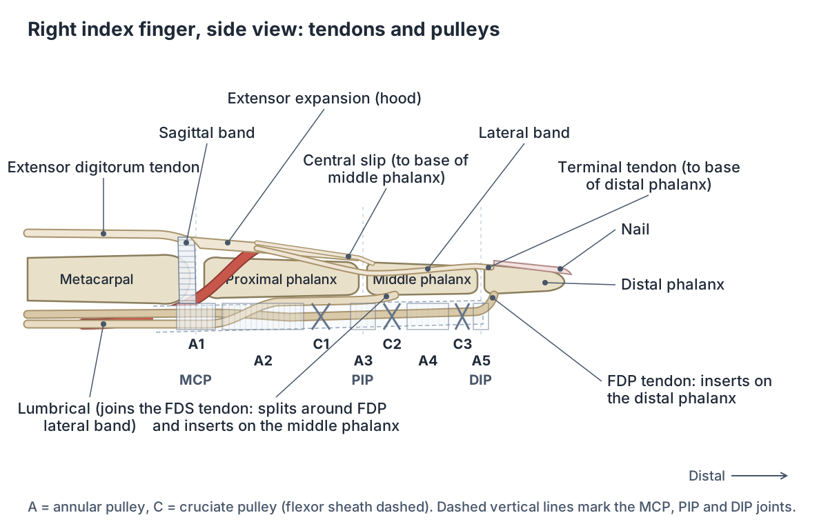
In each finger the tendons run in a fibrous flexor sheath. At the base of the proximal phalanx the superficialis tendon splits in two, the profundus tendon passes through the gap, and the two halves of the superficialis attach to the middle phalanx while the profundus continues to the distal phalanx. (TeachMeAnatomy: The Flexor Pulley System of the Hand.)
The sheath is reinforced by five annular pulleys and three cruciate pulleys. A1 lies over the MCP joint, A2 over the proximal part of the proximal phalanx, A3 over the PIP joint, A4 over the middle of the middle phalanx and A5 over the DIP joint, with C1 to C3 between them. A2 and A4 are thought to be the most important for stopping the tendons from bowstringing away from the bone. (TeachMeAnatomy: The Flexor Pulley System of the Hand.)
The thumb has three pulleys: A1 over the MCP joint, an oblique pulley over the upper half of the proximal phalanx and A2 over its lower half. Trigger finger, in which a thickened tendon catches and the digit locks in flexion, most often involves the A1 pulley. (TeachMeAnatomy: The Flexor Pulley System of the Hand.)
The forearm flexors and extensors whose tendons move the fingers and thumb. Forearm muscles.
Extensor tendons and the extensor mechanism of the finger
On the back of the hand, the tendon of extensor digitorum splits into four and inserts into the extensor hood of each finger. Extensor indicis adds a separate extensor to the index finger and extensor digiti minimi one to the little finger. In the thumb, extensor pollicis longus reaches the distal phalanx and extensor pollicis brevis the proximal phalanx. (TeachMeAnatomy: Muscles of the Posterior Forearm.)
The extensor expansion (dorsal digital expansion, extensor hood) is a triangular aponeurosis through which the extensor tendons attach to the phalanges. It divides into a central slip, which inserts on the base of the middle phalanx, and two lateral slips, which pass around it to insert on the base of the distal phalanx. The tendons of the interossei reinforce these slips. (Radiopaedia: Extensor expansion.)
Because the lumbricals and interossei join the expansion, these small muscles can extend the PIP and DIP joints. The thumb has no true extensor expansion; instead its extensor pollicis longus tendon receives fibrous slips from abductor pollicis brevis and adductor pollicis. (Radiopaedia: Extensor expansion.)
At the MCP joint the best-defined part of the hood is the sagittal band. The lumbricals insert on the radial side of the expansion and the interossei on its lateral bands, and the oblique retinacular ligament links movement of the PIP and DIP joints. (TeachMeAnatomy: The Extensor Mechanism.)
Which tendon does what in a finger?
| Tendon | Inserts on | Main action in the finger | Clinical link |
|---|---|---|---|
| Flexor digitorum superficialis | Middle phalanx (two slips) | Flexes PIP and MCP joints | Runs through the carpal tunnel and A1 pulley (trigger finger) |
| Flexor digitorum profundus | Base of distal phalanx | Only flexor of the DIP joint | Torn off in jersey finger |
| Extensor expansion, central slip | Base of middle phalanx | Extends PIP joint | Injury causes boutonnière deformity |
| Extensor expansion, lateral slips | Base of distal phalanx | Extend DIP joint | Injured in mallet finger |
| Flexor pollicis longus | Base of distal phalanx of thumb | Flexes thumb IP and MCP joints | Has its own sheath in the carpal tunnel |
| Extensor pollicis longus | Distal phalanx of thumb | Extends all thumb joints | Ulnar border of the anatomical snuffbox |
Based on TeachMeAnatomy (forearm muscles, flexor pulley system, extensor mechanism, carpal tunnel, anatomical snuffbox), Radiopaedia (extensor expansion, mallet and jersey finger) and Kenhub (phalanges).
Nerves of the hand: median, ulnar and radial
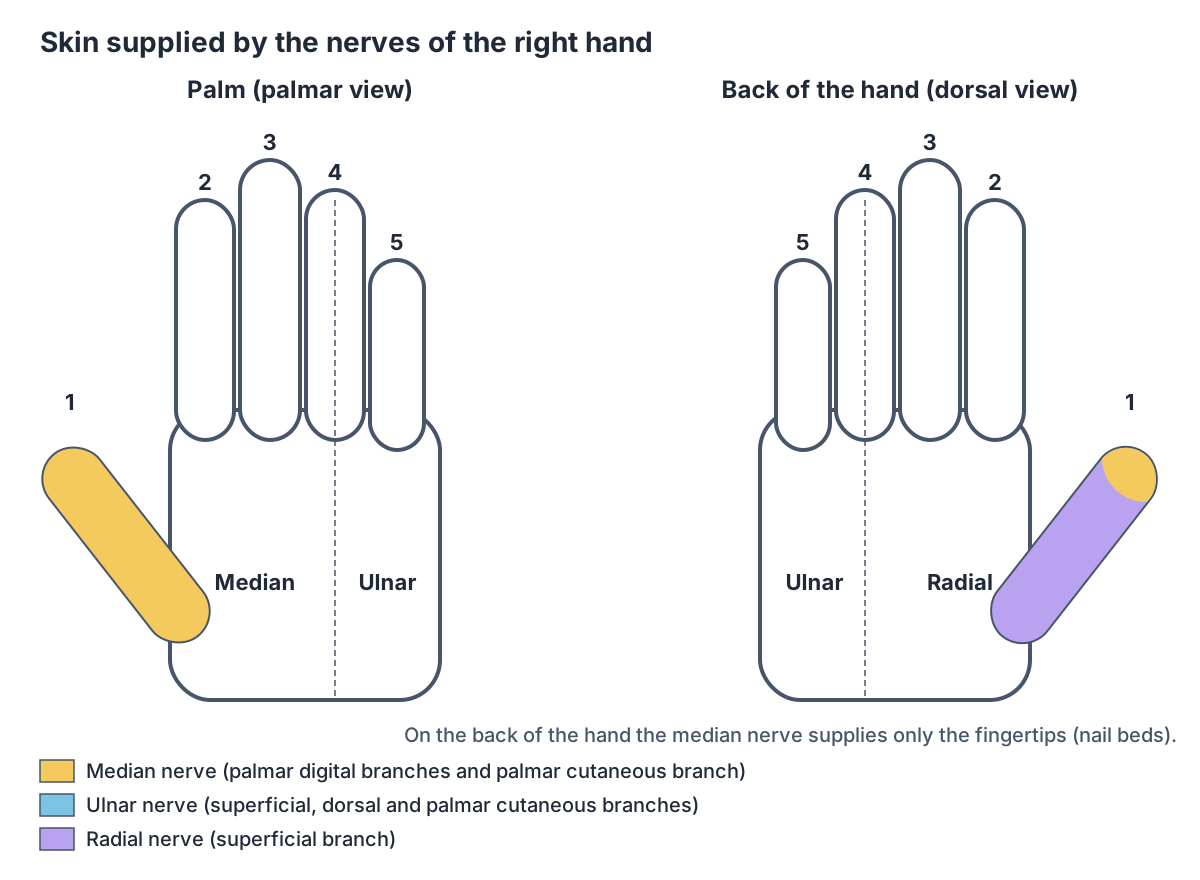
The median nerve enters the hand through the carpal tunnel. Its recurrent branch supplies the thenar muscles and its palmar digital branches supply the lateral two lumbricals. It carries sensation from the palmar surface and fingertips of the lateral three and a half digits, and its palmar cutaneous branch, which does not pass through the tunnel, supplies the skin of the lateral palm. (TeachMeAnatomy: The Median Nerve.)
TeachMeAnatomy adds that the palmar digital branches of the median nerve also supply the dorsal nail beds of those lateral three and a half digits. (TeachMeAnatomy: The Carpal Tunnel.)
The ulnar nerve enters the hand superficial to the flexor retinaculum through Guyon's canal and divides into superficial and deep branches. The deep branch supplies most of the intrinsic hand muscles. For sensation, its dorsal cutaneous branch supplies the back of the medial one and a half fingers, its superficial branch the palmar surface of those fingers, and its palmar cutaneous branch the medial half of the palm. (TeachMeAnatomy: The Ulnar Nerve.)
The radial nerve reaches the hand as its superficial branch, a sensory nerve that supplies the dorsal surface of the lateral three and a half digits and the adjoining back of the hand. (TeachMeAnatomy: The Radial Nerve.)
The brachial plexus and the full course of the median, ulnar and radial nerves. Nerves of the upper limb.
Arteries and veins of the hand
The ulnar artery enters the hand in front of the flexor retinaculum and continues across the palm as the superficial palmar arch, which gives the digital arteries to the four fingers. The radial artery enters the hand on its back, crossing the floor of the anatomical snuffbox, and continues as the deep palmar arch. Each artery also contributes a branch to the other's arch, so the two arches are linked. (TeachMeAnatomy: Arteries of the Upper Limb.)
The radial artery also gives branches to the thumb and the index finger, and its pulse is felt in the distal forearm just lateral to the tendon of flexor carpi radialis. (TeachMeAnatomy: Arteries of the Upper Limb.)
Superficial veins drain into the dorsal venous network on the back of the hand. The cephalic vein arises from this network and runs up the lateral side of the limb, and the basilic vein arises from it and ascends the medial side. Deep veins run in pairs beside the arteries as venae comitantes. (TeachMeAnatomy: Veins of the Upper Limb.)
The radial and ulnar arteries, palmar arches and the superficial veins of the hand and forearm. Arteries and veins of the arm.
Anatomy of the fingertip and nail
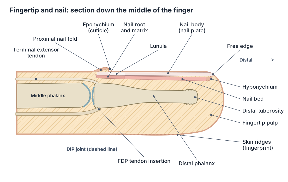
The skeleton of the fingertip is the distal phalanx. Its distal tuberosity anchors the pulp of the fingertip, and two forearm extensors and one flexor (flexor digitorum profundus, flexor pollicis longus and extensor pollicis longus) insert directly into the bases of the distal phalanges. (Kenhub: Phalanges of the hand.)
The nail body is made of densely packed dead keratinocytes. It forms at the nail root, which contains a matrix of dividing cells, and rests on the nail bed, a specialised part of the epidermis rich in blood vessels, which is why nails look pink. The lateral nail folds overlap the sides of the nail. (OpenStax Anatomy and Physiology 2e, 5.2: Accessory Structures of the Skin.)
The fold at the base of the nail forms the cuticle (eponychium), the pale crescent near the base is the lunula, a thick layer of epithelium over the matrix, and the area under the free edge of the nail is the hyponychium. The nail protects the fingertip and helps with picking up small objects. (OpenStax Anatomy and Physiology 2e, 5.2: Accessory Structures of the Skin.)
Fingerprints form where the stratum basale meets the papillae of the underlying dermis. The dermal papillae strengthen the bond between the epidermis and dermis, and the papillary layer contains touch receptors called Meissner corpuscles. (OpenStax Anatomy and Physiology 2e, 5.1: Layers of the Skin.)
The fingertip bone: its tuberosity, tendon insertions and mallet and jersey finger injuries. Distal phalanx of the index finger.
Common hand and finger injuries and conditions
Mallet finger is an injury of the extensor mechanism at the DIP joint, usually from sudden forced flexion of the fingertip, such as a ball striking an extended finger. The fingertip droops and cannot be actively straightened, and a small fragment of the distal phalanx may be pulled off with the tendon. (Radiopaedia: Mallet finger.)
Jersey finger is the opposite injury: the flexor digitorum profundus tendon is pulled off the base of the distal phalanx when a bent finger is forced straight, for example when grabbing an opponent's shirt. It most commonly affects the ring finger. (Radiopaedia: Jersey finger.)
Damage to the central slip over the PIP joint weakens extension there and lets the lateral bands slip towards the palm, producing a boutonnière deformity: the PIP joint is flexed and the DIP joint hyperextended. (TeachMeAnatomy: The Extensor Mechanism.)
A boxer's fracture is a fracture of the neck of the fifth metacarpal. A Bennett's fracture is a fracture of the base of the first metacarpal, caused by forced hyperabduction of the thumb, and it often needs surgical repair. (TeachMeAnatomy: Bones of the Hand.)
Dupuytren contracture is a progressive fibrosis of the palmar fascia that forms lumps and cords and pulls the fingers into flexion. The ring finger ray is most often involved, followed by the middle and little fingers, and it mainly affects people of northern European descent. (Radiopaedia: Dupuytren contracture.)
Hand anatomy in the 3D model
- Abductor digiti minimi of hand
- The muscle forming the ulnar (little-finger) side of the hypothenar eminence.
- Abductor pollicis brevis
- A thenar muscle on the lateral side of the thenar eminence.
- Capitate
- The largest carpal bone, with a rounded head.
- Deep palmar arch
- The deep palmar arch is an arterial arch in the palm, mainly formed by the radial artery.
- Distal phalanx of index finger
- The smallest, end bone of the index finger (second digit), supporting the fingertip and nail.
- Distal phalanx of little finger
- The smallest, end bone of the little finger (fifth digit), supporting the fingertip and nail.
- Distal phalanx of middle finger
- The smallest, end bone of the middle finger (third digit), supporting the fingertip and nail.
- Distal phalanx of ring finger
- The smallest, end bone of the ring finger (fourth digit), supporting the fingertip and nail.
- Distal phalanx of thumb
- The end bone of the thumb, supporting the nail and pulp.
- Dorsal venous network of hand
- The dorsal venous network is a superficial venous plexus on the back of the hand.
- Fifth metacarpal bone
- The little finger metacarpal. One of the five long bones of the palm.
- First metacarpal bone
- The thumb metacarpal, shorter and stouter than the others. One of the five long bones of the palm.
- Flexor digiti minimi brevis of hand
- A small hypothenar muscle.
- Flexor retinaculum of wrist
- A strong fibrous band across the front of the wrist forming the roof of the carpal tunnel.
- Fourth metacarpal bone
- The ring finger metacarpal. One of the five long bones of the palm.
- Hamate
- A wedge-shaped carpal bone of the distal row with a hook.
- Lunate
- A crescent-shaped carpal bone in the middle of the proximal row.
- Middle phalanx of index finger
- The middle of the three phalanges of the index finger (second digit).
- Middle phalanx of little finger
- The middle of the three phalanges of the little finger (fifth digit).
- Middle phalanx of middle finger
- The middle of the three phalanges of the middle finger (third digit).
- Middle phalanx of ring finger
- The middle of the three phalanges of the ring finger (fourth digit).
- Oblique head of adductor pollicis
- The larger head of adductor pollicis in the palm.
- Opponens digiti minimi of hand
- A hypothenar muscle that deepens the hollow of the palm on the little finger side.
- Opponens pollicis
- A thenar muscle lying deep to abductor pollicis brevis that allows the thumb to oppose.
- Pisiform
- A small, pea-shaped carpal bone on the palmar side of the triquetrum.
- Proximal phalanx of index finger
- The longest of the three phalanges of the index finger (second digit), nearest the palm.
- Proximal phalanx of little finger
- The longest of the three phalanges of the little finger (fifth digit), nearest the palm.
- Proximal phalanx of middle finger
- The longest of the three phalanges of the middle finger (third digit), nearest the palm.
- Proximal phalanx of ring finger
- The longest of the three phalanges of the ring finger (fourth digit), nearest the palm.
- Proximal phalanx of thumb
- The first of the thumb's two phalanges.
- Scaphoid
- The largest bone of the proximal carpal row, shaped like a boat.
- Second metacarpal bone
- The index finger metacarpal, usually the longest. One of the five long bones of the palm.
- Dorsal interossei of hand
- Four bipennate muscles between the metacarpals that spread the fingers.
- Lumbricals of hand
- Four small muscles of the palm that arise from the flexor digitorum profundus tendons.
- Palmar interossei of hand
- Small unipennate muscles in the palm that bring the fingers together.
- Superficial head of flexor pollicis brevis
- The larger, lateral head of flexor pollicis brevis in the thenar eminence.
- Superficial palmar arterial arch
- An arterial arch in the palm formed mainly by the ulnar artery.
- Third metacarpal bone
- The middle finger metacarpal. One of the five long bones of the palm.
- Transverse head of adductor pollicis
- The transverse head of adductor pollicis in the palm.
- Trapezium
- A four-sided carpal bone at the thumb side of the distal row.
- Trapezoid
- A small wedge-shaped carpal bone of the distal row.
- Triquetral
- A pyramid-shaped carpal bone on the ulnar side of the proximal row.
Frequently asked questions
How many bones are in the human hand?
The hand and wrist contain 27 bones: 8 carpal bones in the wrist, 5 metacarpals in the palm and 14 phalanges in the fingers and thumb. Both hands together have 54. Small sesamoid bones, such as the two in the thumb's knuckle joint, are not included in this count, and their number varies between people.
What are the joints in the fingers called?
Each finger has three joints. The metacarpophalangeal (MCP) joint is the knuckle where the finger meets the palm. The proximal interphalangeal (PIP) joint is the middle joint, and the distal interphalangeal (DIP) joint is the one nearest the fingertip. The thumb has an MCP joint and only one interphalangeal joint, plus a mobile carpometacarpal joint at its base.
Why can the thumb touch the other fingers?
The thumb's carpometacarpal joint is a saddle joint between the trapezium and the first metacarpal. It allows flexion, extension, abduction, adduction and some rotation, and these combine in opposition, which brings the thumb tip to meet each fingertip. The first metacarpal is also separated from the others, so the thumb moves independently of the palm.
What tendons are in the hand?
On the palm side, two tendons reach each finger, flexor digitorum superficialis to the middle phalanx and flexor digitorum profundus to the distal phalanx, and flexor pollicis longus reaches the thumb. On the back, extensor digitorum, extensor indicis and extensor digiti minimi join the extensor hood of the fingers, while the long and short thumb extensors reach the thumb.
What are finger pulleys?
Pulleys are thickened bands of the fibrous flexor sheath that hold the flexor tendons close to the finger bones. Each finger has five annular pulleys (A1 to A5) and three cruciate pulleys (C1 to C3). A2 and A4 are the most important for preventing bowstringing. The A1 pulley at the base of the finger is where trigger finger usually occurs.
Which nerves supply the fingers?
The median nerve supplies the palm side of the thumb, index, middle and half of the ring finger, including their nail beds. The ulnar nerve supplies the little finger and the other half of the ring finger on both sides. The radial nerve supplies the back of the thumb side of the hand and fingers, apart from the fingertips. Borders vary between people.
What is under a fingernail?
The nail body rests on the nail bed, a part of the epidermis rich in blood vessels that gives the nail its pink colour. The nail grows from the nail root and its matrix under the fold at the base, where the cuticle (eponychium) and the pale lunula are seen. Beneath the free edge lies the hyponychium, and under the nail bed is the distal phalanx.
References
- OpenStax. Anatomy and Physiology 2e, §8.2 Bones of the Upper Limb. Rice University, 2022.
- OpenStax. Anatomy and Physiology 2e, §11.5 Muscles of the Pectoral Girdle and Upper Limbs. Rice University, 2022.
- OpenStax. Anatomy and Physiology 2e, §9.5 Types of Body Movements. Rice University, 2022.
- OpenStax. Anatomy and Physiology 2e, §5.1 Layers of the Skin. Rice University, 2022.
- OpenStax. Anatomy and Physiology 2e, §5.2 Accessory Structures of the Skin. Rice University, 2022.
- TeachMeAnatomy. Bones of the Hand: Carpals, Metacarpals and Phalanges.
- TeachMeAnatomy. Muscles of the Hand.
- TeachMeAnatomy. Muscles of the Anterior Forearm.
- TeachMeAnatomy. Muscles of the Posterior Forearm.
- TeachMeAnatomy. The Flexor Pulley System of the Hand.
- TeachMeAnatomy. The Extensor Mechanism.
- TeachMeAnatomy. The Carpal Tunnel.
- TeachMeAnatomy. The Median Nerve.
- TeachMeAnatomy. The Ulnar Nerve.
- TeachMeAnatomy. The Radial Nerve.
- TeachMeAnatomy. Arteries of the Upper Limb.
- TeachMeAnatomy. Veins of the Upper Limb.
- Kenhub. Phalanges of the hand: anatomy and function.
- Kenhub. Metacarpophalangeal (MCP) joints: bones and ligaments.
- Kenhub. Interphalangeal joints of the hand: bones, ligaments, movements.
- Kenhub. Trapeziometacarpal joint: anatomy and movements.
- Radiopaedia. Extensor expansion.
- Radiopaedia. Mallet finger.
- Radiopaedia. Jersey finger.
- Radiopaedia. Dupuytren contracture.
- American Academy of Orthopaedic Surgeons, OrthoInfo. Arthritis of the Thumb.
- eOrthopod. Hand Anatomy.
- TeachMeAnatomy. The Anatomical Snuffbox.
- Mitsuhashi N. et al. (2009). BodyParts3D: 3D structure database for anatomical concepts. Nucleic Acids Research 37, D782–D785. Data © The Database Center for Life Science, CC BY 4.0.
Related anatomy
- Hand bonesHow many bones are in the human hand? Learn the 8 carpal bones, 5 metacarpals and 14 phalanges by name, plus the thumb…
- Hand musclesHand muscles anatomy: thenar eminence, hypothenar, adductor pollicis, lumbricals and palmar and dorsal interossei…
- Wrist jointWrist anatomy with labelled diagrams and a 3D model: 8 carpal bones, radiocarpal and midcarpal joints, wrist…
- Forearm musclesMuscles of the forearm explained: every anterior flexor and posterior extensor, with origin, insertion, nerve and…
- Upper limb nervesBrachial plexus diagram with roots, trunks, cords and branches; axillary, musculocutaneous, median, ulnar and radial…
- Nail anatomyNail anatomy with labelled diagrams: nail plate, matrix, lunula, cuticle vs eponychium, nail bed, hyponychium and…
Explore the whole body in the interactive 3D human anatomy model, or browse the A–Z list of human structures.