

Parts of the foot: hindfoot, midfoot and forefoot
Clinicians divide the foot into three regions. The hindfoot is the talus and calcaneus; the midfoot is the navicular, cuboid and three cuneiforms; and the forefoot is the metatarsals and phalanges. The bones give mechanical support to the soft tissues and let the foot bear the body's weight at rest and in movement. (TeachMeAnatomy: Bones of the Foot: Tarsals, Metatarsals and Phalanges.)
Seven tarsal bones form the back half of the foot and five metatarsals the front half, and the toes contain 14 phalanges. The toes are numbered 1 to 5, starting with the big toe, the hallux. Together the bones form three arches: a transverse arch and medial and lateral longitudinal arches. (OpenStax Anatomy and Physiology 2e, 8.4: Bones of the Lower Limb.)
The top of the foot is the dorsum (dorsal surface) and the bottom is the sole (plantar surface). Only two small muscles lie on the dorsum; the other intrinsic muscles of the foot lie in four layers in the sole. (TeachMeAnatomy: The Muscles of the Foot.)
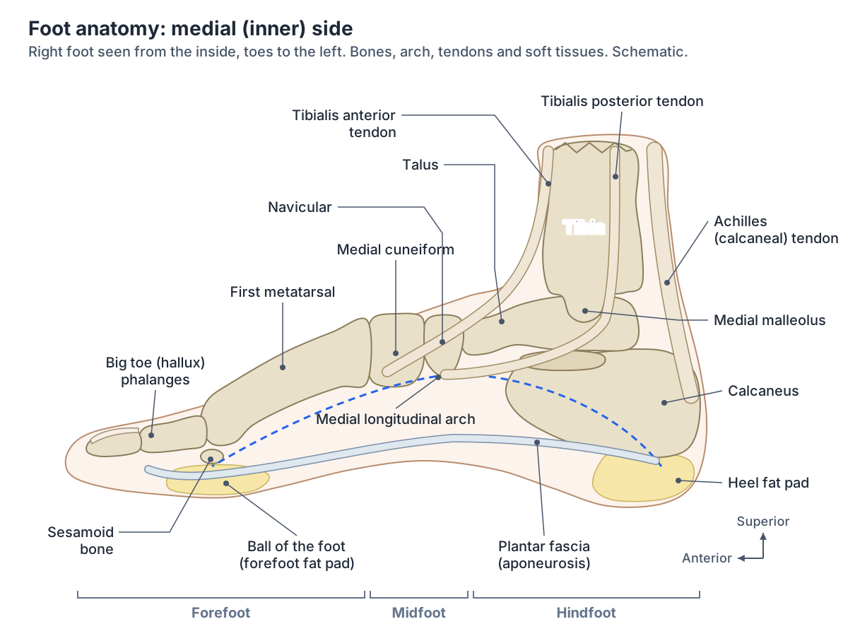
Foot anatomy at a glance
| Part | What it includes | Key fact |
|---|---|---|
| Bones | 7 tarsals, 5 metatarsals, 14 phalanges (26), plus 2 sesamoids | The calcaneus is the largest; the talus has no muscle attachments |
| Joints | Ankle, subtalar, intertarsal, tarsometatarsal, metatarsophalangeal, interphalangeal | Inversion and eversion happen mainly at the subtalar joint |
| Arches | Medial longitudinal, lateral longitudinal, transverse | Form by about 2 to 3 years of age |
| Muscles | 2 dorsal intrinsic muscles, 4 plantar layers, plus extrinsic leg muscles | Intrinsic muscles stabilise the arches and move the toes |
| Tendons | Achilles, tibialis anterior and posterior, fibularis longus and brevis, long toe flexors and extensors | The Achilles tendon is the strongest in the body |
| Sole | Thick skin, fat pads, plantar fascia, four muscle layers | The plantar fascia works as a windlass at push-off |
| Nerves | Tibial (plantar), superficial and deep fibular, sural, saphenous | Deep fibular nerve supplies the first web space |
| Vessels | Dorsalis pedis, medial and lateral plantar arteries, plantar arch; dorsal venous arch, saphenous veins | Two pulses: dorsalis pedis and posterior tibial |
Summary of the sections below; each row is covered in detail on this page or the linked guides.
Bones of the foot
The talus transmits body weight from the leg and has no muscles attached to it, which puts its blood supply at risk after fractures. The calcaneus is the largest tarsal bone and forms the heel. The navicular, on the medial side, receives part of the tibialis posterior tendon; the cuboid, on the lateral side, has a groove for the fibularis longus tendon; and the three wedge-shaped cuneiforms help form the transverse arch. (TeachMeAnatomy: Bones of the Foot: Tarsals, Metatarsals and Phalanges.)
The first metatarsal is the shortest and stoutest and carries most of the load; the second is the longest; and the fifth has a tuberosity at its base that you can see and feel on the outer border of the foot. Two sesamoid bones are always present under the head of the first metatarsal. (Kenhub: Metatarsal bones.)
The big toe has two phalanges (proximal and distal) and each of the other toes has three (proximal, middle and distal). The tarsal bones correspond to the carpal bones of the wrist, and the metatarsals to the metacarpals. (Kenhub: Ankle and foot anatomy.)
All 26 bones of the foot labelled, with joints and fractures. Foot bones.
Joints of the foot and ankle
The foot's joints fall into four groups: intertarsal joints (subtalar, talocalcaneonavicular, calcaneocuboid, cuneonavicular, cuboideonavicular and intercuneiform), tarsometatarsal joints, metatarsophalangeal joints between the metatarsal heads and the toes, and interphalangeal joints. The big toe has one interphalangeal joint and each other toe has two. (Kenhub: Ankle and foot anatomy.)
Above them, the ankle (talocrural) joint between the tibia, fibula and talus moves the foot up and down. Turning the sole inwards and outwards (inversion and eversion) happens mainly at the subtalar joint below it. (TeachMeAnatomy: The Ankle Joint.)
Cleveland Clinic counts 26 bones and 30 joints in each foot, held together by dozens of ligaments. (Cleveland Clinic: Foot Ligaments.)
The ankle joint, its ligaments and sprain anatomy. Ankle anatomy.
Arches of the foot
The foot has two longitudinal arches and one transverse arch, which act like a spring, bearing weight and absorbing shock. The medial longitudinal arch, the higher one, is made of the calcaneus, talus, navicular, three cuneiforms and first three metatarsals. The lateral longitudinal arch (calcaneus, cuboid, fourth and fifth metatarsals) is flatter and rests on the ground when standing. The transverse arch runs across the metatarsal bases, cuboid and cuneiforms. (TeachMeAnatomy: The Arches of the Foot.)
The arches are held up by the wedge shape of the bones, by ligaments (the long plantar, short plantar and plantar calcaneonavicular ligaments), by the plantar aponeurosis and by muscles: tibialis anterior and posterior, fibularis longus, the long toe flexors and the intrinsic foot muscles. Fibularis longus and tibialis posterior also support the transverse arch. (TeachMeAnatomy: The Arches of the Foot.)
The spring (plantar calcaneonavicular) ligament runs from the sustentaculum tali to the navicular and forms a sling under the head of the talus. With the tibialis posterior tendon it is the main stabiliser of the medial arch; when these fail, the hindfoot drifts outwards and the foot becomes flat (pes planus). (Radiopaedia: Spring ligament complex.)
The arches do not form until about 2 to 3 years of age, so flat feet in infants are normal. Pes planus (flat foot) is loss of the longitudinal arches; pes cavus is an unusually high medial arch, which absorbs shock less well. (TeachMeAnatomy: The Arches of the Foot.)
Ligaments of the foot
The spring ligament is the strongest ligament in the foot. Other important ligaments include the calcaneocuboid ligament, which links the heel bone to the bones of the arch, and the Lisfranc ligaments in the midfoot, which are torn in a Lisfranc injury. (Cleveland Clinic: Foot Ligaments.)
The long plantar ligament, which you can find in the 3D model, runs from the underside of the calcaneus to a ridge on the cuboid and on to the bases of the second to fourth or fifth metatarsals. It lies over the short plantar ligament and roofs the tunnel for the fibularis longus tendon, and it stabilises the calcaneocuboid and midtarsal joints. (Radiopaedia: Long plantar ligament.)
The longest ligament of the sole, in 3D. Long plantar ligament.
Bottom of the foot (plantar anatomy)
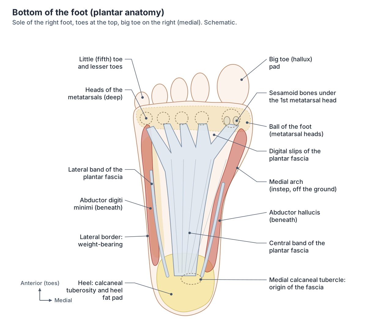
The bottom of the foot, the sole, has thick, hairless skin over its weight-bearing areas (heel, outer border and ball of the foot) and many sweat glands. Beneath the skin, a thick layer of fat divided by fibrous septa cushions and insulates the foot, and under that lies a strong band of deep fascia, the plantar aponeurosis. (Kenhub: Sole of foot.)
The plantar aponeurosis, or plantar fascia, runs from the base of the calcaneus to the phalanges. Its tension helps stop the medial longitudinal arch from collapsing. (Bolgla LA, Malone TR. Plantar fasciitis and the windlass mechanism: a biomechanical link to clinical practice. J Athl Train. 2004.)
With the spring and long plantar ligaments, the plantar fascia supports the longitudinal arch, and it gives attachment to the short muscles of the foot. (Gutteck N, Schilde S, Delank KS. Pain on the plantar surface of the foot. Dtsch Arztebl Int. 2019.)
The fascia also works as a windlass. In 1954 Hicks described it as the tie-rod of an arched truss running from the calcaneus to the phalanges. When the toes bend upwards at push-off, the fascia winds around the metatarsal heads, shortening the distance between the heel and the metatarsals and raising the medial arch, which turns the foot into a firmer lever. (Bolgla LA, Malone TR. Plantar fasciitis and the windlass mechanism: a biomechanical link to clinical practice. J Athl Train. 2004.)
Deep to the fascia, the intrinsic muscles of the sole lie in four layers. The flexor digitorum longus tendons run between the first and second layers, and the medial and lateral plantar nerves and arteries, branches of the tibial nerve and posterior tibial artery, run in planes between the layers. (Kenhub: Sole of foot.)
Under the ball of the foot, the forefoot touches the ground at five points: the head of the first metatarsal with its two sesamoids and the heads of the second to fifth metatarsals. (Kenhub: Metatarsal bones.)
The four layers of muscles in the sole, with nerves and actions. Foot muscles.
Heel anatomy
The heel is formed by the calcaneus, the largest tarsal bone. Its back projects as the calcaneal tuberosity, where the Achilles (calcaneal) tendon attaches. (TeachMeAnatomy: Bones of the Foot: Tarsals, Metatarsals and Phalanges.)
Under the calcaneus lies the heel fat pad, whose main job is to absorb shock in walking and running. It has two layers separated by fibrous septa: superficial microchambers and deep macrochambers. Under load, the deep layer accounted for about 98.6% of the change in pad thickness. The pad degenerates with age and with conditions such as diabetes. (Maemichi T et al. Functional morphologic changes of the heel fat pad and plantar fascia in patients with heel pain. Foot Ankle Orthop. 2024.)
The heel pad is a fibroadipose tissue in which collagen and elastin septa enclose fat cells. During walking it compressed by about 10.3 mm, close to the 10.7 mm predicted as the limit of pain tolerance, so it works near its limit even in normal gait. (Wearing SC, Smeathers JE. The heel fat pad: mechanical properties and clinical applications. J Foot Ankle Res. 2011.)
The skin of the medial heel is supplied by the medial calcaneal nerve, a branch of the tibial nerve. The lateral side of the heel lies in the S1 dermatome. (TeachMeAnatomy: The Cutaneous Innervation of the Lower Limb.)
The heel bone, in 3D. Calcaneus.
Muscles of the foot
Two sets of muscles move the foot. Extrinsic muscles arise in the anterior, lateral and posterior compartments of the leg and mainly produce dorsiflexion, plantarflexion, inversion and eversion. Intrinsic muscles lie within the foot and control fine movements such as moving individual toes. (TeachMeAnatomy: The Muscles of the Foot.)
On the dorsum are extensor digitorum brevis and extensor hallucis brevis, supplied by the deep fibular nerve. The sole has four layers: abductor hallucis, flexor digitorum brevis and abductor digiti minimi; quadratus plantae and the lumbricals; flexor hallucis brevis, adductor hallucis and flexor digiti minimi brevis; and the plantar and dorsal interossei. They are supplied by the medial and lateral plantar nerves and act together to stabilise the arches. (TeachMeAnatomy: The Muscles of the Foot.)
Kenhub also groups the plantar muscles by function: a medial group acting on the big toe, a lateral group acting on the little toe and a central group acting on the lateral four toes. (Kenhub: Ankle and foot anatomy.)
Every intrinsic foot muscle with origin, insertion and nerve. Foot muscles.
Foot tendons
The Achilles (calcaneal) tendon is the thickest and strongest tendon in the body. It connects the calf muscles to the calcaneus, is usually 15 to 26 cm (6 to 10 inches) long in adults, and lifts the heel for walking, running, jumping, climbing stairs and standing on tiptoe. (Cleveland Clinic: Achilles Tendon.)
Tibialis posterior enters the foot behind the medial malleolus, inserts on the plantar surfaces of the medial tarsal bones, inverts and plantarflexes the foot and supports the medial arch. Flexor digitorum longus goes to the lateral four toes and flexor hallucis longus to the big toe. (TeachMeAnatomy: Muscles in the Posterior Compartment of the Leg.)
Behind the lateral malleolus run the two fibularis (peroneus) tendons. Fibularis longus crosses under the foot to the medial cuneiform and the base of the first metatarsal, everts and plantarflexes the foot and supports the lateral and transverse arches. Fibularis brevis inserts on a tubercle of the fifth metatarsal and everts the foot. (TeachMeAnatomy: Muscles in the Lateral Compartment of the Leg.)
Across the front of the ankle, tibialis anterior inserts on the medial cuneiform and first metatarsal base and dorsiflexes and inverts the foot. Extensor hallucis longus goes to the distal phalanx of the big toe, extensor digitorum longus splits into four tendons for the lateral four toes, and fibularis tertius, which not everyone has, goes to the fifth metatarsal. (TeachMeAnatomy: Muscles in the Anterior Compartment of the Leg.)
The calf and shin muscles that send these tendons into the foot. Leg muscles.
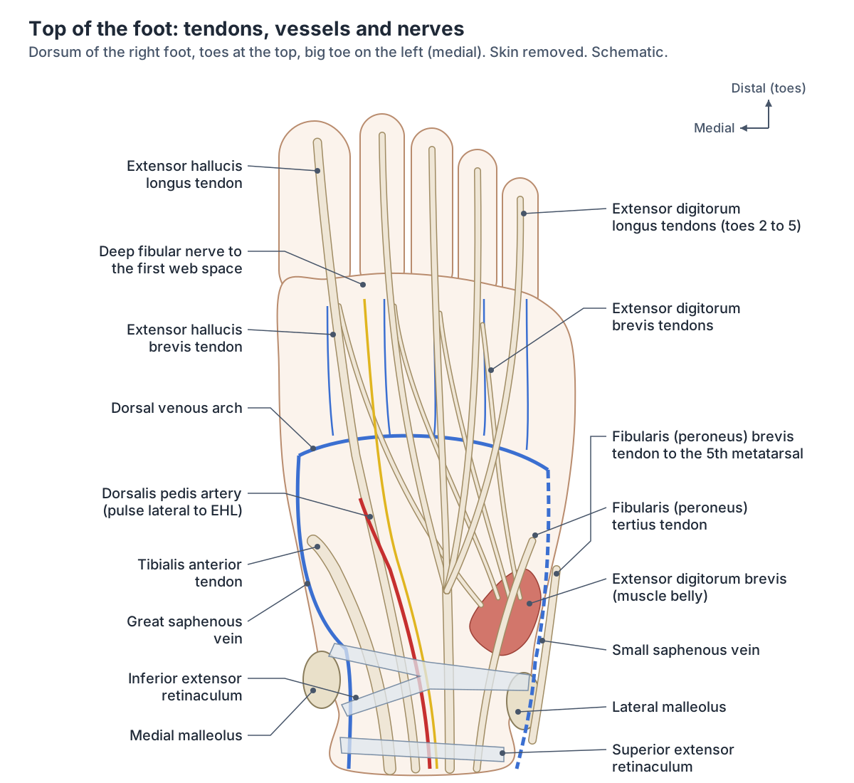
Main tendons of the foot
| Tendon | Passes | Inserts on | Main action |
|---|---|---|---|
| Achilles (calcaneal) | Back of the ankle | Calcaneal tuberosity | Plantarflexes (lifts the heel) |
| Tibialis posterior | Behind the medial malleolus | Navicular tuberosity and other medial tarsal bones | Inverts, plantarflexes, supports the medial arch |
| Flexor digitorum longus | Behind the medial malleolus | Lateral four toes | Flexes toes 2 to 5 |
| Flexor hallucis longus | Behind the medial malleolus | Big toe | Flexes the big toe |
| Fibularis (peroneus) longus | Behind the lateral malleolus, then across the sole | Medial cuneiform and first metatarsal base | Everts, plantarflexes, supports the lateral and transverse arches |
| Fibularis (peroneus) brevis | Behind the lateral malleolus | Tuberosity of the fifth metatarsal | Everts |
| Tibialis anterior | Front of the ankle | Medial cuneiform and first metatarsal base | Dorsiflexes and inverts |
| Extensor hallucis longus | Front of the ankle | Distal phalanx of the big toe | Extends the big toe, dorsiflexes |
| Extensor digitorum longus | Front of the ankle | Lateral four toes | Extends toes 2 to 5, dorsiflexes |
| Fibularis tertius (not always present) | Front of the ankle | Fifth metatarsal | Dorsiflexes and everts |
Sources: TeachMeAnatomy (leg compartments, foot bones), Cleveland Clinic (Achilles tendon).
Toe anatomy: the big toe and the lesser toes
The toes are numbered 1 to 5 from the big toe (hallux) to the little toe, and together they contain 14 phalanges. (OpenStax Anatomy and Physiology 2e, 8.4: Bones of the Lower Limb.)
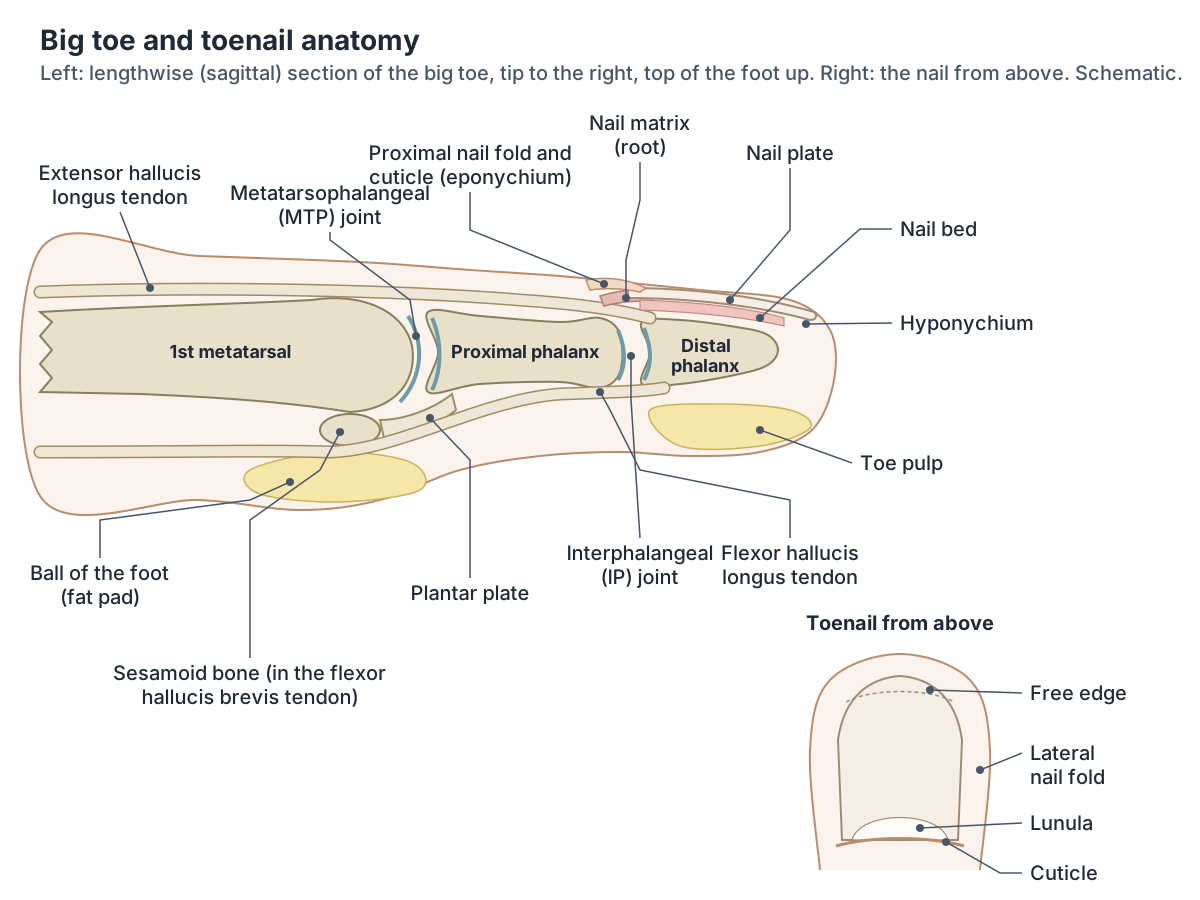
Under the head of the first metatarsal lie two sesamoid bones, a medial (tibial) and a lateral (fibular) one, embedded in the two slips of the flexor hallucis brevis tendon. They protect the underside of the metatarsal head and act as a pulley that improves the leverage of the flexor hallucis brevis and longus tendons. The medial sesamoid is usually the larger. (Radiopaedia: Hallux sesamoids.)
Turf toe is a sprain or tear of the plantar plate under the first metatarsophalangeal joint, usually from forced bending of the big toe upwards (hyperextension). The sesamoids are sometimes fractured as well. (Radiopaedia: Turf toe.)
In hallux valgus the big toe angles towards the second toe while the first metatarsal drifts medially; a painful hallux valgus is called a bunion. The first metatarsophalangeal joint is also where gout often first appears, a presentation called podagra. (Kenhub: Metatarsal bones.)
Hallux valgus is common: it affects about 23% of people aged 18 to 65 and 35% of those over 65. (Gutteck N, Schilde S, Delank KS. Pain on the plantar surface of the foot. Dtsch Arztebl Int. 2019.)
The lesser toes can bend into fixed shapes. In hammer toe the proximal interphalangeal joint is bent down and the distal one usually straightened; in claw toe the metatarsophalangeal joint is bent up and both interphalangeal joints bent down; in mallet toe only the distal interphalangeal joint is bent. Causes include hallux valgus, ill-fitting shoes, inflammatory arthritis and neurological conditions. (Radiopaedia: Hammer toe.)
The big toe bone that takes the most force at push-off, in 3D. Proximal phalanx of the big toe.
Toenail anatomy
A toenail is a plate of densely packed dead keratinocytes that protects the tip of the toe. It grows from the nail root at its base, which has a matrix of dividing cells. The visible nail body rests on the nail bed, which is rich in blood vessels and looks pink. (OpenStax Anatomy and Physiology 2e, 5.2: Accessory Structures of the Skin.)
The lateral nail folds overlap the sides of the nail and anchor it. The fold at the base forms the cuticle (eponychium), the pale crescent near the base is the lunula, lying over the matrix, and the thickened skin under the free edge is the hyponychium. (OpenStax Anatomy and Physiology 2e, 5.2: Accessory Structures of the Skin.)
Nail plate, matrix, lunula, cuticle and eponychium, with fingernail and toenail growth rates. Nail anatomy.
Nerves of the foot
Five nerves supply the skin of the foot. The superficial fibular nerve covers most of the dorsum, except the web space between the first and second toes, which is supplied by the deep fibular nerve. The sural nerve supplies the lateral margin and the saphenous nerve the medial margin of the hindfoot and midfoot. On the sole, the medial plantar nerve supplies the medial two-thirds and the lateral plantar nerve the lateral third. (TeachMeAnatomy: The Cutaneous Innervation of the Lower Limb.)
These territories vary between people. A study of the dorsum of the foot describes the superficial peroneal nerve dividing into medial and lateral dorsal cutaneous nerves, with the saphenous nerve reaching the medial margin as far as the base of the big toe. (Nayak VS et al. Anatomical variations in the cutaneous innervation on the dorsum of the foot. Anat Cell Biol. 2019.)
The tibial nerve enters the sole through the tarsal tunnel behind the medial malleolus. Compression there, tarsal tunnel syndrome, changes sensation over the sole and in severe cases weakens and wastes the intrinsic foot muscles. (TeachMeAnatomy: The Tarsal Tunnel.)
Tibial, fibular, sural and saphenous nerves from the spine to the foot. Nerves of the leg.
Skin nerve supply of the foot
| Area of skin | Nerve | Comes from |
|---|---|---|
| Most of the top of the foot | Superficial fibular nerve | Common fibular nerve |
| Web space between toes 1 and 2 | Deep fibular nerve | Common fibular nerve |
| Outer (lateral) margin | Sural nerve | Tibial and common fibular contributions |
| Inner (medial) margin | Saphenous nerve | Femoral nerve |
| Medial two-thirds of the sole | Medial plantar nerve | Tibial nerve |
| Lateral third of the sole | Lateral plantar nerve | Tibial nerve |
| Medial heel | Medial calcaneal nerve | Tibial nerve |
Source: TeachMeAnatomy, cutaneous innervation of the lower limb. Boundaries vary between people.
Arteries and veins of the foot
The anterior tibial artery becomes the dorsalis pedis artery on the top of the foot, where its pulse is felt just lateral to the extensor hallucis longus tendon. The posterior tibial artery reaches the sole through the tarsal tunnel, where its pulse is felt below and behind the medial malleolus, and divides into the medial and lateral plantar arteries. The dorsalis pedis and lateral plantar arteries join to form the deep plantar arch, which supplies the toes. (TeachMeAnatomy: Arterial Supply to the Lower Limb.)
Superficial veins drain into the dorsal venous arch on the top of the foot. Its medial end, with the dorsal vein of the big toe, forms the great saphenous vein, which passes in front of the medial malleolus; its lateral end, with the dorsal vein of the little toe, forms the small saphenous vein, which passes behind the lateral malleolus. Deep plantar veins form the posterior tibial and fibular veins. (TeachMeAnatomy: Venous Drainage of the Lower Limb.)
Lymph from the medial side of the foot follows the great saphenous vein, and from the lateral side follows the small saphenous vein to the popliteal lymph nodes behind the knee. (Kenhub: Sole of foot.)
From the iliac arteries to the plantar arch, with pulse points. Arteries of the leg.
Foot anatomy and pain: which structure is where?
Knowing the anatomy helps explain common foot problems; this section is educational and not a guide to diagnosis. Pain under the inner heel that is worst with the first steps in the morning is typical of plantar fasciitis, which peaks between 45 and 65 years of age and affects 5.2 to 17.5% of active runners. Heel spurs are seen on X-rays of 27% of people without symptoms, and their size does not match the pain. (Gutteck N, Schilde S, Delank KS. Pain on the plantar surface of the foot. Dtsch Arztebl Int. 2019.)
At the back of the heel, the Achilles tendon can be affected by tendinitis, rupture, heel bursitis and Haglund's deformity. (Cleveland Clinic: Achilles Tendon.)
Pain in the ball of the foot spreading into the toes is called metatarsalgia. It has many causes, including thinning of the plantar fat pad, Morton neuroma, Köhler disease II and rheumatoid arthritis. (Gutteck N, Schilde S, Delank KS. Pain on the plantar surface of the foot. Dtsch Arztebl Int. 2019.)
A Morton neuroma is fibrous thickening around a common plantar digital nerve, most often in the third web space between the third and fourth metatarsal heads. It is much more common in women and in middle age. (Radiopaedia: Morton neuroma.)
Where foot pain is felt and the anatomy there
| Location | Structures | Examples of problems |
|---|---|---|
| Under the heel | Plantar fascia origin, heel fat pad, calcaneus | Plantar fasciitis, heel spur, fat pad changes |
| Back of the heel | Achilles tendon insertion, bursa | Achilles tendinitis or rupture, heel bursitis, Haglund's deformity |
| Ball of the foot | Metatarsal heads, fat pad, plantar digital nerves | Metatarsalgia, Morton neuroma |
| Big toe joint | First metatarsophalangeal joint, sesamoids, plantar plate | Bunion (hallux valgus), gout, turf toe |
| Inner ankle and sole | Tibial nerve in the tarsal tunnel, tibialis posterior, spring ligament | Tarsal tunnel syndrome, flat foot |
| Outer ankle | Anterior talofibular and calcaneofibular ligaments | Inversion ankle sprain |
Educational overview from Gutteck et al. 2019, Radiopaedia, Cleveland Clinic and TeachMeAnatomy; not for self-diagnosis.
Foot anatomy in the 3D model
- Abductor digiti minimi of foot
- A muscle along the lateral border of the sole of the foot.
- Abductor hallucis
- A muscle along the medial border of the sole of the foot.
- Calcaneal tendon
- The thick tendon joining gastrocnemius and soleus to the heel bone.
- Calcaneus
- The largest tarsal bone, forming the heel.
- Cuboid bone
- A cube-shaped tarsal bone on the lateral side of the foot.
- Distal phalanx of big toe
- The end bone of the big toe (hallux), broad and stout.
- Distal phalanx of fourth toe
- The small end bone of the fourth toe (fourth digit of the foot).
- Distal phalanx of little toe
- The small end bone of the little toe (fifth digit of the foot).
- Distal phalanx of second toe
- The small end bone of the second toe (second digit of the foot).
- Distal phalanx of third toe
- The small end bone of the third toe (third digit of the foot).
- Dorsal venous arch of foot
- The dorsal venous arch is a superficial venous arch on the top of the foot.
- Dorsalis pedis artery
- The dorsalis pedis artery is the continuation of the anterior tibial artery onto the top of the foot.
- Extensor hallucis brevis
- A small muscle on the dorsum of the foot.
- Fifth metatarsal bone
- The metatarsal of the little toe. One of the five long bones of the forefoot.
- First lumbrical of foot
- A small muscle in the sole arising from the flexor digitorum longus tendon to the 2nd toe.
- First metatarsal bone
- The shortest, thickest metatarsal, supporting the big toe. One of the five long bones of the forefoot.
- First plantar interosseous of foot
- A small muscle between the metatarsals on the sole of the foot.
- Flexor accessorius
- A muscle of the sole that assists the long toe flexor.
- Flexor digiti minimi brevis of foot
- A small muscle of the lateral sole.
- Flexor digitorum brevis
- A central muscle of the sole.
- Fourth lumbrical of foot
- A small muscle in the sole acting on the 5th toe.
- Fourth metatarsal bone
- A lesser metatarsal on the lateral side of the foot. One of the five long bones of the forefoot.
- Intermediate cuneiform bone
- The smallest of the three cuneiform bones.
- Lateral cuneiform bone
- A wedge-shaped bone of the midfoot.
- Lateral head of flexor hallucis brevis
- The lateral part of flexor hallucis brevis in the sole.
- Lateral plantar artery
- The lateral plantar artery is the larger terminal branch of the posterior tibial artery in the sole.
- Long plantar ligament
- The longest ligament of the sole of the foot.
- Medial cuneiform bone
- The largest of the three cuneiform bones.
- Medial head of flexor hallucis brevis
- The medial part of flexor hallucis brevis in the sole.
- Medial plantar artery
- The smaller terminal branch of the posterior tibial artery in the sole of the foot.
- Middle phalanx of fourth toe
- The middle phalanx of the fourth toe (fourth digit of the foot).
- Middle phalanx of little toe
- The middle phalanx of the little toe (fifth digit of the foot).
- Middle phalanx of second toe
- The middle phalanx of the second toe (second digit of the foot).
- Middle phalanx of third toe
- The middle phalanx of the third toe (third digit of the foot).
- Navicular bone of foot
- A boat-shaped tarsal bone on the medial side of the foot.
- Oblique head of adductor hallucis
- The larger head of adductor hallucis in the sole.
- Plantar arch
- The arterial arch of the sole formed mainly by the lateral plantar artery.
- Plantar venous arch of foot
- A deep venous arch in the sole of the foot.
- Proximal phalanx of big toe
- The proximal of the two phalanges of the big toe (hallux).
- Proximal phalanx of fourth toe
- The first and longest phalanx of the fourth toe (fourth digit of the foot).
- Proximal phalanx of little toe
- The first and longest phalanx of the little toe (fifth digit of the foot).
- Proximal phalanx of second toe
- The first and longest phalanx of the second toe (second digit of the foot).
- Proximal phalanx of third toe
- The first and longest phalanx of the third toe (third digit of the foot).
- Second lumbrical of foot
- A small intrinsic muscle of the sole attached to the flexor digitorum longus tendons.
- Second metatarsal bone
- The longest metatarsal in most people. One of the five long bones of the forefoot.
- Second plantar interosseous of foot
- One of three plantar interossei, small muscles beneath the metatarsals in the fourth layer of the sole.
- Sesamoid bone of foot
- Small bone embedded in a tendon under the head of the first metatarsal.
- Talus
- A tarsal bone that sits between the tibia and fibula above and the calcaneus below.
- Third lumbrical of foot
- A small intrinsic muscle of the sole attached to the flexor digitorum longus tendons.
- Third metatarsal bone
- The middle of the lesser metatarsals. One of the five long bones of the forefoot.
- Third plantar interosseous of foot
- One of three plantar interossei, small muscles beneath the metatarsals in the fourth layer of the sole.
- Transverse head of adductor hallucis
- The transverse part of adductor hallucis in the sole.
Frequently asked questions
How many bones are in the foot?
Each foot has 26 bones: 7 tarsals, 5 metatarsals and 14 phalanges, or 52 in both feet. Two sesamoid bones under the head of the first metatarsal are usually counted separately. The tarsals form the hindfoot and midfoot, and the metatarsals and phalanges form the forefoot.
What is the bottom of the foot called?
The bottom of the foot is the sole, or plantar surface; the top is the dorsum. The sole has thick, hairless skin over the heel, outer border and ball of the foot, a cushioning fat layer, the tough plantar fascia and four layers of small muscles. The arch on the inner side does not touch the ground.
What are the main parts of the foot?
Anatomists divide the foot into the hindfoot (talus and calcaneus), the midfoot (navicular, cuboid and three cuneiforms) and the forefoot (five metatarsals and the phalanges of the toes). Everyday names include the heel, the arch or instep, the ball of the foot under the metatarsal heads, the toes and the top of the foot.
What tendons are in the foot?
The Achilles tendon inserts on the heel. Tibialis posterior, flexor digitorum longus and flexor hallucis longus pass behind the inner ankle; fibularis longus and brevis pass behind the outer ankle; and tibialis anterior, extensor hallucis longus, extensor digitorum longus and fibularis tertius cross the front of the ankle to the top of the foot.
What is the plantar fascia?
The plantar fascia, or plantar aponeurosis, is a thick band of connective tissue under the sole that runs from the calcaneus to the toes. It helps hold up the longitudinal arch and works like a windlass: when the toes bend up at push-off, it tightens and raises the arch. Inflammation of its heel attachment causes plantar fasciitis.
What is the ball of the foot?
The ball of the foot is the padded area of the sole under the heads of the metatarsal bones, just behind the toes. The head of the first metatarsal, with its two sesamoid bones, and the heads of the second to fifth metatarsals are its contact points with the ground. Pain here is called metatarsalgia.
What is the big toe called in anatomy?
The big toe is the hallux, or toe number 1; the toes are numbered 1 to 5 from the big toe to the little toe. The hallux has two phalanges and one interphalangeal joint, while each of the other toes has three phalanges and two interphalangeal joints. Two sesamoid bones lie under its metatarsal head.
References
- OpenStax. Anatomy and Physiology 2e, §8.4 Bones of the Lower Limb. Rice University, 2022.
- OpenStax. Anatomy and Physiology 2e, §5.2 Accessory Structures of the Skin. Rice University, 2022.
- TeachMeAnatomy. Bones of the Foot: Tarsals, Metatarsals and Phalanges.
- TeachMeAnatomy. The Arches of the Foot.
- TeachMeAnatomy. The Muscles of the Foot.
- TeachMeAnatomy. The Ankle Joint.
- TeachMeAnatomy. Muscles in the Anterior Compartment of the Leg.
- TeachMeAnatomy. Muscles in the Posterior Compartment of the Leg.
- TeachMeAnatomy. Muscles in the Lateral Compartment of the Leg.
- TeachMeAnatomy. The Cutaneous Innervation of the Lower Limb.
- TeachMeAnatomy. Arterial Supply to the Lower Limb.
- TeachMeAnatomy. Venous Drainage of the Lower Limb.
- TeachMeAnatomy. The Tarsal Tunnel.
- Kenhub. Ankle and foot anatomy.
- Kenhub. Sole of foot.
- Kenhub. Metatarsal bones.
- Cleveland Clinic. Foot Ligaments: What They Are, Anatomy & Function.
- Cleveland Clinic. Achilles Tendon.
- Radiopaedia. Spring ligament complex.
- Radiopaedia. Long plantar ligament.
- Radiopaedia. Hallux sesamoids.
- Radiopaedia. Turf toe.
- Radiopaedia. Hammer toe.
- Radiopaedia. Morton neuroma.
- Bolgla LA, Malone TR. Plantar fasciitis and the windlass mechanism: a biomechanical link to clinical practice. J Athl Train. 2004;39(1):77-82.
- Gutteck N, Schilde S, Delank KS. Pain on the plantar surface of the foot. Dtsch Arztebl Int. 2019;116(6):83-88.
- Maemichi T, Matsumoto M, Tsutsui T, et al. Functional morphologic changes of the heel fat pad and plantar fascia in patients with heel pain during weightbearing and nonweightbearing. Foot Ankle Orthop. 2024;9(2):24730114241247824.
- Wearing SC, Smeathers JE. The heel fat pad: mechanical properties and clinical applications. J Foot Ankle Res. 2011;4(Suppl 1):I14.
- Nayak VS, Bhat N, Nayak SS, Sumalatha S. Anatomical variations in the cutaneous innervation on the dorsum of the foot. Anat Cell Biol. 2019;52(1):34-37.
- Mitsuhashi N. et al. (2009). BodyParts3D: 3D structure database for anatomical concepts. Nucleic Acids Research 37, D782–D785. Data © The Database Center for Life Science, CC BY 4.0.
Related anatomy
- Foot bonesFoot bone anatomy with a labeled diagram and 3D model: all 26 bones of the foot, the 7 tarsals, 5 metatarsals and 14…
- Foot musclesFoot muscle anatomy: extensor digitorum brevis on the dorsum and the four plantar layers, from abductor hallucis to…
- Ankle jointAnkle anatomy with labelled diagrams and a 3D model: tibia, fibula and talus, the lateral and deltoid ligaments…
- Leg musclesLeg muscle anatomy with a 3D model and labeled diagrams: all leg muscle groups, the calf muscles, tibialis anterior…
- Leg anatomyLeg anatomy explained: lower limb regions, 30 leg bones, hip, knee and ankle joints, muscle groups, nerves and…
- Nail anatomyNail anatomy with labelled diagrams: nail plate, matrix, lunula, cuticle vs eponychium, nail bed, hyponychium and…
Explore the whole body in the interactive 3D human anatomy model, or browse the A–Z list of human structures.