

What is hair?
Hair is a keratinous filament that grows out of the epidermis and is made mostly of dead, keratinised cells. Hair follicles, like sweat glands, sebaceous glands and nails, originate from the epidermis during development and can extend down through the dermis into the hypodermis. (OpenStax Anatomy and Physiology 2e, 5.2: Accessory Structures of the Skin.)
Keratin is an intracellular fibrous protein that gives hair, nails and skin their hardness and water-resistant properties. (OpenStax Anatomy and Physiology 2e, 5.1: Layers of the Skin.)
Hair follicles are present everywhere on the skin except the lips, the palms of the hands and the soles of the feet. They lie in the dermis, the layer between the epidermis and the fat layer, and new hair is made in the hair matrix at the base of each follicle. (Merck Manual Consumer Version: Overview of Hair Growth.)
The epidermis, dermis and hypodermis in 3D, the layers a hair follicle passes through. Layers of the skin.
Parts of a hair: shaft, root and bulb
The hair shaft is the part of a hair that projects above the skin, and the hair root is the part anchored in the follicle below the surface. The root ends deep in the dermis at the hair bulb, which contains the hair matrix, a layer of dividing basal cells, and surrounds the hair papilla, a peg of connective tissue containing blood capillaries and nerve endings. (OpenStax Anatomy and Physiology 2e, 5.2: Accessory Structures of the Skin.)
The visible hair is completely dead, so cutting or shaving it causes no sensation and does not damage the root. Electrolysis and plucking aim to destroy or remove the bulb so that the hair cannot grow back, while most chemical hair removers act only on the surface. (OpenStax Anatomy and Physiology 2e, 5.2: Accessory Structures of the Skin.)
Layers of the hair shaft: cuticle, cortex and medulla
A hair shaft has three layers. The medulla forms the central core and is surrounded by the cortex, a layer of compressed keratinised cells, which is covered by an outer cuticle of very hard keratinised cells. Not all hairs have a medulla. Hair texture, straight or curly, depends on the shape and structure of the cortex and, where present, the medulla, which in turn follow the shape of the follicle. (OpenStax Anatomy and Physiology 2e, 5.2: Accessory Structures of the Skin.)
In Kenhub's histology account the cuticle is a thin, hard layer of overlapping keratin plates, the cortex is the main body of the hair and the medulla is its core. Cut across, straight hair is round, wavy hair oval and curly hair kidney-shaped, but the basic structure is the same in all of them. (Kenhub: Scalp and hair histology.)
The shaft is produced by dividing matrix cells in the hair bulb. As these cells differentiate and move upwards they build the growing shaft, whose cortex is made of intermediate filaments and proteins. (Natarelli N, Gahoonia N, Sivamani RK. Integrative and Mechanistic Approach to the Hair Growth Cycle and Hair Loss. J Clin Med 2023.)
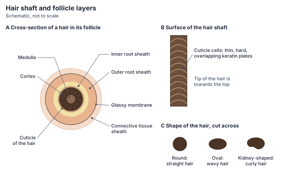
Hair follicle anatomy: the root sheaths
The wall of the hair follicle is made of three concentric layers of cells. The inner (internal) root sheath, formed from the basal cells of the hair matrix, surrounds the root of the growing hair. The outer (external) root sheath is an extension of the epidermis that encloses the root. The glassy membrane is the basement membrane between this epithelium and the surrounding connective tissue. (OpenStax Anatomy and Physiology 2e, 5.2: Accessory Structures of the Skin.)
Kenhub describes the same layers from the inside out. The medulla, cortex and cuticle keratinise to form the hair. The inner root sheath is mildly keratinised and breaks down at the level of the sebaceous gland ducts, leaving space for sebum around the hair. The outer root sheath does not form hair and is separated from the surrounding connective tissue by the glassy membrane, a specialised basement membrane. (Kenhub: Scalp and hair histology.)
Outside the epithelial sheaths is a fibrous root sheath of dermal connective tissue that supplies blood to the follicle. Each follicle has an arrector pili muscle, several sebaceous glands that release sebum into it, and free nerve endings that sense movement and bending of the hair. (Kenhub: Hair follicle.)
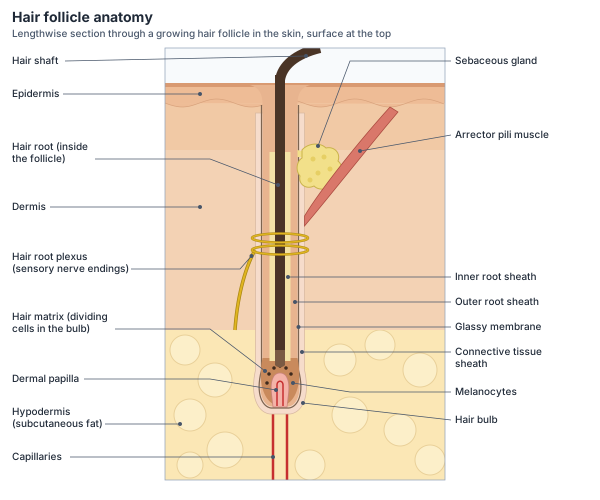
Layers of a hair and its follicle, from the centre outwards
| Layer | Part of | Notes |
|---|---|---|
| Medulla | Hair | Central core; not present in every hair |
| Cortex | Hair | Main body of the hair; its shape and that of the medulla set the texture |
| Cuticle of the hair | Hair | Thin, hard layer of overlapping keratin plates on the surface |
| Inner root sheath | Follicle (epithelium) | Formed from matrix cells; mildly keratinised; breaks down at the sebaceous duct |
| Outer root sheath | Follicle (epithelium) | Extension of the epidermis around the root; does not form hair |
| Glassy membrane | Follicle | Basement membrane between the outer root sheath and the connective tissue |
| Connective tissue sheath | Follicle (dermis) | Fibrous outer sheath of dermal connective tissue that carries blood to the follicle |
Compiled from OpenStax §5.2 and Kenhub (scalp and hair histology; hair follicle).
Hair bulb, matrix and dermal papilla
Hair grows from the bulb. The basal cells of the hair matrix divide to produce keratinocytes, which push the hair up through the follicle and become keratinised on the way. Matrix cells near the centre of the bulb differentiate into the cells of the inner root sheath. (OpenStax Anatomy and Physiology 2e, 5.2: Accessory Structures of the Skin.)
The dermal papilla, a group of mesenchymal cells at the base of the follicle, controls the number of matrix cells and so the size of the hair. Melanocytes scattered among the matrix cells of the bulb pass pigment into the growing hair. (Natarelli N, Gahoonia N, Sivamani RK. Integrative and Mechanistic Approach to the Hair Growth Cycle and Hair Loss. J Clin Med 2023.)
Kenhub describes the papilla as containing small blood vessels and nerve projections, with the mass of dividing cells around it forming the hair root and the hair matrix that produces the shaft. (Kenhub: Scalp and hair histology.)
Arrector pili muscle and sebaceous gland
Each hair root is connected to a smooth muscle, the arrector pili, which contracts in response to signals from the sympathetic nervous system. Contraction makes the hair stand up, trapping a layer of air that adds insulation; this produces goose bumps. (OpenStax Anatomy and Physiology 2e, 5.2: Accessory Structures of the Skin.)
The arrector pili muscles lie at an angle to the follicles and are attached to the papillary layer of the dermis, so when they contract they pull the hair upright. (Kenhub: Scalp and hair histology.)
A sebaceous gland is an oil gland. These glands are found all over the body, most are associated with hair follicles, and they secrete sebum, a mixture of lipids, onto the skin surface. (OpenStax Anatomy and Physiology 2e, 5.2: Accessory Structures of the Skin.)
Sebum released into the follicle keeps the hair soft and has a bactericidal (bacteria-killing) action. (Kenhub: Hair follicle.)
Hair colour and why hair turns grey
Hair colour comes from melanin made by melanocytes in the hair papilla region of the bulb. The type of melanin, and so the colour, is genetically determined. Melanin production falls with age, so hair turns grey or white. (OpenStax Anatomy and Physiology 2e, 5.2: Accessory Structures of the Skin.)
Melanocytes in the follicle incorporate melanin into the hair shaft. Melanin comes in black, brown and yellow forms, and their combinations give the range of natural hair colours. (Kenhub: Scalp and hair histology.)
Greying happens when hair follicles make less melanin. It often begins in the 30s, typically at the temples, and its timing is largely determined by genes. With age, strands also become finer and many follicles stop producing new hairs. (MedlinePlus Medical Encyclopedia: Aging changes in hair and nails.)
Types of hair: lanugo, vellus and terminal hair
Lanugo is unpigmented, fine, soft and silky hair that covers the fetus. It is usually shed at around 8 months of gestation and replaced by fine vellus hair and terminal scalp hair in preparation for birth. (DermNet: Hypertrichosis.)
OpenStax's timeline of fetal development agrees: the scalp begins to grow hair in weeks 13 to 16, a silky hair called lanugo covers the skin during weeks 17 to 20, and the lanugo is shed in the last weeks before birth. (OpenStax Anatomy and Physiology 2e, 28.3: Fetal Development.)
Vellus follicles produce tiny, virtually invisible hairs. Under the influence of androgens, vellus follicles in androgen-sensitive sites, such as the face, chest, lower abdomen, upper back and inner thighs, become larger intermediate and terminal follicles that make bigger, pigmented hairs. Androgens also change how long a hair grows and the size and activity of the dermal papilla. (DermNet: Hirsutism.)
Hair growth is regulated by androgens (male hormones). Testosterone stimulates hair growth in the pubic area and armpits, and dihydrotestosterone stimulates hair growth in the beard area and hair loss on the scalp. (Merck Manual Consumer Version: Overview of Hair Growth.)
Types of hair
| Type | What it is like | Where and when |
|---|---|---|
| Lanugo | Fine, soft, silky and unpigmented | Covers the fetus; shed before birth, at around 8 months of gestation |
| Vellus | Tiny, virtually invisible, usually unpigmented | Most of the body surface |
| Terminal | Larger, pigmented hairs from larger follicles | Scalp from before birth; under the influence of androgens also the pubic area, armpits and other androgen-sensitive sites such as the beard |
From DermNet (hypertrichosis; hirsutism), OpenStax §28.3 and the Merck Manual.
The hair growth cycle: anagen, catagen, telogen and exogen
Hair grows in a continuous cycle of four phases: anagen (growth), catagen (regression), telogen (rest) and exogen (shedding). Each follicle cycles independently of its neighbours and goes through ten to thirty cycles in a lifetime. (Natarelli N, Gahoonia N, Sivamani RK. Integrative and Mechanistic Approach to the Hair Growth Cycle and Hair Loss. J Clin Med 2023.)
In anagen the cells at the root divide rapidly and push the hair out; OpenStax gives this phase 2 to 7 years. Catagen, the transition, lasts about 2 to 3 weeks and growth stops. Telogen, the rest, lasts about 2 to 4 months, after which a new anagen phase begins and pushes the old hair out. (OpenStax Anatomy and Physiology 2e, 5.2: Accessory Structures of the Skin.)
Natarelli and colleagues give scalp anagen as two to eight years and note that hair length matches the length of anagen, which is why eyebrow hair, with an anagen of only two to three months, stays short. In catagen, which lasts about two weeks, the follicle regresses and detaches from the dermal papilla, cells of the bulb die by apoptosis, and the papilla moves up towards the hair-follicle bulge; if it cannot reach the bulge, cycling stops and the hair is lost. Telogen lasts about two to three months. (Natarelli N, Gahoonia N, Sivamani RK. Integrative and Mechanistic Approach to the Hair Growth Cycle and Hair Loss. J Clin Med 2023.)
The Merck Manual gives a growing phase of 2 to 6 years for scalp hair, a transitional phase of about 3 weeks and a resting phase of 2 to 3 months, at the end of which the hair falls out and a new hair starts growing in the same follicle. (Merck Manual Consumer Version: Alopecia (Hair Loss).)
For eyebrows and eyelashes the Merck Manual gives a growing phase of 1 to 6 months, much shorter than on the scalp. (Merck Manual Consumer Version: Overview of Hair Growth.)
DermNet estimates that about 85% of scalp follicles are growing (anagen) and about 15% resting (telogen) at any one time, with a follicle usually growing for about 4 years and resting for about 4 months. Natarelli and colleagues put the resting fraction of scalp hair at about 9%, compared with 40 to 50% of hair on the trunk. (DermNet: Telogen effluvium.)
The five layers of the scalp, where about 100,000 hair follicles sit. Scalp anatomy.
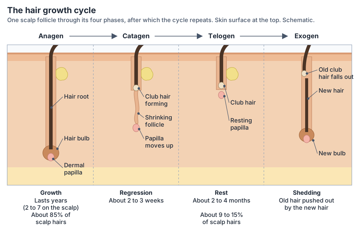
Phases of the hair growth cycle on the scalp
| Phase | What happens | OpenStax | Natarelli et al. 2023 | Merck Manual |
|---|---|---|---|---|
| Anagen | Growth: matrix cells divide and push the hair out | 2 to 7 years | 2 to 8 years | 2 to 6 years |
| Catagen | Regression: the follicle shrinks and the bulb detaches from the papilla | 2 to 3 weeks | About 2 weeks | About 3 weeks |
| Telogen | Rest: no growth; a club hair sits in a short follicle | 2 to 4 months | 2 to 3 months | 2 to 3 months |
| Exogen | Shedding: the old hair is pushed out as a new hair grows | Old hair pushed out by the new anagen hair | Named as a separate phase | Hair falls out at the end of rest |
DermNet gives about 4 years of anagen and about 4 months of telogen as typical. The ranges differ between sources; all agree that anagen lasts years and catagen only weeks.
How fast does hair grow, and how many hairs do you lose a day?
During anagen hair typically grows about 0.3 mm a day, and on average about 50 hairs are lost and replaced each day. (OpenStax Anatomy and Physiology 2e, 5.2: Accessory Structures of the Skin.)
Natarelli and colleagues give an average growth rate of about 1 cm a month, which matches 0.3 mm a day. They estimate that most people have about 100,000 scalp hairs and that normal shedding is 100 to 150 telogen hairs a day. (Natarelli N, Gahoonia N, Sivamani RK. Integrative and Mechanistic Approach to the Hair Growth Cycle and Hair Loss. J Clin Med 2023.)
The NHS says we can lose between 50 and 100 hairs a day, often without noticing. (NHS: Hair loss.)
The Merck Manual also gives about 50 to 100 scalp hairs a day as normal. It counts losing more than 100 a day as abnormal, except after shampooing, when up to 250 may come out. (Merck Manual Consumer Version: Alopecia (Hair Loss).)
What does hair do?
Hair protects: scalp hair shields the skull from the sun, and hairs in the nose and ears and the eyelashes trap dust and particles that may carry allergens and microbes; eyebrows keep sweat and particles out of the eyes. A hair root plexus of sensory nerves around each follicle makes hair sensitive to air movement and to anything touching the skin, and the arrector pili help keep in heat by raising the hairs. (OpenStax Anatomy and Physiology 2e, 5.2: Accessory Structures of the Skin.)
Kenhub adds that hair cushions the skin, protects against ultraviolet light, filters air, takes part in temperature regulation and sensation, and is part of the secondary sexual characteristics. (Kenhub: Hair follicle.)
Hair in the 3D model
The 3D viewer highlights the three hair surfaces in the BodyParts3D model, an adult male built from MRI: the hair of the head, the eyebrows and the pubic hair. The hair of the head and the pubic hair belong to the skin system, which starts hidden, so raise the Skin slider to see them; the eyebrows show at once. The model shows where hair grows but not the follicles, which are microscopic, so the diagrams on this page show those. (Mitsuhashi N et al. BodyParts3D: 3D structure database for anatomical concepts. Nucleic Acids Res 2009.)
The other keratin appendage of the skin: nail plate, matrix, lunula and nail bed. Nail anatomy.
Hair anatomy in the 3D model
- Eyebrow
- The eyebrow is a band of hairs on the ridge above the orbit.
- Hair of head
- The hair of the head is made of keratinised hair shafts growing from follicles in the scalp.
- Pubic hair
- The pubic hair is terminal hair growing over the pubic region after puberty.
Frequently asked questions
What are the 3 layers of a hair?
A hair shaft has three layers. The cuticle is the thin, hard outer layer of overlapping keratin plates. The cortex, made of compressed keratinised cells, forms the main body of the hair. The medulla is the central core, which some hairs do not have.
What are the parts of a hair follicle?
From the inside out, a follicle has an inner root sheath, an outer root sheath continuous with the epidermis, a glassy membrane and a connective tissue sheath. At its base the hair bulb holds the dividing matrix cells and melanocytes around the dermal papilla. A sebaceous gland, an arrector pili muscle and sensory nerve endings are attached to each follicle.
What are the 4 stages of hair growth?
Anagen is the growth phase and lasts years on the scalp (about 2 to 8, depending on the source). Catagen is a short regression phase of about 2 to 3 weeks. Telogen is a resting phase of about 2 to 4 months. Exogen is shedding, when the old club hair falls out as a new hair grows in the same follicle.
How many hairs do you lose a day?
Losing about 50 to 100 scalp hairs a day is normal, according to the NHS and the Merck Manual; one review gives 100 to 150. These are hairs at the end of their resting phase. More may come out on days you wash your hair, up to about 250 after shampooing.
Is hair alive?
The hair you can see is dead: it is made of keratinised cells, so cutting it does not hurt. The living part is in the follicle, where matrix cells in the hair bulb divide around the dermal papilla, which contains blood vessels and nerve endings, and a plexus of sensory nerves wraps around each hair root.
Why does hair turn grey?
Hair colour comes from melanin that melanocytes in the hair bulb add to the growing hair. With age the follicles make less melanin, so new hairs grow in grey or white. Greying often starts in the 30s, usually at the temples, and when it starts is largely decided by genes.
What is the difference between vellus and terminal hair?
Vellus hairs are tiny, fine and usually unpigmented, and cover most of the body. Terminal hairs are larger and pigmented, like scalp hair. Androgens (male hormones) turn vellus follicles in sites such as the armpits, pubic area and beard into terminal follicles. Lanugo is a third type that covers the fetus before birth.
References
- Betts JG, et al. Anatomy and Physiology 2e, §5.2 Accessory Structures of the Skin. OpenStax, Rice University, 2022.
- Betts JG, et al. Anatomy and Physiology 2e, §5.1 Layers of the Skin. OpenStax, Rice University, 2022.
- Betts JG, et al. Anatomy and Physiology 2e, §28.3 Fetal Development. OpenStax, Rice University, 2022.
- Kenhub. Scalp and hair histology. Accessed 2026-10-24.
- Kenhub. Hair follicle. Accessed 2026-10-24.
- Natarelli N, Gahoonia N, Sivamani RK. Integrative and Mechanistic Approach to the Hair Growth Cycle and Hair Loss. J Clin Med. 2023;12(3):893.
- DermNet. Hypertrichosis. Accessed 2026-10-24.
- DermNet. Hirsutism. Accessed 2026-10-24.
- DermNet. Telogen effluvium. Accessed 2026-10-24.
- Levinbook WS. Overview of Hair Growth. Merck Manual Consumer Version, reviewed September 2026.
- Levinbook WS. Alopecia (Hair Loss). Merck Manual Consumer Version, reviewed September 2026.
- NHS. Hair loss. Accessed 2026-10-24.
- MedlinePlus Medical Encyclopedia. Aging changes in hair and nails. US National Library of Medicine.
- Mitsuhashi N, et al. BodyParts3D: 3D structure database for anatomical concepts. Nucleic Acids Res. 2009;37:D782-D785.
- Mitsuhashi N. et al. (2009). BodyParts3D: 3D structure database for anatomical concepts. Nucleic Acids Research 37, D782–D785. Data © The Database Center for Life Science, CC BY 4.0.
Related anatomy
- ScalpScalp anatomy with labelled diagrams: the five SCALP layers, arteries, veins and danger area, nerves, lymph, why scalp…
- Nail anatomyNail anatomy with labelled diagrams: nail plate, matrix, lunula, cuticle vs eponychium, nail bed, hyponychium and…
- Hair of headHair of head: The hair of the head is made of keratinised hair shafts growing from follicles in the scalp. See where…
- SkinSkin: The skin is the body's largest organ, forming a protective covering. See where it is in an interactive 3D human…
- Head anatomyHead anatomy guide: skull, brain, face, sense organs and ENT structures, head and neck muscles, vessels, nerves and…
Explore the whole body in the interactive 3D human anatomy model, or browse the A–Z list of human structures.