

What are the buttocks? The gluteal region
In anatomy the buttocks belong to the gluteal region, the most proximal region on the back of the lower limb. Kenhub defines it as the prominence behind the pelvis that extends from the iliac crest above to the gluteal sulcus (the fold of the buttock) below, and out to the posterior margin of the greater trochanter of the femur. (Kenhub: Regions of the lower limb.)
The region lies below the lower back and outside the pelvic cavity. The two buttocks are separated in the midline by the intergluteal cleft, and laterally the gluteal region is bordered by the hip region. Its muscles bear weight and help keep the pelvis level. (Kenhub: Gluteal region.)
IMAIOS describes the buttock itself as the rounded bulge in the lower part of the gluteal region. Its rounded shape comes from the gluteus maximus muscle, and the skin and fascia over it are thick and contain abundant fat. (IMAIOS e-Anatomy: Buttock.)
The hip joint at the side of the gluteal region: bones, ligaments and movements. Hip anatomy.
Surface landmarks: iliac crest, dimples, cleft and gluteal fold
The iliac crest, the curved upper margin of the ilium, is the upper edge of the region and can be felt along the waistline when you put your hands on your waist. (OpenStax Anatomy and Physiology 2e, 8.3: The Pelvic Girdle and Pelvis.)
The posterior superior iliac spine (PSIS) is harder to feel than the front spine, but it is often marked by a dimple (the dimple of Venus) above the medial part of the buttock, about 4 cm lateral to the spinous process of the second sacral vertebra. (Kenhub: Posterior superior iliac spine.)
The intergluteal cleft, also called the natal cleft, is the groove that separates the two buttocks. (Kenhub: Regions of the lower limb.)
The gluteal fold is the horizontal skin crease below the buttock that separates it from the upper thigh. It forms because the gluteal skin is attached by fibrous tissue to the deep fascia. It is not the lower border of gluteus maximus: that border lies deeper and crosses the fold diagonally, downwards and outwards. (IMAIOS e-Anatomy: Gluteal fold.)
The ischial tuberosity, the 'sitting bone', carries the weight of the body when you sit, and you can feel it by shifting your weight on a chair. It is also the attachment of the hamstring muscles. (OpenStax Anatomy and Physiology 2e, 8.3: The Pelvic Girdle and Pelvis.)
The hip bones and sacrum that form the bony frame of the buttocks. Pelvis.
Fat and skin of the buttocks
Much of the size and shape of the buttocks comes from subcutaneous fat. Women store relatively more fat in the hips and thighs (the gluteofemoral region) than men, mostly in the superficial layer, and for the same body mass index they typically carry about 10% more body fat. Men store relatively more fat in the abdomen and around the organs. (Karastergiou K et al. Sex differences in human adipose tissues: the biology of pear shape. Biol Sex Differ. 2012.)
This pattern changes with age and hormones. A 2022 review notes that women tend to accumulate fat in the lower body and men in the central trunk, and that menopause is followed by more fat inside the abdomen and less in the gluteofemoral depot. (Boulet N et al. The sexual dimorphism of human adipose depots. Biomedicines. 2022.)
Gluteofemoral fat appears to be metabolically protective: the review by Karastergiou and colleagues links this 'pear shape' fat pattern with a lower risk of diabetes, although how it lowers risk is not yet clear. (Karastergiou K et al. Sex differences in human adipose tissues: the biology of pear shape. Biol Sex Differ. 2012.)
Muscles of the buttocks
The gluteal muscles, gluteus maximus, gluteus medius, gluteus minimus and tensor fasciae latae, form the bulk of the gluteal region. Beneath the lower half of gluteus maximus lies a deeper group of smaller muscles. (Kenhub: Regions of the lower limb.)
Their Latin names describe their size: OpenStax uses the buttock muscles as its example of muscles named for size, gluteus maximus being the largest and gluteus minimus the smallest. (OpenStax Anatomy and Physiology 2e, 11.2: Naming Skeletal Muscles.)
The three lie in layers. Deep to gluteus maximus is gluteus medius, and deep to gluteus medius is gluteus minimus, the smallest of the trio. OpenStax counts the gluteal group among the largest and most powerful muscles in the body. (OpenStax Anatomy and Physiology 2e, 11.6: Appendicular Muscles of the Pelvic Girdle and Lower Limbs.)
Gluteus maximus arises from the back of the ilium behind the posterior gluteal line, the sacrum and coccyx and the sacrotuberous ligament. Its superficial three-quarters insert into the iliotibial tract and its deep quarter into the gluteal tuberosity of the femur. It is supplied by the inferior gluteal nerve (L5 to S2) and by both gluteal arteries. (Kenhub: Gluteus maximus muscle.)
Gluteus maximus is the main extensor of the hip and brings the trunk upright from a bent position. It works intermittently when walking or climbing but not when standing still, and when sitting it contracts to ease the pressure of the trunk on the ischial tuberosities. Bursae separate it from the greater trochanter and the ischial tuberosity. (Kenhub: Gluteus maximus muscle.)
Gluteus medius and gluteus minimus lie on the outer surface of the ilium and insert on the greater trochanter. Both abduct and medially rotate the thigh and keep the pelvis level when walking; both are supplied by the superior gluteal nerve. If they are paralysed, the pelvis drops on the side of the lifted leg when the person stands on one leg, a positive Trendelenburg sign. (TeachMeAnatomy: Muscles of the Gluteal Region.)
The deep group, piriformis, the superior and inferior gemelli, obturator internus and quadratus femoris, lies under gluteus maximus. All of them laterally rotate the thigh, and piriformis, obturator internus and the gemelli also help abduct it. OpenStax adds obturator externus to the lateral rotators deep to gluteus maximus. (TeachMeAnatomy: Muscles of the Gluteal Region.)
Every gluteal and deep rotator muscle with attachments, actions and nerve supply. Hip muscles.
Muscles of the buttocks: origin, insertion, nerve and action
| Muscle | Origin | Insertion | Nerve (roots) | Main action |
|---|---|---|---|---|
| Gluteus maximus | Ilium behind the posterior gluteal line, sacrum, coccyx, sacrotuberous ligament | Iliotibial tract (superficial three-quarters), gluteal tuberosity of femur (deep quarter) | Inferior gluteal nerve (L5 to S2) | Extends the hip; assists lateral rotation |
| Gluteus medius | Outer ilium between the anterior and posterior gluteal lines | Lateral surface of the greater trochanter | Superior gluteal nerve (L4 to S1) | Abducts and medially rotates the thigh; keeps the pelvis level |
| Gluteus minimus | Outer ilium between the anterior and inferior gluteal lines | Anterior surface of the greater trochanter | Superior gluteal nerve (L4 to S1) | Abducts and medially rotates the thigh; keeps the pelvis level |
| Tensor fasciae latae | Anterior superior iliac spine, outer lip of the iliac crest | Iliotibial tract | Superior gluteal nerve (L4 to S1) | Tightens the fascia lata; abducts and medially rotates the thigh |
| Piriformis | Anterior surface of the sacrum | Greater trochanter | Nerve to piriformis | Lateral rotation and abduction |
| Superior gemellus | Ischial spine | Greater trochanter | Nerve to obturator internus | Lateral rotation and abduction |
| Obturator internus | Pubis and ischium around the obturator foramen | Greater trochanter | Nerve to obturator internus | Lateral rotation and abduction |
| Inferior gemellus | Ischial tuberosity | Greater trochanter | Nerve to quadratus femoris | Lateral rotation and abduction |
| Quadratus femoris | Lateral ischial tuberosity | Quadrate tubercle on the intertrochanteric crest | Nerve to quadratus femoris | Lateral rotation |
Based on Kenhub (gluteal muscles, gluteus maximus) and TeachMeAnatomy (muscles of the gluteal region, sacral plexus). Roots are given where the sources state them.
Piriformis and the sciatic foramina: how nerves reach the buttock
Nerves and vessels reach the buttock from the pelvis through two openings. The greater sciatic foramen is formed by the greater sciatic notch of the hip bone, the sacrum and the sacrospinous ligament. The smaller lesser sciatic foramen lies below it, bounded by the lesser sciatic notch and the sacrospinous and sacrotuberous ligaments. (OpenStax Anatomy and Physiology 2e, 8.3: The Pelvic Girdle and Pelvis.)
Piriformis, which runs from the front of the sacrum to the greater trochanter, is the key landmark. It divides the gluteal region into upper and lower parts, and vessels and nerves are named by whether they pass above or below it. (TeachMeAnatomy: Muscles of the Gluteal Region.)
Above piriformis (the suprapiriform foramen) pass the superior gluteal artery, vein and nerve. Below piriformis (the infrapiriform foramen) pass the sciatic nerve, the pudendal nerve, the inferior gluteal artery, vein and nerve, the posterior femoral cutaneous nerve, the nerve to obturator internus and the nerve to quadratus femoris. (TeachMeAnatomy: The Greater and Lesser Sciatic Foramina.)
The lesser sciatic foramen carries the tendon of obturator internus, its nerve, and the internal pudendal artery, vein and pudendal nerve. The pudendal nerve leaves the pelvis through the lower part of the greater sciatic foramen and re-enters it through the lesser foramen to supply the perineum. (TeachMeAnatomy: The Greater and Lesser Sciatic Foramina.)
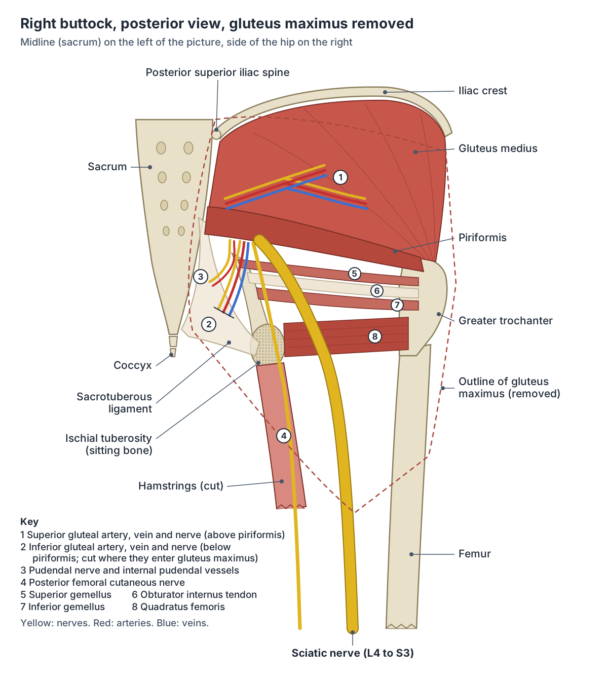
What passes through the sciatic foramina
| Opening | Structures |
|---|---|
| Greater sciatic foramen, above piriformis | Superior gluteal artery, vein and nerve |
| Greater sciatic foramen, below piriformis | Sciatic nerve; pudendal nerve; inferior gluteal artery, vein and nerve; posterior femoral cutaneous nerve; nerve to obturator internus; nerve to quadratus femoris |
| Lesser sciatic foramen | Tendon of obturator internus; nerve to obturator internus; internal pudendal artery and vein; pudendal nerve |
From TeachMeAnatomy (greater and lesser sciatic foramina).
Where is the sciatic nerve in the buttock?
The sciatic nerve (roots L4 to S3) enters the gluteal region through the greater sciatic foramen directly below piriformis and runs down the back of the thigh. It ends at the top of the popliteal fossa by dividing into the tibial and common fibular nerves. It passes through the lower inner quadrant of the buttock, which is why intramuscular injections are given only in the upper outer quadrant. (TeachMeAnatomy: The Sciatic Nerve.)
IMAIOS describes the sciatic nerve as the largest branch of the sacral plexus and places it underneath gluteus maximus in the buttock. (IMAIOS e-Anatomy: Buttock.)
The relationship to piriformis varies. In a 2022 study of 40 sciatic nerves, 37 (92.5%) left the pelvis below piriformis and 3 passed partly between its fibres. The same paper cites a larger series of 294 limbs in which 93.6% had the typical pattern, and a meta-analysis that found variants in about 31% of East Asian populations. (Reynoso JP et al. Anatomical variations of the sciatic nerve exit from the pelvis and its relationship with the piriformis muscle. Neurol Int. 2022.)
In about 12% of people the tibial and common fibular parts are already separate as they leave the pelvis. Compression of the nerve by piriformis, piriformis syndrome, causes pain radiating down the leg, numbness, weakness and tenderness in the buttock. (TeachMeAnatomy: The Sciatic Nerve.)
The full course of the sciatic, tibial and common fibular nerves down the lower limb. Nerves of the leg.
Nerves of the buttock
The superior gluteal nerve (L4, L5, S1) enters the buttock above piriformis and supplies gluteus medius, gluteus minimus and tensor fasciae latae. The inferior gluteal nerve (L5, S1, S2) enters below piriformis and supplies only gluteus maximus. (TeachMeAnatomy: The Sacral Plexus.)
The posterior femoral cutaneous nerve (S1 to S3) supplies the skin on the back of the thigh and leg and part of the perineum. The pudendal nerve (S2 to S4) supplies the perineum. Small direct branches, the nerves to piriformis, obturator internus and quadratus femoris, supply the deep rotators. (TeachMeAnatomy: The Sacral Plexus.)
All the muscles of the gluteal region are supplied by branches of the sacral plexus, and most of them receive blood from branches of the internal iliac artery. (Kenhub: Gluteal region.)
Superior and inferior gluteal arteries and veins
The superior gluteal artery is the terminal branch of the posterior trunk of the internal iliac artery. It passes between the lumbosacral trunk and the S1 nerve root and leaves the pelvis through the greater sciatic foramen above piriformis. (TeachMeAnatomy: Superior Gluteal Artery.)
In the buttock it divides. Its superficial branch runs between gluteus medius and gluteus maximus; its deep branch runs under gluteus medius and splits into superior and inferior divisions that cross gluteus minimus and join the deep circumflex iliac and lateral circumflex femoral arteries. It supplies the gluteal muscles, tensor fasciae latae, the skin of the region and the head of the femur. (TeachMeAnatomy: Superior Gluteal Artery.)
The inferior gluteal artery is the terminal branch of the anterior trunk of the internal iliac artery. It runs between the S2 and S3 nerve roots, leaves the pelvis below piriformis and descends between the greater trochanter and the ischial tuberosity. It supplies the gluteal muscles, the head of the femur, the sciatic nerve and the skin and muscles of the upper back of the thigh. (TeachMeAnatomy: Inferior Gluteal Artery.)
Each artery is accompanied by a vein of the same name through the greater sciatic foramen: the superior gluteal vein above piriformis and the inferior gluteal vein below it. The internal pudendal vessels pass through the lesser sciatic foramen. (TeachMeAnatomy: The Greater and Lesser Sciatic Foramina.)
The internal and external iliac arteries and their branches to the buttock and lower limb. Arteries of the leg.
Intramuscular injections in the buttock: the safe quadrant
For a dorsogluteal injection the buttock is divided into four quadrants by a vertical line from the highest point of the iliac crest and a horizontal line through the midpoint between the ischial tuberosity and the top of the iliac crest. The upper outer quadrant is regarded as the safest place for a dorsogluteal injection; as described above, the sciatic nerve runs through the lower inner part. (Cornwall J. Are nursing students safe when choosing gluteal intramuscular injection locations? Australas Med J. 2011.)
Even so, the dorsogluteal area lies close to the sciatic nerve and the gluteal arteries, and it has more subcutaneous fat. A 2018 review summarised imaging studies in which only 32 to 58% of intended intramuscular gluteal injections actually reached muscle, with women more often missing because their subcutaneous fat is thicker (in one CT study an average of 33.2 mm in women and 23.1 mm in men). (Soliman E et al. A narrative review of the success of intramuscular gluteal injections and its impact in psychiatry. Biodes Manuf. 2018.)
The ventrogluteal site, proposed by the Swiss anatomist von Hochstetter in 1954 to avoid sciatic nerve injury, lies at the side of the hip in an inverted triangle between the iliac crest, the anterior superior iliac spine and the greater trochanter, over gluteus medius and gluteus minimus. (Roldán-Chicano MT et al. Adverse effects of dorsogluteal intramuscular injection versus ventrogluteal intramuscular injection: a systematic review and meta-analysis. Nurs Open. 2023.)
In the 'V method' the base of the hand rests on the greater trochanter, the index finger points to the anterior superior iliac spine and the middle finger to the iliac crest, and the site is in the middle of the triangle formed. A 2023 meta-analysis found subcutaneous tissue about 20 mm thick at this site, against about 43 mm at the dorsogluteal site, with muscle about 38 mm thick at both. (Gulnar E et al. Gluteal muscle and subcutaneous tissue thicknesses in adults: a systematic review and meta-analysis. J Res Nurs. 2023.)
A second 2023 meta-analysis of 17 studies (1,429 participants) found less pain, bleeding and bruising and thinner subcutaneous fat at the ventrogluteal site, and concluded that it is safer and produces fewer adverse effects than the dorsogluteal site, though most included studies were not randomised. (Roldán-Chicano MT et al. Adverse effects of dorsogluteal intramuscular injection versus ventrogluteal intramuscular injection: a systematic review and meta-analysis. Nurs Open. 2023.)
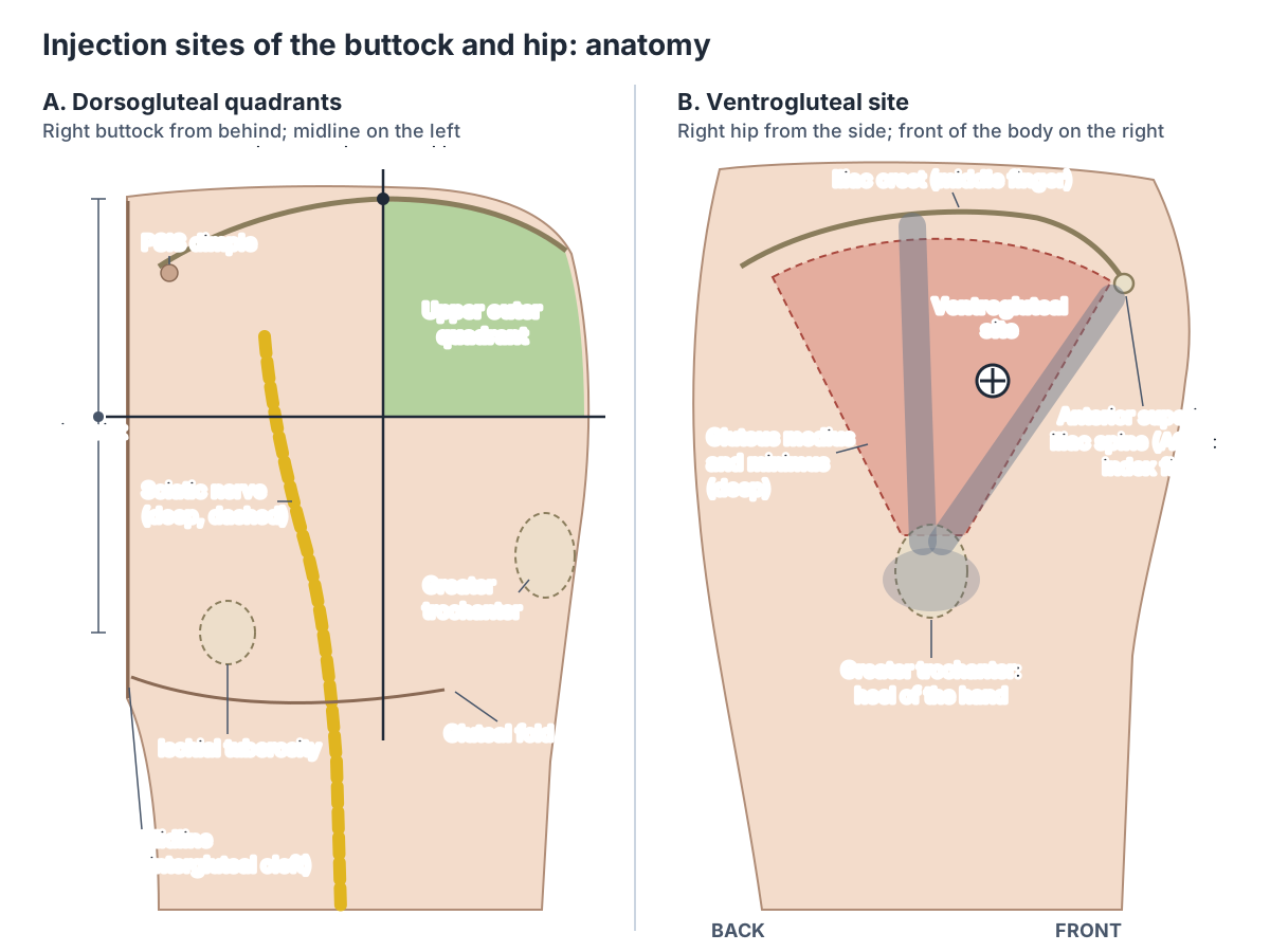
Dorsogluteal vs ventrogluteal injection site
| Feature | Dorsogluteal (upper outer quadrant) | Ventrogluteal |
|---|---|---|
| Where | Back of the buttock, upper outer quadrant | Side of the hip, triangle between iliac crest, anterior superior iliac spine and greater trochanter |
| Muscles | Gluteus maximus and gluteus medius | Gluteus medius and gluteus minimus |
| Nearby structures | Sciatic nerve, superior gluteal artery and nerve | Away from the sciatic nerve |
| Subcutaneous tissue (pooled mean) | About 43 mm | About 20 mm |
| Adverse effects (meta-analysis) | More pain, bleeding and bruising | Fewer; judged the safer site |
From Roldán-Chicano et al. 2023, Gulnar et al. 2023 and Soliman et al. 2018. Educational summary only.
The buttocks in the 3D model
The model opens from behind and highlights gluteus maximus, gluteus medius and minimus, tensor fasciae latae, the deep lateral rotators (piriformis, the gemelli, obturator internus and externus, quadratus femoris) and the superior gluteal, inferior gluteal and internal pudendal veins. Zoom in below gluteus maximus to find the deep rotators, or rotate to the side to see tensor fasciae latae and gluteus medius over the hip. The model, BodyParts3D from one adult male, has no sciatic nerve, gluteal arteries, sacrotuberous ligament or fat, so the size and shape of the buttocks in life are larger than the muscles alone suggest.
Use the veins as markers of the sciatic foramina: the superior gluteal vein leaves the pelvis above piriformis and the inferior gluteal vein below it, alongside the sciatic nerve. (TeachMeAnatomy: The Greater and Lesser Sciatic Foramina.)
Buttocks in the 3D model
- Gemellus inferior
- A small lateral rotator of the hip, below obturator internus.
- Gemellus superior
- A small lateral rotator of the hip, above obturator internus.
- Gluteus maximus
- The largest and most superficial buttock muscle.
- Gluteus medius
- A fan-shaped muscle under gluteus maximus on the outer ilium.
- Gluteus minimus
- The smallest and deepest gluteal muscle.
- Inferior gluteal vein
- The inferior gluteal vein drains the buttock.
- Internal pudendal vein
- The internal pudendal vein drains the perineum and external genitalia.
- Obturator externus
- A flat, fan-shaped deep muscle of the medial thigh that wraps behind the neck of the femur.
- Obturator internus
- A lateral rotator of the hip that arises inside the pelvis and leaves through the lesser sciatic foramen.
- Piriformis
- A pear-shaped lateral rotator of the hip that passes through the greater sciatic foramen.
- Quadratus femoris
- A flat quadrangular deep gluteal muscle below the inferior gemellus.
- Superior gluteal vein
- A vein of the gluteal region draining into the internal iliac vein.
- Tensor fasciae latae
- A small hip muscle at the front and side of the thigh.
Frequently asked questions
What muscles make up the buttocks?
Three gluteal muscles make up most of each buttock: the large gluteus maximus on the surface, gluteus medius beneath its upper part and gluteus minimus beneath that. Tensor fasciae latae sits at the front of the region. Under the lower half of gluteus maximus lie the small lateral rotators: piriformis, the two gemelli, obturator internus and quadratus femoris.
What is the crease under the buttocks called?
It is the gluteal fold, also called the gluteal sulcus. It is a horizontal skin crease where the gluteal skin is tethered by fibrous tissue to the deep fascia, and it marks the lower edge of the buttock. It is not the lower border of gluteus maximus, which lies deeper and crosses the fold at a slant. The groove between the buttocks is the intergluteal (natal) cleft.
What bone do you sit on?
You sit on the ischial tuberosities, the rough lower parts of the ischium on each side of the pelvis, often called the sitting bones. They carry the weight of the body when sitting and you can feel them by rocking on a firm chair. The hamstring muscles attach to them, and gluteus maximus contracts during sitting to ease the pressure on them.
Where is the sciatic nerve in the buttock?
The sciatic nerve enters the buttock through the greater sciatic foramen just below piriformis, lies deep to gluteus maximus and runs down into the back of the thigh. It crosses the lower inner part of the buttock. In two cadaver series, about 6 to 8% of nerves passed partly through or above piriformis instead of entirely below it.
Where is it safe to inject into the buttock?
For a dorsogluteal injection the upper outer quadrant is used, because the sciatic nerve runs through the lower inner part of the buttock. The ventrogluteal site at the side of the hip, between the anterior superior iliac spine, the iliac crest and the greater trochanter, is now often suggested as the safer choice: its fat layer is thinner and it lies away from the sciatic nerve. This is anatomy for learning, not injection training.
Why do women have more fat on their buttocks?
Women store relatively more fat under the skin of the hips and thighs, the gluteofemoral region, while men store more around the abdomen. For the same body mass index women typically have about 10% more body fat. Sex hormones are involved: after menopause fat tends to shift away from the gluteofemoral region towards the abdomen. Gluteofemoral fat is linked with a lower diabetes risk.
What are the dimples above the buttocks?
The dimples of Venus are small hollows in the skin over the posterior superior iliac spines, the back ends of the iliac crests. They lie about 4 cm to each side of the spinous process of the second sacral vertebra, where muscles and ligaments surround the bony spine without covering it, so the skin dips slightly.
References
- OpenStax. Anatomy and Physiology 2e, §8.3 The Pelvic Girdle and Pelvis. Rice University, 2022.
- OpenStax. Anatomy and Physiology 2e, §11.2 Naming Skeletal Muscles. Rice University, 2022.
- OpenStax. Anatomy and Physiology 2e, §11.6 Appendicular Muscles of the Pelvic Girdle and Lower Limbs. Rice University, 2022.
- Kenhub. Regions of the lower limb.
- Kenhub. Gluteal region.
- Kenhub. Gluteal muscles: attachments, supply and function.
- Kenhub. Gluteus maximus muscle.
- Kenhub. Posterior superior iliac spine.
- IMAIOS e-Anatomy. Buttock.
- IMAIOS e-Anatomy. Gluteal fold.
- TeachMeAnatomy. Muscles of the Gluteal Region.
- TeachMeAnatomy. The Greater Sciatic Foramen and Lesser Sciatic Foramen.
- TeachMeAnatomy. The Sciatic Nerve.
- TeachMeAnatomy. The Sacral Plexus.
- TeachMeAnatomy. Superior Gluteal Artery.
- TeachMeAnatomy. Inferior Gluteal Artery.
- Reynoso JP, De Jesus Encarnacion M, Nurmukhametov R, et al. Anatomical variations of the sciatic nerve exit from the pelvis and its relationship with the piriformis muscle: a cadaveric study. Neurol Int. 2022;14(4):894-902.
- Cornwall J. Are nursing students safe when choosing gluteal intramuscular injection locations? Australas Med J. 2011;4(6):315-321.
- Roldán-Chicano MT, Rodríguez-Tello J, Cebrián-López R, Moore JR, García-López MM. Adverse effects of dorsogluteal intramuscular injection versus ventrogluteal intramuscular injection: a systematic review and meta-analysis. Nurs Open. 2023;10(9):5975-5988.
- Gulnar E, Ordu Y, Biyik Bayram S, Çalışkan N. Gluteal muscle and subcutaneous tissue thicknesses in adults: a systematic review and meta-analysis. J Res Nurs. 2023;28(3):181-196.
- Soliman E, Ranjan S, Xu T, et al. A narrative review of the success of intramuscular gluteal injections and its impact in psychiatry. Biodes Manuf. 2018;1(3):161-170.
- Karastergiou K, Smith SR, Greenberg AS, Fried SK. Sex differences in human adipose tissues: the biology of pear shape. Biol Sex Differ. 2012;3:13.
- Boulet N, Briot A, Galitzky J, Bouloumié A. The sexual dimorphism of human adipose depots. Biomedicines. 2022;10(10):2615.
- Mitsuhashi N. et al. (2009). BodyParts3D: 3D structure database for anatomical concepts. Nucleic Acids Research 37, D782–D785. Data © The Database Center for Life Science, CC BY 4.0.
Related anatomy
- Hip musclesMuscles of the hip explained: gluteus maximus, medius and minimus, TFL, piriformis, obturator internus and the other…
- Hip jointHip anatomy explained: the ball-and-socket joint, acetabulum and labrum, iliofemoral and other ligaments, movements…
- PelvisPelvis and hip bone anatomy in 3D: ilium, ischium and pubis, ASIS and PSIS, ligaments, female pelvic anatomy and a…
- Lower limb nervesLower limb nerves explained: lumbar and sacral plexus diagram, femoral, saphenous, sciatic, tibial, common peroneal…
- Gluteus maximusGluteus maximus: The largest and most superficial buttock muscle. See where it is in an interactive 3D human body…
- PiriformisPiriformis: A pear-shaped lateral rotator of the hip that passes through the greater sciatic foramen. See where it is…
Explore the whole body in the interactive 3D human anatomy model, or browse the A–Z list of human structures.