

Where is the hip joint?
The hip joint lies deep in the groin, not at the waist. In front of it, only muscles separate the capsule from the main vessels of the thigh: iliopsoas, with its bursa, lies between the joint and the femoral artery and femoral nerve, and pectineus between the joint and the femoral vein. (Radiopaedia: Hip joint.)
The bony "hips" you feel when you put your hands on your waist are the iliac crests, the curved upper margins of the ilium. The anterior superior iliac spine, the front end of the crest, can be felt at the front and side of the hip. (OpenStax Anatomy and Physiology 2e, 8.3: The Pelvic Girdle and Pelvis.)
On the side of the upper thigh, the greater trochanter of the femur is the most lateral bony point you can feel. It lies just lateral to the femoral neck and gives attachment to gluteus medius, gluteus minimus and piriformis. (TeachMeAnatomy: The Femur.)
The femoral pulse is felt just below the inguinal ligament at the mid-inguinal point, halfway between the pubic symphysis and the anterior superior iliac spine, where the femoral artery enters the thigh. (TeachMeAnatomy: The Femoral Triangle.)
Hip joint anatomy: a ball-and-socket joint
The hip is a ball-and-socket synovial joint between the head of the femur and the acetabulum of the pelvis. The femoral head is hemispherical and fits deeply into the cup-like acetabulum on the lower outer side of the hip bone. Both surfaces are covered by articular cartilage, which is thicker over the weight-bearing areas. (TeachMeAnatomy: The Hip Joint.)
It is a multiaxial joint. Its range of motion is smaller than the shoulder's because the hip is built for stability and weight bearing: it carries the weight of the body and needs strength and stability for standing and walking. (OpenStax Anatomy and Physiology 2e, 9.6: Anatomy of Selected Synovial Joints.)
Kenhub calls the hip the most stable joint in the body, sacrificing mobility for weight bearing. The surfaces fit together best when the hip is partly flexed and abducted. (Kenhub: Hip joint.)
Hip and pelvis anatomy: the bones of the hip
Each hip bone (coxal bone) is formed by three bones, the ilium, ischium and pubis, which fuse during the late teenage years. They meet in the centre of the hip bone to form the acetabulum. Through its joint with the sacrum, each hip bone attaches a lower limb to the axial skeleton. (OpenStax Anatomy and Physiology 2e, 8.3: The Pelvic Girdle and Pelvis.)
Radiopaedia notes that the Y-shaped triradiate cartilage between the three parts of the acetabulum is usually fused by age 14 to 16. (Radiopaedia: Hip joint.)
The pelvic girdle is made of the two hip bones, the sacrum and the coccyx. The hip joint is where this ring meets each femur. (Kenhub: Hip and thigh anatomy.)
On the femur, the rounded head is joined to the shaft by the narrower neck, a common fracture site. The fovea capitis is a small pit on the head, and the greater trochanter is the large projection above the base of the neck. (OpenStax Anatomy and Physiology 2e, 8.4: Bones of the Lower Limb.)
The lesser trochanter projects from the back and inner side of the femur, just below the neck, and receives iliopsoas. In front, the intertrochanteric line joins the two trochanters and marks the attachment of the hip capsule and iliofemoral ligament; behind, the intertrochanteric crest joins them. (TeachMeAnatomy: The Femur.)
Ilium, ischium, pubis and male vs female pelvis, in 3D. Pelvis bones.
Acetabulum, labrum and articular cartilage
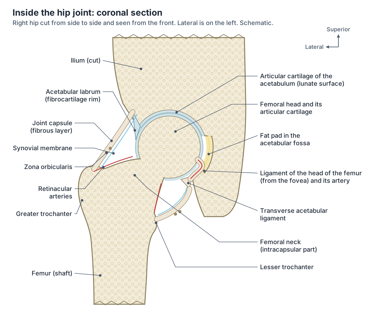
Only part of the acetabulum is cartilage-covered. The articular lunate surface is a C-shaped, incomplete ring covering the upper and outer parts of the socket. It is broadest at the front and top, where most body weight is borne. Its gap at the bottom is the acetabular notch, and the deep central floor, the acetabular fossa, has no cartilage and holds a fat pad. (Kenhub: Hip joint.)
The acetabular labrum is a ring of fibrocartilage attached to the rim of the acetabulum. It deepens the socket, increases the articular surface by about 10% and helps more than half of the femoral head fit inside the socket. It also seals the joint and helps keep synovial fluid evenly spread. (Kenhub: Acetabular labrum.)
The transverse acetabular ligament bridges the acetabular notch, turning it into a foramen through which vessels and nerves enter the joint. The labrum continues along its outer edge. (Kenhub: Hip joint.)
The fat pad in the acetabular fossa is also called the Haversian fat pad or pulvinar. The femoral head is covered in hyaline cartilage everywhere except the fovea. (Radiopaedia: Hip joint.)
Hip joint capsule, zona orbicularis and bursa
The strong fibrous capsule attaches to the rim of the acetabulum and the transverse ligament. On the femur it attaches to the intertrochanteric line in front, the base of the neck above, about 1 cm above the intertrochanteric crest behind, and near the lesser trochanter below. So part of the femoral neck is inside the capsule and part is outside. (Kenhub: Hip joint.)
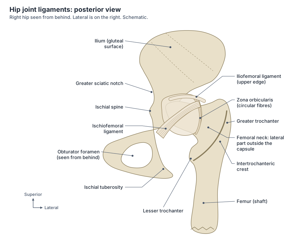
The capsule is thickest at the front and top, where stress is greatest, and thin and loose at the back and bottom. Its deep circular fibres form the zona orbicularis, a collar around the femoral neck with no bony attachment. (Kenhub: Hip joint.)
Anatomy studies show that the capsule's attachment below the anterior inferior iliac spine is wider than many textbooks suggest, and that the gluteus minimus tendon and the deep layer of iliopsoas connect to the capsule, which is thickened at those points. (Tsutsumi M, Nimura A, Akita K. Clinical anatomy of the musculoskeletal system in the hip region. Anat Sci Int. 2022.)
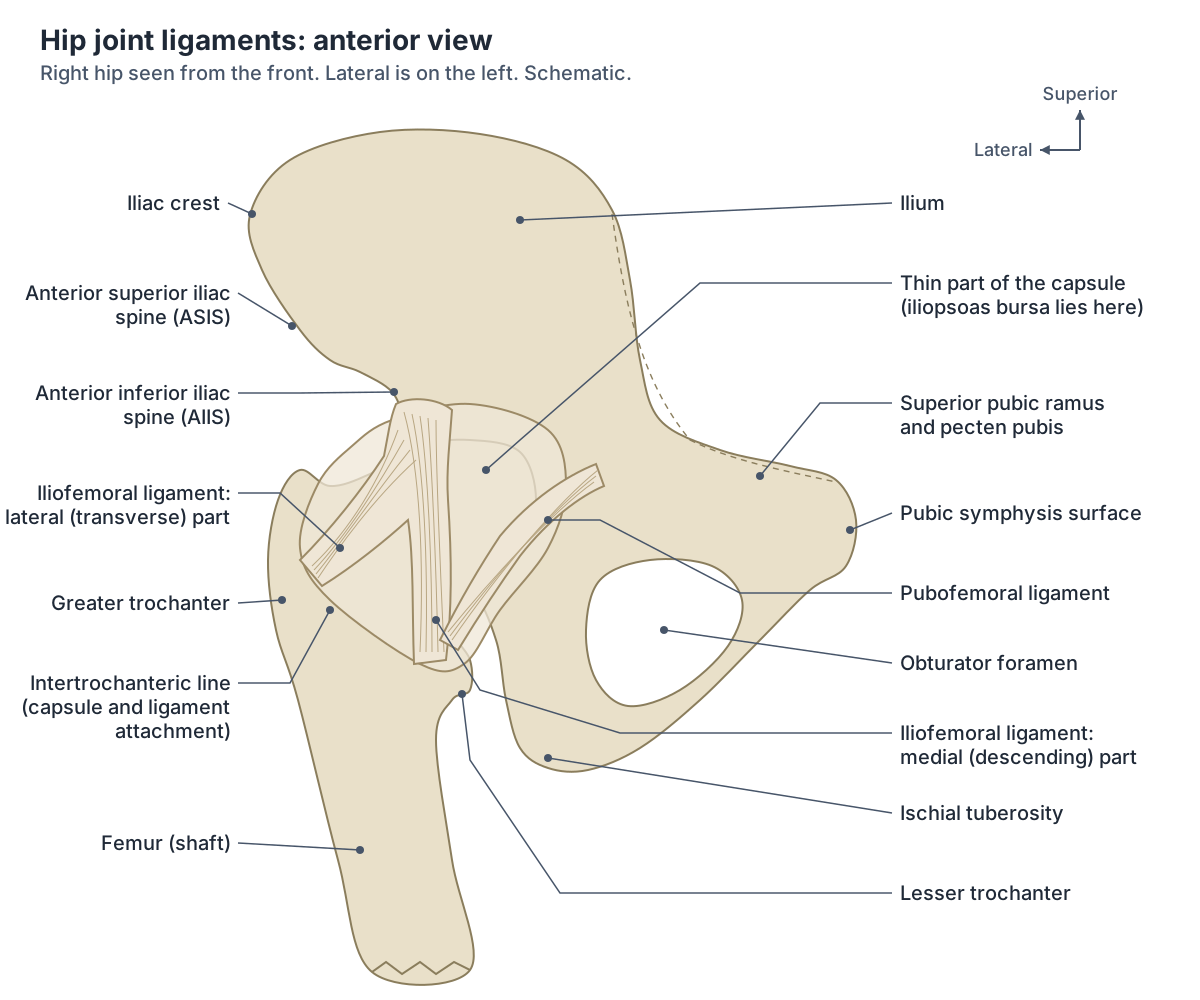
The iliopectineal (psoas) bursa lies between the iliopsoas tendon and the thin part of the capsule between the iliofemoral and pubofemoral ligaments. It communicates with the joint cavity. (Kenhub: Hip joint.)
Ligaments of the hip: iliofemoral, pubofemoral and ischiofemoral
Three ligaments thicken the capsule. The iliofemoral ligament runs from the anterior inferior iliac spine to the intertrochanteric line; the pubofemoral ligament from the superior pubic ramus to the intertrochanteric line; and the ischiofemoral ligament from the body of the ischium to the greater trochanter region. All three spiral around the neck, so they tighten in extension. (TeachMeAnatomy: The Hip Joint.)
The iliofemoral ligament, also called the Y-ligament of Bigelow, is shaped like an inverted Y. Its thicker descending (medial) part runs to the lower end of the intertrochanteric line and its transverse part to the upper end. It is said to be the strongest ligament in the body and stops the hip overextending when you stand. (Kenhub: Iliofemoral ligament.)
Kenhub adds that the iliofemoral ligament limits extension to about 10 to 20 degrees beyond vertical, that the pubofemoral ligament prevents excessive abduction, and that the ischiofemoral ligament is the weakest of the three and limits internal rotation. (Kenhub: Hip joint.)
Inside the joint, the ligament of the head of the femur (ligamentum teres) runs between the acetabulum and the femoral head. It is normally slack and gives no significant support, but it carries a small artery to the femoral head. (OpenStax Anatomy and Physiology 2e, 9.6: Anatomy of Selected Synovial Joints.)
Head, neck, trochanters and the attachments around the hip, in 3D. Femur.
Ligaments of the hip joint
| Ligament | From | To | Main role |
|---|---|---|---|
| Iliofemoral (Y-ligament of Bigelow) | Anterior inferior iliac spine and acetabular rim | Intertrochanteric line | Strongest; prevents hyperextension when standing |
| Pubofemoral | Superior pubic ramus and obturator crest | Lower capsule and intertrochanteric line | Prevents excessive abduction and extension |
| Ischiofemoral | Body of the ischium, behind and below the acetabulum | Base of the greater trochanter and back of the neck | Weakest; limits internal rotation and hyperextension |
| Ligament of the head of the femur (ligamentum teres) | Acetabular notch and transverse ligament | Fovea of the femoral head | Carries a small artery; little mechanical role |
| Transverse acetabular ligament | One side of the acetabular notch | The other side of the notch | Completes the acetabular rim; vessels pass beneath it |
Sources: TeachMeAnatomy, Kenhub, Radiopaedia, OpenStax.
What keeps the hip joint stable?
The deep acetabulum surrounds almost all of the femoral head, and the labrum makes it deeper still. The thick capsule and its three ligaments add most of the remaining stability, and because they spiral, they tighten as the hip extends. (TeachMeAnatomy: The Hip Joint.)
Muscles balance the ligaments. At the front, where the ligaments are strongest, the hip flexors are fewer and weaker; at the back, where the ligaments are weakest, the rotator muscles are more numerous and stronger and pull the femoral head into the socket. (TeachMeAnatomy: The Hip Joint.)
Compared with the shoulder, the hip allows less movement but is considerably more stable. The labrum adds stability but also limits range of movement. (Radiopaedia: Hip joint.)
Hip movements and range of motion
The hip moves in three planes: flexion and extension, abduction and adduction, and internal (medial) and external (lateral) rotation, which combine as circumduction. Kenhub gives about 120 degrees of passive flexion, up to about 30 degrees of extension beyond vertical, about 45 degrees each of abduction and adduction, about 35 degrees of internal rotation and about 45 degrees of external rotation with the hip extended. (Kenhub: Hip joint.)
Measured values are often lower. In 105 healthy men aged 30 to 40, passive hip flexion averaged 120 degrees, extension about 9.5 degrees, abduction about 38.5 degrees, adduction about 30.5 degrees, internal rotation about 32.5 degrees and external rotation about 33.6 degrees, with wide differences between people. (Roaas A, Andersson GB. Normal range of motion of the hip, knee and ankle joints in male subjects, 30-40 years of age. Acta Orthop Scand. 1982.)
Flexion is greater with the knee bent because knee flexion relaxes the hamstrings, and extension is limited by the capsule and iliofemoral ligament. (TeachMeAnatomy: The Hip Joint.)
Hip movements, ranges and main muscles
| Movement | Typical range (Kenhub) | Measured mean in healthy men (Roaas 1982) | Main muscles |
|---|---|---|---|
| Flexion | About 120 degrees (passive) | 120 degrees | Iliopsoas, rectus femoris, sartorius, pectineus |
| Extension | Up to about 30 degrees beyond vertical | About 9.5 degrees | Gluteus maximus, hamstrings |
| Abduction | About 45 degrees | About 38.5 degrees | Gluteus medius and minimus, tensor fasciae latae, piriformis |
| Adduction | About 45 degrees | About 30.5 degrees | Adductors longus, brevis and magnus, gracilis, pectineus |
| Internal (medial) rotation | About 35 degrees | About 32.5 degrees | Anterior gluteus medius and minimus, tensor fasciae latae |
| External (lateral) rotation | About 45 degrees | About 33.6 degrees | Gluteus maximus, piriformis, obturators, gemelli, quadratus femoris |
Passive ranges differ with method, age and sex; the 1982 study measured 105 men aged 30 to 40.
Which muscles move the hip?
Flexion is produced by iliopsoas, rectus femoris, sartorius and pectineus; extension by gluteus maximus and the hamstrings (semimembranosus, semitendinosus, biceps femoris); abduction by gluteus medius, gluteus minimus, piriformis and tensor fasciae latae; and adduction by adductors longus, brevis and magnus, pectineus and gracilis. (TeachMeAnatomy: The Hip Joint.)
Lateral rotation is produced by gluteus maximus, piriformis and biceps femoris, assisted by the obturators, gemelli and quadratus femoris. Medial rotation is produced by the anterior fibres of gluteus medius and minimus and by tensor fasciae latae. (TeachMeAnatomy: The Hip Joint.)
Gluteus medius is the largest hip abductor and is important for hip stability. It has a long, thick posterior part inserting on the back of the greater trochanter and a thinner anterolateral part, which tears more often. (Tsutsumi M, Nimura A, Akita K. Clinical anatomy of the musculoskeletal system in the hip region. Anat Sci Int. 2022.)
Gluteal muscles, deep rotators and tensor fasciae latae. Hip muscles.
Blood supply of the hip and femoral head
The medial and lateral circumflex femoral arteries, branches of the deep femoral artery, join around the base of the femoral neck to form a ring. The medial circumflex femoral artery supplies most of the joint; the lateral one has to pierce the iliofemoral ligament. The artery to the head of the femur and the superior and inferior gluteal arteries add a smaller supply. (TeachMeAnatomy: The Hip Joint.)
The arteries around the hip give off retinacular arteries, which run along the neck and carry most of the blood to the femoral head and neck. The artery of the head of the femur, inside the ligamentum teres, comes from the obturator artery. (Kenhub: Hip joint.)
Radiopaedia describes two anastomoses: the cruciate anastomosis (medial and lateral circumflex femoral, first perforating and inferior gluteal arteries) and the trochanteric anastomosis (superior gluteal and the ascending branches of both circumflex femoral arteries, often with the inferior gluteal). (Radiopaedia: Hip joint.)
Because the head depends on vessels running along the neck, damage to the medial circumflex femoral artery can cause avascular necrosis of the femoral head. (TeachMeAnatomy: The Hip Joint.)
The main artery of the thigh and its deep femoral branch. Femoral artery.
Nerve supply: why hip pain can be felt in the knee
TeachMeAnatomy lists the sciatic, femoral and obturator nerves as the nerves of the hip joint. The same nerves supply the knee, which is why pain from the hip can be felt in the knee and the other way round. (TeachMeAnatomy: The Hip Joint.)
Kenhub describes the supply in regions, from roots L2 to S1: the femoral nerve to the front, the obturator nerve below, the superior gluteal nerve above and the nerve to quadratus femoris behind. It notes that pain from the spine can also be referred to the hip. (Kenhub: Hip joint.)
Radiopaedia gives the nerve to rectus femoris (from the femoral nerve), the nerve to quadratus femoris, the anterior division of the obturator nerve and, variably, the superior gluteal nerve. (Radiopaedia: Hip joint.)
Femoral, obturator, sciatic and gluteal nerves. Nerves of the leg.
Hip fractures, dislocation and developmental dysplasia
A broken hip is usually a fracture of the femoral neck. Hip fractures are common in older people with osteoporosis, and damage to the blood supply of the femoral neck or head can lead to necrosis of the bone. (OpenStax Anatomy and Physiology 2e, 9.6: Anatomy of Selected Synovial Joints.)
Femoral neck fractures usually follow a low-energy fall in older people, more often women. The lower fragment is pulled up and rotated outwards, so the leg looks shortened and externally rotated. Intracapsular fractures can damage the medial circumflex femoral artery and cause avascular necrosis of the head; in extracapsular fractures the head's blood supply stays intact. (TeachMeAnatomy: The Femur.)
Traumatic dislocation is relatively uncommon. About 90% are posterior: the femoral head tears through the back and bottom of the capsule, the leg is shortened and medially rotated, and the sciatic nerve is injured in 10 to 20% of cases. Anterior dislocation is rare. (TeachMeAnatomy: The Hip Joint.)
In developmental dysplasia of the hip (DDH), the acetabulum is too shallow. Signs include limited abduction, a shorter leg and uneven skin folds on the thigh or buttock, and it is usually treated with a Pavlik harness, with surgery if that fails. (TeachMeAnatomy: The Hip Joint.)
Hip impingement (FAI) and labral tears
Femoroacetabular impingement (FAI) is abnormal contact between the femoral head or head-neck junction and the acetabulum. In cam impingement a bony bump at the head-neck junction jams into the front and top of the acetabulum when the hip flexes; in pincer impingement the acetabular rim covers too much of the head. Many people have both. (Macfarlane RJ, Haddad FS. The diagnosis and management of femoro-acetabular impingement. Ann R Coll Surg Engl. 2010.)
FAI causes a large share of acetabular labral tears, a major cause of hip pain in young adults. Groin pain is the main symptom (in 83% of patients in one series), often with stiffness in flexion and internal rotation and clicking, and FAI may lead to osteoarthritis of the hip. (Macfarlane RJ, Haddad FS. The diagnosis and management of femoro-acetabular impingement. Ann R Coll Surg Engl. 2010.)
Hip joint in the 3D model
Frequently asked questions
Where exactly is the hip joint?
The hip joint is deep in the groin, where the head of the femur sits in the acetabulum of the hip bone. It lies deep to the femoral vessels, with only the iliopsoas and pectineus muscles in between. The bony points people call their hips are the iliac crests at the waist, which are higher.
What type of joint is the hip?
The hip is a synovial ball-and-socket joint. The ball is the head of the femur and the socket is the acetabulum, deepened by the fibrocartilage labrum. It is multiaxial, allowing flexion, extension, abduction, adduction and rotation, but it moves less than the shoulder because it is built for stability and weight bearing.
What are the three ligaments of the hip?
The three capsular ligaments are the iliofemoral, pubofemoral and ischiofemoral ligaments. The iliofemoral ligament runs from the anterior inferior iliac spine to the intertrochanteric line and stops overextension; the pubofemoral limits abduction; the ischiofemoral, at the back, limits internal rotation. Inside the joint is the ligament of the head of the femur.
What is the strongest ligament in the body?
The iliofemoral ligament of the hip, also called the Y-ligament of Bigelow, is usually described as the strongest ligament in the body. It is an inverted Y-shaped band on the front of the hip joint. It tightens as the hip extends, pulling the femoral head into the socket when you stand upright, and it limits extension to about 10 to 20 degrees beyond vertical.
What is the hip labrum?
The acetabular labrum is a ring of fibrocartilage around the rim of the hip socket. It deepens the acetabulum, increases the joint surface by about 10%, and seals the joint so synovial fluid stays evenly spread. It can tear, often because of femoroacetabular impingement, causing groin pain, stiffness and clicking.
Why can hip problems cause knee pain?
The hip and knee share their nerve supply: the femoral, obturator and sciatic nerves send branches to both joints. So pain arising in the hip can be felt in the knee, and the other way round. Pain from the spine can also be referred to the hip.
References
- TeachMeAnatomy. The Hip Joint.
- Kenhub. Hip joint.
- Kenhub. Acetabular labrum.
- Kenhub. Iliofemoral ligament.
- Kenhub. Hip and thigh anatomy.
- Radiopaedia. Hip joint.
- OpenStax. Anatomy and Physiology 2e, §9.6 Anatomy of Selected Synovial Joints. Rice University, 2022.
- OpenStax. Anatomy and Physiology 2e, §8.3 The Pelvic Girdle and Pelvis. Rice University, 2022.
- OpenStax. Anatomy and Physiology 2e, §8.4 Bones of the Lower Limb. Rice University, 2022.
- TeachMeAnatomy. The Femur.
- TeachMeAnatomy. The Femoral Triangle.
- Macfarlane RJ, Haddad FS. The diagnosis and management of femoro-acetabular impingement. Ann R Coll Surg Engl. 2010;92(5):363-367.
- Tsutsumi M, Nimura A, Akita K. Clinical anatomy of the musculoskeletal system in the hip region. Anat Sci Int. 2022;97(2):157-164.
- Roaas A, Andersson GB. Normal range of motion of the hip, knee and ankle joints in male subjects, 30-40 years of age. Acta Orthop Scand. 1982;53(2):205-208.
- Mitsuhashi N. et al. (2009). BodyParts3D: 3D structure database for anatomical concepts. Nucleic Acids Research 37, D782–D785. Data © The Database Center for Life Science, CC BY 4.0.
Related anatomy
- PelvisPelvis and hip bone anatomy in 3D: ilium, ischium and pubis, ASIS and PSIS, ligaments, female pelvic anatomy and a…
- Hip musclesMuscles of the hip explained: gluteus maximus, medius and minimus, TFL, piriformis, obturator internus and the other…
- FemurFemur bone labelled: head, neck, trochanters, linea aspera, condyles and epicondyles, with muscle attachments, blood…
- Thigh musclesThigh muscles explained: quadriceps and sartorius at the front, adductor muscles of the inner thigh, hamstrings at the…
- ButtocksButtocks anatomy with a 3D model and labelled diagrams: surface landmarks, gluteal muscles, sciatic nerve, gluteal…
- Knee jointKnee anatomy with labelled diagrams and a 3D model: femur, tibia and patella, the ACL, PCL, MCL and LCL, menisci…
Explore the whole body in the interactive 3D human anatomy model, or browse the A–Z list of human structures.