

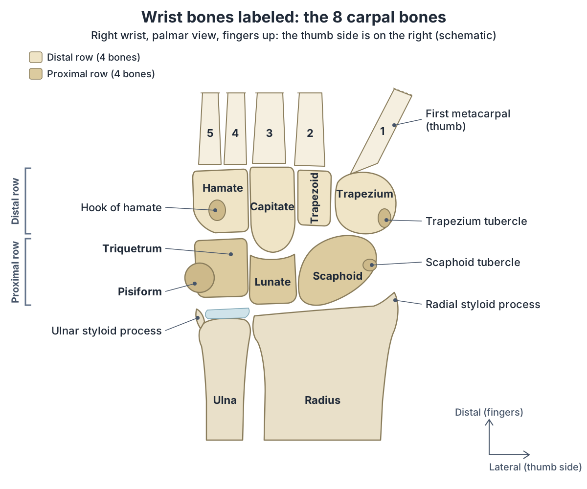
How many bones are in the wrist?
The wrist has eight carpal bones, the bones that form the articulation of the forearm with the hand. They are usually divided into two rows of four. The word carpus can mean these eight bones, and it is also used for the wrist joint as a whole. (Radiopaedia: Carpal bones.)
In the body-region names that anatomists use, the wrist is the carpal region, and the forearm above it is the antebrachial region. (OpenStax Anatomy and Physiology 2e, 1.6: Anatomical Terminology.)
The lower ends of the radius and ulna lie at the top of the wrist but are forearm bones, not carpal bones, and only the radius meets the carpals directly. The scaphoid and lunate articulate with the end of the radius, while the triquetrum articulates with a pad of fibrocartilage that spans the radius and the styloid process of the ulna, so the ulna does not articulate directly with any carpal bone. (OpenStax Anatomy and Physiology 2e, 8.2: Bones of the Upper Limb.)
For the wrist joint itself, its ligaments, the TFCC and the tendons that cross it, see wrist anatomy.
The 8 carpal bones named, row by row
The carpal bones are arranged in two rows. Listed from the lateral (thumb) side to the medial (little-finger) side, the proximal row is the scaphoid, lunate, triquetrum and pisiform, and the distal row is the trapezium, trapezoid, capitate and hamate. (OpenStax Anatomy and Physiology 2e, 8.2: Bones of the Upper Limb.)
Most of the names describe shapes. OpenStax translates them as boat-shaped (scaphoid), moon-shaped (lunate), three-cornered (triquetrum), pea-shaped (pisiform), table (trapezium), resembles a table (trapezoid), head-shaped (capitate) and hooked bone (hamate). (OpenStax Anatomy and Physiology 2e, 8.2: Bones of the Upper Limb.)
The proximal row faces the forearm and the distal row faces the palm. In the proximal row, the scaphoid and lunate articulate with the radius to form the wrist (radiocarpal) joint, and the distal row articulates with the metacarpals at the carpometacarpal joints. (TeachMeAnatomy: The Bones of the Hand: Carpals, Metacarpals and Phalanges.)
For the carpals together with the metacarpals and phalanges of the hand, see the hand bones guide.
The 8 carpal bones
| Row | Bone | Articulates with | Notable feature |
|---|---|---|---|
| Proximal | Scaphoid | Radius, lunate, capitate, trapezium, trapezoid | Largest bone of the proximal row; palmar tubercle; floor of the anatomical snuffbox; the carpal bone most often fractured |
| Proximal | Lunate | Radius, capitate, scaphoid, triquetrum; also the hamate in many wrists (type II lunate) | Crescent (moon) shape; loss of its blood supply is Kienböck disease |
| Proximal | Triquetrum | Lunate, hamate, pisiform; the articular disc at the wrist | Three-cornered (pyramid) shape; facet for the pisiform on its palmar surface |
| Proximal | Pisiform | Triquetrum (pisotriquetral joint) | Pea-shaped sesamoid bone in the tendon of flexor carpi ulnaris; usually the last carpal bone to ossify |
| Distal | Trapezium | Scaphoid, trapezoid, 1st and 2nd metacarpals | Base of the thumb; saddle joint with the 1st metacarpal; tubercle and groove for flexor carpi radialis |
| Distal | Trapezoid | Scaphoid, trapezium, capitate, 2nd metacarpal | Small and wedge-shaped |
| Distal | Capitate | Mainly the 3rd metacarpal; scaphoid, lunate, trapezoid, hamate | Largest carpal bone; head-shaped; first to ossify |
| Distal | Hamate | 4th and 5th metacarpals, capitate, triquetrum; the lunate in wrists with a type II lunate | Hook (hamulus) in the palm: medial wall of the carpal tunnel and attachment of the flexor retinaculum |
Each row runs from the thumb side to the little-finger side. Sources: Kenhub, Carpal bones; Radiopaedia bone articles; OpenStax Anatomy and Physiology 2e, 8.2; TeachMeAnatomy.
Wrist bones labeled: how to read a palmar view
The labelled wrist bones diagram on this page shows a right wrist from the palm (palmar) side with the fingers pointing up. The thumb is on the lateral side, so in this view the thumb side is on the viewer's right: the scaphoid and trapezium are on the right, and the pisiform and hamate on the left. The same view of a left wrist is a mirror image, with the thumb side on the viewer's left. (OpenStax Anatomy and Physiology 2e, 8.2: Bones of the Upper Limb.)
Two landmarks show that a picture is a palmar view and not a view of the back of the hand. One is the pisiform, a tiny pea-shaped bone on the distal palmar surface of the triquetrum; the other is the hook of the hamate (hamulus), which projects from the palmar surface of the hamate. (Kenhub: Carpal bones.)
The diagram also marks the two tubercles on the thumb side, the tubercle of the scaphoid and the tubercle of the trapezium. With the hook of the hamate and the pisiform on the little-finger side, they are the four bony points that the flexor retinaculum spans. (TeachMeAnatomy: The Carpal Tunnel.)
Scaphoid
The scaphoid is the largest bone of the proximal row and sits on the radial (thumb) side of the lunate. It is boat-shaped, lies obliquely, and articulates with five bones: the radius, trapezoid, trapezium, lunate and capitate. The scaphoid tubercle is a bony prominence on the palmar surface of its distal pole, and its narrow middle, the waist, is the commonest site of scaphoid fracture. (Radiopaedia: Scaphoid.)
The scaphoid and the trapezium form the floor of the anatomical snuffbox, and in a scaphoid fracture the characteristic clinical feature is pain and tenderness in the snuffbox. (TeachMeAnatomy: The Anatomical Snuffbox.)
For the scaphoid's poles, blood supply, ossification and fractures in detail, see the scaphoid guide.
Lunate bone
The lunate, named for its moon-shaped appearance, sits in the middle of the proximal row between the scaphoid and the triquetrum. Proximally it articulates with the radius and distally with the capitate. (Kenhub: Carpal bones.)
The lunate is crescent-shaped. Along with the scaphoid and triquetrum it forms the carpal surface of the radiocarpal (wrist) joint, it has small joints with its neighbours, the scaphoid, capitate and triquetrum, and in many wrists (a type II lunate) a separate facet for the hamate, and it articulates with the triangular fibrocartilage complex. It is the bone affected in Kienböck disease, described in the injuries section below. (Radiopaedia: Lunate.)
Triquetrum (triquetral bone)
The triquetrum is a pyramid-shaped bone, its name meaning three-cornered bone, on the medial (little-finger) side of the proximal row. It articulates with the lunate laterally and the hamate distally, and an isolated oval facet on its palmar surface articulates with the pisiform. (Kenhub: Carpal bones.)
The triquetrum does not reach the ulna. It articulates with a fibrocartilaginous pad that spans the radius and the styloid process of the ulna, which is why the end of the ulna does not directly articulate with any carpal bone. (OpenStax Anatomy and Physiology 2e, 8.2: Bones of the Upper Limb.)
Pisiform
The pisiform sits on the palm side of the proximal row, in a plane in front of the other carpal bones, and articulates with the triquetrum, forming the pisotriquetral joint. It is considered a sesamoid bone within the tendon of flexor carpi ulnaris, and it is often the last carpal bone to ossify. (Radiopaedia: Pisiform.)
For why the pisiform is a sesamoid, every attachment on it and its place beside Guyon's canal, see the pisiform guide.
Trapezium bone
The trapezium is the most lateral (radial) bone of the distal row, located between the scaphoid and the first metacarpal at the base of the thumb. It articulates with the scaphoid proximally, the trapezoid medially, and the thumb and index finger metacarpals distally. (Radiopaedia: Trapezium.)
Its joint with the first metacarpal is a sellar (saddle-shaped) joint. The main feature of the bone is a tubercle on its palmar surface, next to a groove that contains the tendon of flexor carpi radialis, and it gives proximal attachment to the three thenar muscles of the thumb: opponens pollicis, abductor pollicis brevis and flexor pollicis brevis. (Kenhub: Trapezium bone.)
Trapezoid bone, and trapezium vs trapezoid
The trapezoid is a small, wedge-shaped bone in the distal row. It articulates with the scaphoid proximally, the trapezium laterally and the capitate medially, and its distal facet articulates with the second metacarpal, the metacarpal of the index finger. (Kenhub: Carpal bones.)
The two names are easy to mix up, and both mean table-like. The trapezium is the one at the thumb end of the distal row, and the trapezoid sits beside it, nearer the middle of the wrist. In OpenStax's mnemonic, given below, the last word, Thumb, stands for the trapezium. (OpenStax Anatomy and Physiology 2e, 8.2: Bones of the Upper Limb.)
Capitate
The capitate is the largest of all the carpal bones. It sits at the centre of the distal row and is head-shaped. It articulates mainly with the base of the third metacarpal, and also with the scaphoid and lunate proximally, the trapezoid laterally and the hamate medially. (Radiopaedia: Capitate.)
It is generally the first carpal bone to ossify, within the first months of life, as the ossification section below explains. (Radiopaedia: Capitate.)
Hamate and the hook of the hamate
The hamate is wedge-shaped and is the most medial bone of the distal row. It articulates distally with the fourth and fifth metacarpals, laterally with the capitate and proximally with the triquetrum. Its main feature is the hamulus, or hook of the hamate, which forms part of the medial wall of the carpal tunnel and is the attachment point for several muscles and ligaments, including the flexor retinaculum. (Kenhub: Carpal bones.)
The hook lies in the palm, slightly off centre towards the little finger, and the ulnar artery and ulnar nerve pass closely by on its little-finger side. (AAOS OrthoInfo: Hook of the Hamate Fracture of the Wrist.)
The carpal arch and the carpal tunnel
In the articulated hand the carpal bones form an arch that is concave on the palm side. TeachMeAnatomy describes the lateral side of the arch as formed by the scaphoid and trapezium tubercles and the medial side by the hook of the hamate and the pisiform. (TeachMeAnatomy: The Carpal Tunnel.)
A thick band of connective tissue, the flexor retinaculum, spans the arch between the hook of the hamate and pisiform medially and the scaphoid and trapezium laterally. The bones form the base and sides of the carpal tunnel and the retinaculum forms its roof; the tunnel contains 9 tendons, surrounded by synovial sheaths, and the median nerve. (TeachMeAnatomy: The Carpal Tunnel.)
For the carpal tunnel's contents in a cross-section, Guyon's canal and carpal tunnel syndrome, see wrist anatomy.
Joints of the wrist bones
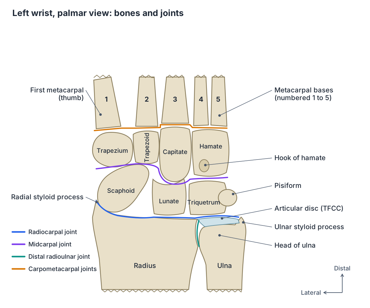
Radiopaedia describes the wrist joint as three separate joints. At the radiocarpal joint, the radius and its attached articular disc meet the proximal row (scaphoid, lunate and triquetrum). At the midcarpal joint, the scaphoid, lunate and triquetrum meet the trapezium, trapezoid, capitate and hamate. The distal radioulnar joint, between the radius and the head of the ulna, lies just above them and does not involve the carpal bones. (Radiopaedia: Wrist joint.)
Together, the radiocarpal and midcarpal joints are responsible for all movements of the hand at the wrist. Beyond the distal row, the proximal end of each metacarpal articulates with one of the distal carpal bones at the carpometacarpal joints. (OpenStax Anatomy and Physiology 2e, 8.2: Bones of the Upper Limb.)
For the joints, tendons and nerves of the palm, fingers and thumb beyond the wrist, see hand anatomy.
Carpal bones mnemonics
OpenStax gives the mnemonic "So Long To Pinky, Here Comes The Thumb". It starts on the lateral (thumb) side and names the proximal bones from lateral to medial (scaphoid, lunate, triquetrum, pisiform), then makes a U-turn to name the distal bones from medial to lateral (hamate, capitate, trapezoid, trapezium). It therefore starts and finishes on the thumb side, and its last word, Thumb, is the trapezium. (OpenStax Anatomy and Physiology 2e, 8.2: Bones of the Upper Limb.)
Kenhub uses "She Likes To Play Try To Catch Her" for scaphoid, lunate, triquetral, pisiform, trapezium, trapezoid, capitate and hamate. This version reads both rows in the same direction, from the thumb side, so the first T of the second half is the trapezium. The two mnemonics give the distal row in opposite orders, so learn the one that matches the order your course uses. (Kenhub: Carpal bones.)
Radiopaedia offers a memory aid for the order in which the carpal bones ossify: start at the capitate and move anticlockwise around the palmar (volar) surface of a right wrist, through the hamate, triquetrum, lunate, scaphoid, trapezium and trapezoid. The pisiform, which ossifies last, is left out of the circle. (Radiopaedia: Ossification centres of the hand.)
When do the carpal bones ossify?
At birth there is no calcification in the carpal bones. They ossify in a predictable sequence, starting with the capitate and ending with the pisiform, although there is great individual variability in the ages. (Radiopaedia: Ossification centres of the hand.)
A radiological study of 279 hand and wrist X-rays of Saudi children found the same sequence. The capitate and hamate appeared first, during the first year of life, followed by the triquetrum and then the lunate. The scaphoid, trapezium and trapezoid came next, appearing together in girls, while in boys the trapezoid followed the other two. The pisiform was last, at the end of the first decade. Some bones appeared earlier than reported in other populations. (Al-Khater KM et al. Time of appearance of ossification centers in carpal bones. A radiological retrospective study on Saudi children. Saudi Med J. 2020.)
Because the carpal bones appear in sequence, they can be used to estimate a child's age from an X-ray. A 2024 review of age estimation gives the upper age limit at which each one appears: the capitate and hamate by 1 year, the triquetrum by 3, the lunate by 5, the scaphoid by 6, the trapezium and trapezoid by 7, and the pisiform by 12 years. (Bhardwaj V et al. Demystifying the Radiography of Age Estimation in Criminal Jurisprudence: A Pictorial Review. Indian J Radiol Imaging. 2024.)
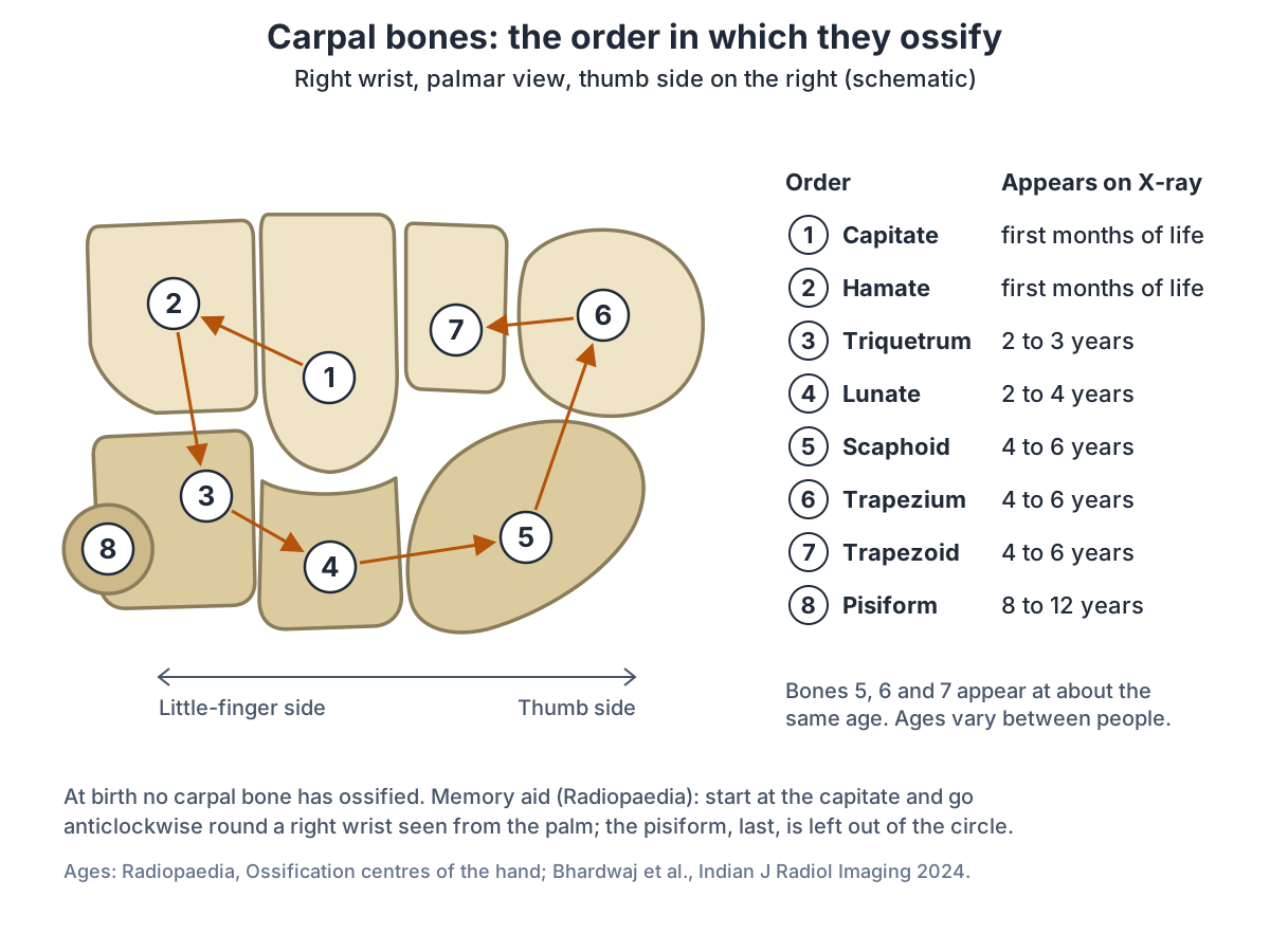
Order in which the carpal bones ossify
| Order | Bone | Approximate age it appears on X-ray (Radiopaedia) | Upper age limit (Bhardwaj et al. 2024) |
|---|---|---|---|
| 1 | Capitate | 1 to 3 months | 1 year |
| 2 | Hamate | 2 to 4 months | 1 year |
| 3 | Triquetrum | 2 to 3 years | 3 years |
| 4 | Lunate | 2 to 4 years | 5 years |
| 5 | Scaphoid | 4 to 6 years | 6 years |
| 6 | Trapezium | 4 to 6 years | 7 years |
| 7 | Trapezoid | 4 to 6 years | 7 years |
| 8 | Pisiform | 8 to 12 years | 12 years |
The scaphoid, trapezium and trapezoid (5 to 7) appear at about the same age. Ages vary between individuals and populations; in a Saudi study some bones appeared earlier (Al-Khater et al. 2020). Sources: Radiopaedia, Ossification centres of the hand; Bhardwaj et al., Indian J Radiol Imaging 2024.
Carpal vs metacarpal bones
The carpal bones are the eight bones of the wrist, at the base of the hand; the metacarpal bones are the five bones of the palm, numbered 1 to 5 beginning at the thumb. The proximal end of each metacarpal articulates with one of the distal carpal bones, and its expanded distal end articulates with a proximal phalanx at the metacarpophalangeal joint. (OpenStax Anatomy and Physiology 2e, 8.2: Bones of the Upper Limb.)
For the metacarpals and phalanges, with all 27 bones of the hand labelled, see the hand bones guide.
Common wrist bone injuries
The scaphoid is the most commonly fractured carpal bone, typically from a fall on an outstretched hand, and pain and tenderness in the anatomical snuffbox is the characteristic sign. Its blood supply runs from distal to proximal, so a fracture across its waist can interrupt the supply to the proximal part, which is then at particular risk of avascular necrosis. (TeachMeAnatomy: The Bones of the Hand: Carpals, Metacarpals and Phalanges.)
A scaphoid fracture does not always show up on an X-ray straight away, and because parts of the scaphoid have a poor blood supply, failure to heal (nonunion) is more common than in other fractures. (AAOS OrthoInfo: Scaphoid Fracture of the Wrist.)
The triquetrum is the second commonest carpal bone to fracture, after the scaphoid, usually from a fall onto an outstretched hand. Most triquetral fractures are small fractures of the back (dorsal) surface of the bone. (Radiopaedia: Triquetral fracture.)
Fractures of the hook of the hamate are rare. They occur mainly in players of golf, baseball and racquet sports, from a direct blow while swinging, and they may cause Guyon's canal syndrome, a compression of the ulnar nerve near the hook. (Radiopaedia: Hook of hamate fracture.)
A forceful backward bend (hyperextension) of the wrist, from a fall on an outstretched hand or high-energy trauma such as a road accident, usually in a young adult, can tear the ligaments around the lunate. (Dvorsky JL et al. A Review of Perilunate Wrist Dislocations. J Hand Surg Glob Online. 2025.)
In a perilunate dislocation the lunate stays in line with the radius while the rest of the carpus is pushed dorsally (towards the back of the hand). In the rarer and more severe lunate dislocation, the lunate itself is pushed and rotated towards the palm, into the carpal tunnel. Swelling and the displaced lunate can compress the median nerve, causing an acute carpal tunnel syndrome, so these injuries need urgent treatment, and even after treatment there is a high risk of later arthritis and wrist instability. (Radiopaedia: Lunate dislocation.)
Kienböck disease is osteonecrosis (death of bone tissue) of the lunate. Overall it affects men and women about equally, and in young adult men it most often affects the dominant wrist. Its symptoms are non-specific: wrist pain, swelling, reduced range of motion and pain with gripping and loading. (Radiopaedia: Kienböck disease.)
In Kienböck's disease the blood supply to the lunate is interrupted, and the cause is not known. These notes explain the anatomy only; anyone with a wrist injury or persistent wrist pain should see a clinician. (AAOS OrthoInfo: Kienböck's Disease.)
How to use the 3D model of the wrist bones
The 3D model uses BodyParts3D, built from an adult male body, and highlights all eight carpal bones of the left wrist from the palm side, with the hand hanging down in the anatomical position. Because the fingers point down, the proximal row is at the top of the view and the distal row below it, and the thumb side, with the scaphoid, trapezium and trapezoid, is on the viewer's right. The Female option uses the same carpal bones. Rotate the wrist to see its back, and zoom out to see the ghosted ends of the radius and ulna and the metacarpals around it.
In the palm-side view the pisiform overlaps the triquetrum, because it sits in a plane in front of the rest of the carpal bones. (Radiopaedia: Pisiform.)
Wrist bones (carpal bones) in the 3D model
- Capitate
- The largest carpal bone, with a rounded head.
- Hamate
- A wedge-shaped carpal bone of the distal row with a hook.
- Lunate
- A crescent-shaped carpal bone in the middle of the proximal row.
- Pisiform
- A small, pea-shaped carpal bone on the palmar side of the triquetrum.
- Scaphoid
- The largest bone of the proximal carpal row, shaped like a boat.
- Trapezium
- A four-sided carpal bone at the thumb side of the distal row.
- Trapezoid
- A small wedge-shaped carpal bone of the distal row.
- Triquetral
- A pyramid-shaped carpal bone on the ulnar side of the proximal row.
Frequently asked questions
How many bones are in the wrist?
The wrist has eight carpal bones, in two rows of four. The proximal row, nearest the forearm, is the scaphoid, lunate, triquetrum and pisiform; the distal row, nearest the palm, is the trapezium, trapezoid, capitate and hamate. The lower ends of the radius and ulna lie just above them but are counted as forearm bones, not wrist bones.
What are the 8 carpal bones in order?
From the thumb side to the little-finger side, the proximal row is the scaphoid, lunate, triquetrum and pisiform, and the distal row is the trapezium, trapezoid, capitate and hamate. Kenhub's mnemonic "She Likes To Play Try To Catch Her" follows this order. OpenStax's "So Long To Pinky, Here Comes The Thumb" names the distal row in the reverse direction.
Where is the trapezium bone?
The trapezium is the carpal bone at the base of the thumb, the most lateral bone of the distal row. It sits between the scaphoid and the first metacarpal, and it also articulates with the trapezoid and the second metacarpal. Its saddle-shaped joint with the first metacarpal is the carpometacarpal joint of the thumb, and the three thenar muscles take attachment from it.
What is the largest carpal bone?
The capitate is the largest of the eight carpal bones. It sits at the centre of the distal row and articulates mainly with the third metacarpal, as well as with the scaphoid, lunate, trapezoid and hamate. It is also generally the first carpal bone to ossify, within the first months of life. The largest bone of the proximal row is the scaphoid.
Which wrist bone is most commonly fractured?
The scaphoid, on the thumb side of the proximal row, is the most commonly fractured carpal bone, usually after a fall onto an outstretched hand. Pain and tenderness in the anatomical snuffbox is the typical sign, and the break may not show on the first X-ray. The triquetrum is the second commonest. Anyone with a wrist injury should see a clinician.
What is the difference between carpal and metacarpal bones?
Carpal bones are the eight bones of the wrist, in two rows. Metacarpal bones are the five bones of the palm, numbered 1 to 5 from the thumb. The base of each metacarpal articulates with the distal row of carpals at the carpometacarpal joints, and its head articulates with a proximal phalanx. With the 14 phalanges, they make up the 27 bones of the wrist and hand.
Which carpal bone is a sesamoid bone?
The pisiform. It is a small, pea-shaped bone considered a sesamoid within the tendon of flexor carpi ulnaris, and it sits on the palmar surface of the triquetrum, with which it forms the pisotriquetral joint. It is also usually the last carpal bone to ossify, appearing on X-rays at about 8 to 12 years of age.
References
- OpenStax. Anatomy and Physiology 2e, §8.2 Bones of the Upper Limb. Rice University, 2022.
- OpenStax. Anatomy and Physiology 2e, §1.6 Anatomical Terminology. Rice University, 2022.
- TeachMeAnatomy. The Bones of the Hand: Carpals, Metacarpals and Phalanges.
- TeachMeAnatomy. The Carpal Tunnel.
- TeachMeAnatomy. The Anatomical Snuffbox.
- Kenhub. Carpal bones.
- Kenhub. Trapezium bone.
- Radiopaedia. Carpal bones.
- Radiopaedia. Wrist joint.
- Radiopaedia. Ossification centres of the hand.
- Radiopaedia. Scaphoid.
- Radiopaedia. Lunate.
- Radiopaedia. Pisiform.
- Radiopaedia. Trapezium.
- Radiopaedia. Trapezoid.
- Radiopaedia. Capitate.
- Radiopaedia. Hamate.
- Radiopaedia. Triquetral fracture.
- Radiopaedia. Hook of hamate fracture.
- Radiopaedia. Kienböck disease.
- Radiopaedia. Lunate dislocation.
- Al-Khater KM, Hegazi TM, Al-Thani HF, Al-Muhanna HT, Al-Hamad BW, Alhuraysi SM, Alsfyani WA, Alessa FW, et al. Time of appearance of ossification centers in carpal bones. A radiological retrospective study on Saudi children. Saudi Med J. 2020;41(9):938–946.
- Bhardwaj V, Kumar I, Aggarwal P, Singh PK, Shukla RC, Verma A. Demystifying the Radiography of Age Estimation in Criminal Jurisprudence: A Pictorial Review. Indian J Radiol Imaging. 2024;34(3):496–510.
- Dvorsky JL, Rosenberry S, Da Costa A, Green A, Fowler J. A review of perilunate wrist dislocations. J Hand Surg Glob Online. 2025;7(5):100803.
- Guo RC, Cardenas JM, Wu CH. Triquetral Fractures Overview. Curr Rev Musculoskelet Med. 2021;14(2):101–106.
- American Academy of Orthopaedic Surgeons. Scaphoid Fracture of the Wrist. OrthoInfo.
- American Academy of Orthopaedic Surgeons. Hook of the Hamate Fracture of the Wrist. OrthoInfo.
- American Academy of Orthopaedic Surgeons. Kienböck's Disease. OrthoInfo.
- Lichtman DM, Pientka WF 2nd. Kienböck Disease: Recent Advances in Understanding and Management. J Bone Joint Surg Am. 2025.
- Mitsuhashi N. et al. (2009). BodyParts3D: 3D structure database for anatomical concepts. Nucleic Acids Research 37, D782–D785. Data © The Database Center for Life Science, CC BY 4.0.
Related anatomy
- Wrist jointWrist anatomy with labelled diagrams and a 3D model: 8 carpal bones, radiocarpal and midcarpal joints, wrist…
- Hand bonesHow many bones are in the hand? A labeled hand bones diagram and 3D model name all 27: the 8 carpals, 5 metacarpals…
- ScaphoidScaphoid bone anatomy: where it is, poles, waist, tubercle, articulations with five bones, retrograde blood supply…
- PisiformPisiform bone anatomy: location, why it is a sesamoid, muscles and ligaments attached, Guyon's canal, ossification and…
- Hand anatomyHand anatomy with labelled diagrams and a 3D model: 27 bones, finger and thumb joints, flexor and extensor tendons…
- Arm bonesArm bones labeled in a diagram and 3D model: the humerus, radius and ulna, their landmarks and joints, how the forearm…
Explore the whole body in the interactive 3D human anatomy model, or browse the A–Z list of human structures.