

What is the wrist? The joints that make it up
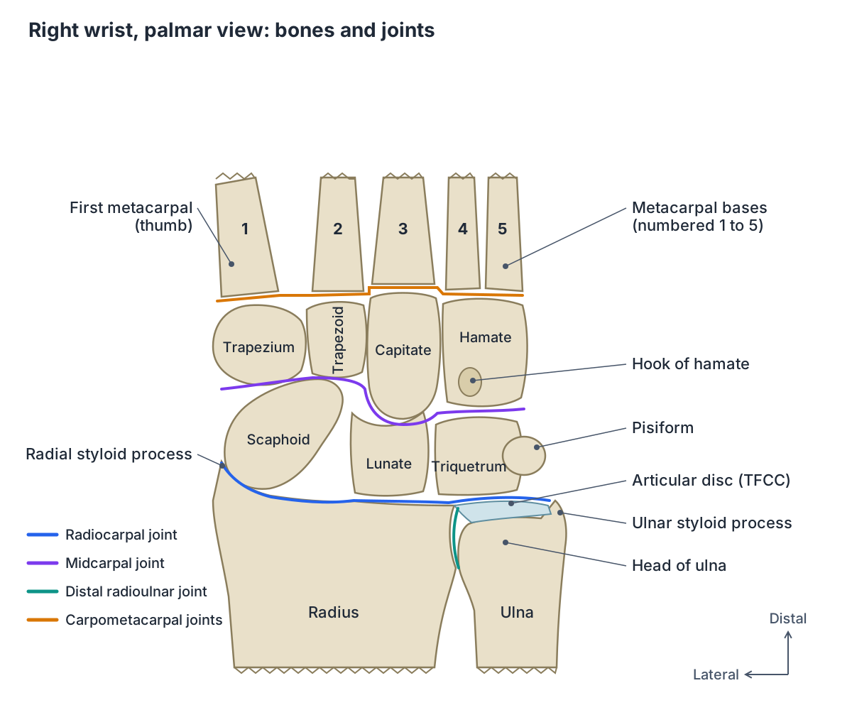
The wrist and the base of the hand are formed by eight small carpal bones. The radiocarpal joint, between the forearm and the first row of carpals, and the midcarpal joint, between the two rows of carpals, are together responsible for all movements of the hand at the wrist. (OpenStax Anatomy and Physiology 2e, 8.2: Bones of the Upper Limb.)
Radiopaedia describes the wrist as really three separate joints: the radiocarpal joint (radius and articular disc against the scaphoid, lunate and triquetrum), the distal radioulnar joint (ulnar notch of the radius against the head of the ulna) and the midcarpal joint (proximal row of carpals against the distal row). The word carpus can mean either the whole wrist joint or only the eight carpal bones. (Radiopaedia: Wrist joint.)
Definitions vary. Kenhub notes that, technically, the radiocarpal joint is considered the only articular component of the wrist joint, so many textbooks use "wrist joint" to mean the radiocarpal joint alone. (Kenhub: Wrist joint.)
The palm, fingers and thumb beyond the wrist: bones, joints, tendons, nerves and vessels. Hand anatomy.
Joints of the wrist region
| Joint | Bones | Type | Main role |
|---|---|---|---|
| Radiocarpal | Distal radius and articular disc with scaphoid, lunate and triquetrum | Condyloid (ellipsoid) synovial | Most of wrist extension; part of flexion and deviation |
| Midcarpal | Proximal row (scaphoid, lunate, triquetrum) with distal row (trapezium, trapezoid, capitate, hamate) | Synovial, between the two carpal rows | Most of wrist flexion and of radial and ulnar deviation |
| Distal radioulnar | Ulnar notch of the radius with the head of the ulna, joined by the articular disc | Pivot | Pronation and supination |
| Carpometacarpal | Distal row of carpals with the bases of the metacarpals | Synovial (thumb: saddle) | Links wrist to hand; thumb CMC allows opposition |
Based on Radiopaedia, OpenStax §8.2, TeachMeAnatomy and Kenhub (wrist joint and trapeziometacarpal joint).
Wrist bones: the carpal bones, radius and ulna
The carpal bones lie in two rows. From the thumb side to the little finger side, the proximal row is the scaphoid, lunate, triquetrum and pisiform, and the distal row is the trapezium, trapezoid, capitate and hamate. In the articulated hand they form a U-shaped grouping that is hollow on the palm side. (OpenStax Anatomy and Physiology 2e, 8.2: Bones of the Upper Limb.)
The pisiform is a sesamoid bone that forms within the tendon of flexor carpi ulnaris. The distal row articulates with the metacarpals at the carpometacarpal joints. (TeachMeAnatomy: Bones of the Hand.)
The distal radius has a smooth surface that articulates with two carpal bones, a pointed styloid process on its lateral side and the ulnar notch on its medial side. The distal ulna ends in a small rounded head with a styloid process projecting from its posterior side. The ulna does not articulate directly with any carpal bone. (OpenStax Anatomy and Physiology 2e, 8.2: Bones of the Upper Limb.)
The articular surface of the distal radius is divided into a scaphoid fossa and a lunate fossa by a ridge running from front to back. (Kenhub: Wrist joint.)
Each of the eight carpal bones in detail, with the metacarpals, phalanges and sesamoid bones. Hand bones.
Radiocarpal joint: the wrist joint proper
The radiocarpal joint is a condyloid (ellipsoid) synovial joint. Its proximal surface is the distal end of the radius and the articular disc; its distal surface is the proximal row of carpal bones except the pisiform. Together these carpals form a convex surface that fits the concave radius and disc. (TeachMeAnatomy: The Wrist Joint.)
The scaphoid and lunate articulate directly with the radius, whereas the triquetrum articulates with a fibrocartilaginous pad that spans the radius and the styloid process of the ulna. (OpenStax Anatomy and Physiology 2e, 8.2: Bones of the Upper Limb.)
A two-layered capsule, an outer fibrous layer and an inner synovial membrane, encloses the joint. It attaches above to the distal radius and ulna and below to the proximal row of carpals. The close-packed position is extension with slight radial deviation. (Kenhub: Wrist joint.)
Wrist ligaments: extrinsic and intrinsic
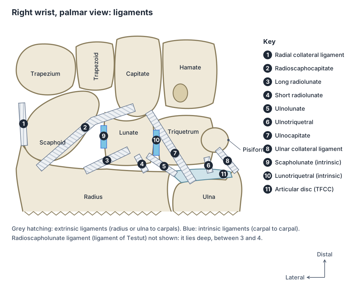
Wrist ligaments are grouped by where they run. Extrinsic ligaments link the radius or ulna to the carpal bones and are the main stabilisers. Intrinsic ligaments are short ligaments between the carpal bones themselves; they are small and easily damaged by excessive force or twisting. (TeachMeAnatomy: The Wrist Joint.)
On the palm side, the radioscaphocapitate ligament forms the lateral part of the capsule, and its most lateral fibres make up the radial collateral ligament. The long radiolunate ligament runs on its ulnar side, and the short radiolunate ligament runs from the rim of the lunate fossa to the palmar lunate. Kenhub notes that the palmar ligaments are stronger than the single dorsal radiocarpal ligament, which limits full flexion. (Kenhub: Wrist joint.)
Radiopaedia lists the palmar ulnocarpal ligaments as the ulnolunate, ulnotriquetral and ulnocapitate ligaments. It describes the radioscapholunate ligament (ligament of Testut) as not a true ligament: it gives no mechanical strength but acts as a conduit for vessels and nerves. (Radiopaedia: Wrist joint.)
The collateral ligaments sit at the sides. The ulnar collateral ligament runs from the ulnar styloid process to the triquetrum and pisiform, and the radial collateral ligament runs from the radial styloid process to the scaphoid and trapezium. The palmar radiocarpal ligaments make the hand follow the forearm in supination, and the dorsal radiocarpal ligament makes it follow in pronation. (TeachMeAnatomy: The Wrist Joint.)
The main intrinsic ligaments of the proximal row are the scapholunate and lunotriquetral ligaments; Radiopaedia also classes the TFCC as an intrinsic stabiliser. (Radiopaedia: Wrist joint.)
Main ligaments of the wrist
| Ligament | Group | Runs between | Key point |
|---|---|---|---|
| Radioscaphocapitate | Extrinsic, palmar | Radius to scaphoid and capitate | Forms the lateral capsule; lateral fibres are the radial collateral ligament |
| Long and short radiolunate | Extrinsic, palmar | Radius to lunate | Hold the lunate to the radius |
| Radioscapholunate (Testut) | Extrinsic, palmar | Radius to scapholunate junction | Not a true ligament; carries vessels and nerves |
| Ulnolunate, ulnotriquetral, ulnocapitate | Extrinsic, palmar ulnocarpal | Ulnar side of TFCC to lunate, triquetrum, capitate | Part of the TFCC; limit ulnar deviation |
| Dorsal radiocarpal | Extrinsic, dorsal | Radius to the back of the carpals | Thinner and weaker than the palmar ligaments; limits full flexion |
| Radial collateral | Extrinsic, collateral | Radial styloid to scaphoid and trapezium | Limits ulnar deviation |
| Ulnar collateral | Extrinsic, collateral | Ulnar styloid to triquetrum and pisiform | Limits radial deviation |
| Scapholunate | Intrinsic | Scaphoid to lunate | Dorsal part strongest; most commonly injured wrist ligament |
| Lunotriquetral | Intrinsic | Lunate to triquetrum | Joins the ulnar side of the proximal row |
Based on Kenhub, Radiopaedia and TeachMeAnatomy. Ligament names and groupings differ slightly between sources.
Scapholunate ligament and wrist instability
The scapholunate ligament is a U-shaped complex joining the scaphoid and lunate. Its dorsal part, about 3 mm thick, is the strongest and controls flexion and extension; the volar part, about 1 mm thick, controls rotation; and the thin fibrocartilage between them is the weakest part. (Radiopaedia: Scapholunate ligament.)
Kenhub calls the scapholunate ligament the most commonly injured ligament of the wrist. Typical symptoms after a fall on an outstretched hand are pain, swelling, and popping or clicking with extension and grip. (Kenhub: Wrist joint.)
A complete tear can lead to scapholunate dissociation: the scaphoid tilts into flexion and the lunate into extension. On a frontal X-ray the gap between the two bones widens to more than 3 to 4 mm (the Terry Thomas sign). Left untreated, it is the leading cause of scapholunate advanced collapse (SLAC), the most common pattern of wrist osteoarthritis. (Radiopaedia: Scapholunate dissociation.)
Triangular fibrocartilage complex (TFCC)
The TFCC sits on the ulnar (little finger) side of the wrist, between the ulna and the lunate and triquetrum, with its apex pointing at the radius. Its parts include the triangular fibrocartilage disc, the dorsal and volar radioulnar ligaments, the ulnolunate, ulnotriquetral and ulnocapitate ligaments, a meniscus homologue, the ulnar collateral ligament and the sheath of the extensor carpi ulnaris tendon. (Radiopaedia: Triangular fibrocartilage complex.)
It is the main stabiliser of the distal radioulnar joint and a buttress supporting the proximal carpal row. About 20% of the axial load passing through the wrist goes to the ulna through the TFCC and about 80% through the radiocarpal joint. Its central and radial parts are avascular. (Radiopaedia: Triangular fibrocartilage complex.)
Kenhub summarises its roles as stabilising the radiocarpal and ulnocarpal joints, transmitting axial load from the carpus to the ulna and assisting complex wrist movements. (Kenhub: Wrist joint.)
The relative length of the radius and ulna (ulnar variance) varies between people: the ulna can end level with the radius, short of it (negative variance) or beyond it (positive variance). The TFCC is thicker with negative variance and thinner with positive variance. (Radiopaedia: Triangular fibrocartilage complex.)
Distal radioulnar joint
The distal radioulnar joint is a pivot joint between the ulnar notch of the radius and the head of the ulna. A fibrocartilaginous articular disc binds the two bones together and separates this joint from the wrist joint. During pronation and supination the ulnar notch of the radius slides over the head of the ulna. (TeachMeAnatomy: The Radioulnar Joints.)
The radius and ulna behave like a ring, so a fracture of one often involves the other. A Galeazzi fracture is a fracture of the distal radius with dislocation of the ulnar head at the distal radioulnar joint. (TeachMeAnatomy: The Radioulnar Joints.)
The head and styloid process of the ulna and how the forearm bones rotate around each other. Ulna.
Carpal tunnel anatomy: borders and contents
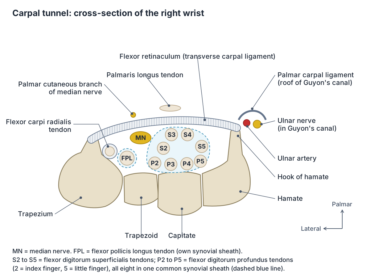
The carpal tunnel is a passage on the palm side of the wrist. Its floor and sides are the concave carpal arch, formed laterally by the tubercles of the scaphoid and trapezium and medially by the hook of the hamate and the pisiform. Its roof is the flexor retinaculum (transverse carpal ligament), which spans between these four bony points and turns the arch into a tunnel. (TeachMeAnatomy: The Carpal Tunnel.)
Ten structures pass through it: the median nerve and nine tendons, namely four of flexor digitorum superficialis, four of flexor digitorum profundus and one of flexor pollicis longus. The eight finger flexor tendons share one synovial sheath, and flexor pollicis longus has its own. The tendon of flexor carpi radialis runs within the flexor retinaculum, not inside the tunnel. (TeachMeAnatomy: The Carpal Tunnel.)
OpenStax describes the same arrangement: the tendons of nine anterior forearm muscles and an important nerve pass through this narrow tunnel, and compression of that nerve, the median nerve, produces carpal tunnel syndrome. (OpenStax Anatomy and Physiology 2e, 8.2: Bones of the Upper Limb.)
The palmar cutaneous branch of the median nerve leaves the nerve before the tunnel and runs superficial to the flexor retinaculum. Beyond the tunnel the median nerve divides into a recurrent branch to the thenar muscles and palmar digital nerves to the skin of the lateral three and a half digits and the lateral two lumbricals. (TeachMeAnatomy: The Carpal Tunnel.)
Carpal tunnel syndrome is described as the most common mononeuropathy. It causes numbness, tingling and pain in the median nerve territory, often worse in the morning, and severe untreated cases weaken and waste the thenar muscles. Tinel's sign (tapping over the nerve) and Phalen's manoeuvre (holding the wrist flexed for 60 seconds) are used to provoke symptoms. (TeachMeAnatomy: The Carpal Tunnel.)
The median, ulnar and radial nerves from the brachial plexus to the hand. Nerves of the upper limb.
Guyon's canal (ulnar canal)
Guyon's canal is a fibro-osseous tunnel about 4 cm long on the little finger side of the wrist, running from the pisiform to the hook of the hamate. Its medial wall is the pisiform, the flexor carpi ulnaris tendon and abductor digiti minimi; its lateral wall is the hook of the hamate; its roof is the palmar carpal ligament; and its floor is the flexor retinaculum, pisohamate ligament and hypothenar muscles. (TeachMeAnatomy: The Ulnar Canal (Guyon's Canal).)
It carries the ulnar nerve and artery, with the artery on the radial side of the nerve. The nerve divides inside the canal into a superficial (sensory) and a deep (motor) branch. Compression here, most often by a ganglion cyst, lipoma or trauma, causes ulnar canal syndrome. (TeachMeAnatomy: The Ulnar Canal (Guyon's Canal).)
Anatomical snuffbox
The anatomical snuffbox is a triangular hollow on the thumb side of the back of the wrist, best seen when the thumb is extended. Its ulnar (medial) border is the tendon of extensor pollicis longus, its radial (lateral) border is the tendons of extensor pollicis brevis and abductor pollicis longus, and its proximal border is the styloid process of the radius. (TeachMeAnatomy: The Anatomical Snuffbox.)
Its floor is formed by the scaphoid and trapezium. The radial artery crosses the floor, the superficial branch of the radial nerve runs in the skin and subcutaneous tissue over it, and the cephalic vein, which arises from the dorsal venous network of the hand, crosses it. Tenderness in the snuffbox after a fall is the classic sign of a scaphoid fracture. (TeachMeAnatomy: The Anatomical Snuffbox.)
Carpal tunnel, Guyon's canal and anatomical snuffbox at a glance
| Space | Boundaries | Contents | Clinical link |
|---|---|---|---|
| Carpal tunnel | Carpal arch (scaphoid and trapezium tubercles, pisiform, hook of hamate); roof: flexor retinaculum | Median nerve; 4 FDS, 4 FDP and 1 FPL tendons | Carpal tunnel syndrome |
| Guyon's canal | Pisiform and hook of hamate; roof: palmar carpal ligament; floor: flexor retinaculum | Ulnar nerve and ulnar artery | Ulnar canal syndrome |
| Anatomical snuffbox | EPL tendon (ulnar side); EPB and APL tendons (radial side); radial styloid (proximal) | Radial artery on its floor; superficial branch of radial nerve; start of cephalic vein | Tenderness suggests scaphoid fracture |
FDS = flexor digitorum superficialis, FDP = flexor digitorum profundus, FPL = flexor pollicis longus, EPL/EPB = extensor pollicis longus/brevis, APL = abductor pollicis longus. Based on TeachMeAnatomy.
Tendons crossing the wrist: retinacula and extensor compartments
Fibrous bands called retinacula hold the tendons against the wrist. The flexor retinaculum stretches across the palm side and the extensor retinaculum across the back of the wrist. (OpenStax Anatomy and Physiology 2e, 11.5: Muscles of the Pectoral Girdle and Upper Limbs.)
Fibrous septa from the extensor retinaculum divide the back of the wrist into six extensor compartments, from radial to ulnar: (1) abductor pollicis longus and extensor pollicis brevis; (2) extensor carpi radialis longus and brevis; (3) extensor pollicis longus; (4) extensor digitorum and extensor indicis; (5) extensor digiti minimi; (6) extensor carpi ulnaris. Lister's tubercle on the radius separates compartments 2 and 3. (Radiopaedia: Extensor compartments of the wrist.)
The flexors and extensors whose tendons cross the wrist, with a diagram of the six extensor compartments. Forearm muscles.
Wrist movements, range of motion and muscles
The wrist moves in four main directions. Flexion is produced mainly by flexor carpi radialis and flexor carpi ulnaris, and extension by extensor carpi radialis longus and brevis and extensor carpi ulnaris. Ulnar deviation (adduction) uses extensor and flexor carpi ulnaris together, and radial deviation (abduction) uses abductor pollicis longus, flexor carpi radialis and the radial wrist extensors. (TeachMeAnatomy: The Wrist Joint.)
Radiopaedia gives a total range of about 80 degrees of flexion and 60 degrees of extension. Most extension happens at the radiocarpal joint, while most flexion and most radial and ulnar deviation happen at the midcarpal joint. (Radiopaedia: Wrist joint.)
Kenhub gives the radiocarpal joint's own share as about 50 degrees of flexion, 35 degrees of extension, 30 degrees of ulnar deviation and 7 degrees of radial deviation, which shows how much the midcarpal joint adds. (Kenhub: Wrist joint.)
Everyday tasks need less. In a study of 40 healthy adults, all the tested activities could be done with 60 degrees of extension, 54 degrees of flexion, 40 degrees of ulnar deviation and 17 degrees of radial deviation, and most needed only about 40 degrees each of flexion and extension. (Ryu J et al. Functional ranges of motion of the wrist joint. J Hand Surg Am. 1991.)
Muscles that move the wrist
| Movement | Main muscles | Assisted by |
|---|---|---|
| Flexion | Flexor carpi radialis, flexor carpi ulnaris | Flexor digitorum superficialis (and palmaris longus, flexor digitorum profundus, flexor pollicis longus) |
| Extension | Extensor carpi radialis longus and brevis, extensor carpi ulnaris | Extensor digitorum |
| Ulnar deviation (adduction) | Extensor carpi ulnaris and flexor carpi ulnaris together | None listed |
| Radial deviation (abduction) | Abductor pollicis longus, flexor carpi radialis, extensor carpi radialis longus and brevis | None listed |
Based on TeachMeAnatomy, Kenhub and Radiopaedia (secondary flexors).
Blood supply and nerves of the wrist
The wrist is supplied by the palmar and dorsal carpal arches. The dorsal arch is formed by the dorsal carpal branches of the radial and ulnar arteries, reinforced by the anterior and posterior interosseous arteries. The palmar arch is formed by the palmar carpal branches of the radial and ulnar arteries, reinforced by the anterior interosseous artery and branches of the deep palmar arch. (Radiopaedia: Wrist joint.)
The joint's nerves are the anterior interosseous nerve (from the median nerve), the posterior interosseous nerve (from the radial nerve) and the deep and dorsal branches of the ulnar nerve. (Kenhub: Wrist joint.)
The radial pulse is felt in the distal forearm just lateral to the tendon of flexor carpi radialis. The radial artery then passes to the back of the wrist across the floor of the anatomical snuffbox, while the ulnar artery enters the hand in front of the flexor retinaculum, lateral to the ulnar nerve. (TeachMeAnatomy: Arteries of the Upper Limb.)
The radial and ulnar arteries, the palmar arches and the veins of the hand and forearm. Arteries and veins of the arm.
Common wrist injuries and conditions
Colles' fracture is the most common fracture of the wrist and follows a fall onto an outstretched hand. The distal radius breaks and its distal fragment is displaced backwards, giving a "dinner fork" deformity; the ulnar styloid process is avulsed in most cases. (TeachMeAnatomy: The Wrist Joint.)
A Smith fracture is the reverse: the distal fragment is angled towards the palm, usually after a fall onto a flexed wrist or a blow to the back of the wrist. It accounts for less than 3% of fractures of the radius and ulna. (Radiopaedia: Smith fracture.)
The scaphoid is the most commonly fractured carpal bone. Its blood supply enters at its distal end, so a fracture through its waist can cut off the proximal part, with a risk of avascular necrosis and, if the fracture is missed, wrist osteoarthritis later in life. (TeachMeAnatomy: The Wrist Joint.)
A fall on a dorsiflexed wrist can push the lunate forwards (anterior dislocation of the lunate) into the carpal tunnel, causing median nerve symptoms and thenar weakness. (TeachMeAnatomy: The Wrist Joint.)
Kienböck disease is osteonecrosis of the lunate. It is most common in young adult men, usually in the dominant wrist, and causes non-specific wrist pain, swelling and stiffness. Negative ulnar variance is present in 60 to 75% of patients, although a causal link has not been proven. (Radiopaedia: Kienböck disease.)
The distal radius, its styloid process and ulnar notch, and Colles and Smith fractures. Radius.
Wrist joint in the 3D model
- Abductor pollicis longus
- A deep forearm extensor that forms the lateral border of the anatomical snuffbox.
- Capitate
- The largest carpal bone, with a rounded head.
- Extensor pollicis brevis
- A deep thumb extensor forming the lateral border of the anatomical snuffbox.
- Extensor pollicis longus
- A thumb extensor whose tendon turns around the dorsal tubercle of the radius.
- Flexor retinaculum of wrist
- A strong fibrous band across the front of the wrist forming the roof of the carpal tunnel.
- Hamate
- A wedge-shaped carpal bone of the distal row with a hook.
- Lunate
- A crescent-shaped carpal bone in the middle of the proximal row.
- Pisiform
- A small, pea-shaped carpal bone on the palmar side of the triquetrum.
- Radius
- The lateral bone of the forearm.
- Scaphoid
- The largest bone of the proximal carpal row, shaped like a boat.
- Trapezium
- A four-sided carpal bone at the thumb side of the distal row.
- Trapezoid
- A small wedge-shaped carpal bone of the distal row.
- Triquetral
- A pyramid-shaped carpal bone on the ulnar side of the proximal row.
- Ulna
- The medial bone of the forearm.
Frequently asked questions
How many bones are in the wrist?
The wrist contains eight carpal bones in two rows: scaphoid, lunate, triquetrum and pisiform in the proximal row, and trapezium, trapezoid, capitate and hamate in the distal row. The wrist joint also involves the lower ends of the radius and ulna, and the distal row of carpals meets the bases of the five metacarpals at the carpometacarpal joints.
What type of joint is the wrist?
The main wrist joint, the radiocarpal joint, is a condyloid (ellipsoid) synovial joint. The rounded proximal row of carpal bones sits in the shallow hollow of the radius and articular disc, allowing flexion, extension, radial and ulnar deviation and circumduction, but not rotation. Turning the palm over happens at the radioulnar joints of the forearm instead.
What are the main ligaments of the wrist?
Extrinsic ligaments join the forearm bones to the carpals: the palmar radiocarpal ligaments (radioscaphocapitate, long and short radiolunate), the ulnocarpal ligaments, the dorsal radiocarpal ligament and the radial and ulnar collateral ligaments. Intrinsic ligaments join carpal bones to each other, the most important being the scapholunate and lunotriquetral ligaments. The TFCC stabilises the ulnar side.
What is the TFCC in the wrist?
The triangular fibrocartilage complex is a group of fibrocartilage and ligaments on the little finger side of the wrist, between the ulna and the lunate and triquetrum. It is the main stabiliser of the distal radioulnar joint and carries about a fifth of the axial load passing through the wrist. Its central and radial parts have no blood supply of their own.
What passes through the carpal tunnel?
Ten structures pass through the carpal tunnel: the median nerve and nine flexor tendons, four from flexor digitorum superficialis, four from flexor digitorum profundus and one from flexor pollicis longus. The flexor carpi radialis tendon runs in its own split of the flexor retinaculum, and the ulnar nerve and artery pass outside the tunnel through Guyon's canal.
Where is the anatomical snuffbox and why does it matter?
The anatomical snuffbox is the triangular hollow on the thumb side of the back of the wrist, seen when you extend your thumb. Tendons of extensor pollicis longus, extensor pollicis brevis and abductor pollicis longus form its borders, and the scaphoid lies in its floor. Tenderness there after a fall is the classic sign of a scaphoid fracture.
How far should the wrist bend?
Radiopaedia gives a total of about 80 degrees of flexion and 60 degrees of extension, with most flexion at the midcarpal joint and most extension at the radiocarpal joint. Daily tasks need much less: a study of 40 healthy adults found that most activities need only about 40 degrees each of flexion and extension. Normal values differ between people.
References
- OpenStax. Anatomy and Physiology 2e, §8.2 Bones of the Upper Limb. Rice University, 2022.
- OpenStax. Anatomy and Physiology 2e, §11.5 Muscles of the Pectoral Girdle and Upper Limbs. Rice University, 2022.
- TeachMeAnatomy. The Wrist Joint: articulations, movements and injuries.
- TeachMeAnatomy. Bones of the Hand: Carpals, Metacarpals and Phalanges.
- TeachMeAnatomy. The Carpal Tunnel: borders, contents and carpal tunnel syndrome.
- TeachMeAnatomy. The Anatomical Snuffbox.
- TeachMeAnatomy. The Ulnar Canal (Guyon's Canal).
- TeachMeAnatomy. The Radioulnar Joints.
- TeachMeAnatomy. Arteries of the Upper Limb.
- Kenhub. Wrist joint (radiocarpal joint): anatomy, ligaments and movements.
- Kenhub. Trapeziometacarpal joint: anatomy and movements.
- Radiopaedia. Wrist joint.
- Radiopaedia. Triangular fibrocartilage complex.
- Radiopaedia. Scapholunate ligament.
- Radiopaedia. Scapholunate dissociation.
- Radiopaedia. Extensor compartments of the wrist.
- Radiopaedia. Smith fracture.
- Radiopaedia. Kienböck disease.
- Ryu J, Cooney WP, Askew LJ, An KN, Chao EYS. Functional ranges of motion of the wrist joint. J Hand Surg Am. 1991;16(3):409-419.
- Mitsuhashi N. et al. (2009). BodyParts3D: 3D structure database for anatomical concepts. Nucleic Acids Research 37, D782–D785. Data © The Database Center for Life Science, CC BY 4.0.
Related anatomy
- Hand bonesHow many bones are in the human hand? Learn the 8 carpal bones, 5 metacarpals and 14 phalanges by name, plus the thumb…
- RadiusRadius labeled: head of radius, neck, radial tuberosity, shaft, Lister's tubercle, ulnar notch and styloid process…
- UlnaUlna labeled: olecranon, trochlear notch, coronoid process, radial notch, shaft, head and styloid process, how the…
- Forearm musclesMuscles of the forearm explained: every anterior flexor and posterior extensor, with origin, insertion, nerve and…
- Hand anatomyHand anatomy with labelled diagrams and a 3D model: 27 bones, finger and thumb joints, flexor and extensor tendons…
- Upper limb nervesBrachial plexus diagram with roots, trunks, cords and branches; axillary, musculocutaneous, median, ulnar and radial…
Explore the whole body in the interactive 3D human anatomy model, or browse the A–Z list of human structures.