

What are the arm bones? Arm vs forearm in anatomy
Anatomists reserve the word arm (brachium) for the upper arm, the region between the shoulder and the elbow, and call the region between the elbow and the wrist the forearm (antebrachium) rather than the lower arm. In that strict sense the arm has a single bone, the humerus. (OpenStax Anatomy and Physiology 2e, 1.6: Anatomical Terminology.)
Outside anatomy, 'arm bones' usually means all three long bones of the limb: the humerus in the upper arm and the paired bones of the forearm, the ulna on the medial side and the radius on the lateral (thumb) side. The bones of the wrist and hand are counted as a separate group. (OpenStax Anatomy and Physiology 2e, 8.2: Bones of the Upper Limb.)
For the muscles, nerves, vessels and regions of the whole upper limb, see the arm anatomy guide.
How many bones are in the arm?
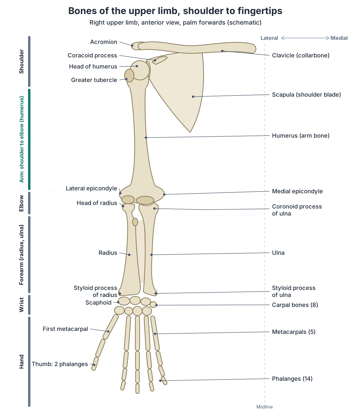
There are 30 bones in each upper limb: the humerus, the radius and ulna, 8 carpal bones in the wrist, 5 metacarpal bones in the palm and 14 phalanges in the fingers and thumb. Three of them, the humerus, radius and ulna, are the long bones of the arm and forearm; the other 27 are in the wrist and hand. (OpenStax Anatomy and Physiology 2e, 8.2: Bones of the Upper Limb.)
Kenhub counts them by region in the same way: one bone in the arm (the humerus), two in the forearm (the radius and ulna), 8 in the carpus, 5 in the metacarpus and 14 in the digits. (Kenhub: Upper extremity anatomy.)
The clavicle (collarbone) and the scapula (shoulder blade) are not counted among the 30. Together they form the pectoral (shoulder) girdle, the bones that attach each upper limb to the axial skeleton. (OpenStax Anatomy and Physiology 2e, 8.1: The Pectoral Girdle.)
For every bone of the body grouped by region, see the skeletal system guide.
Arm bones diagram: labelling the humerus, radius and ulna
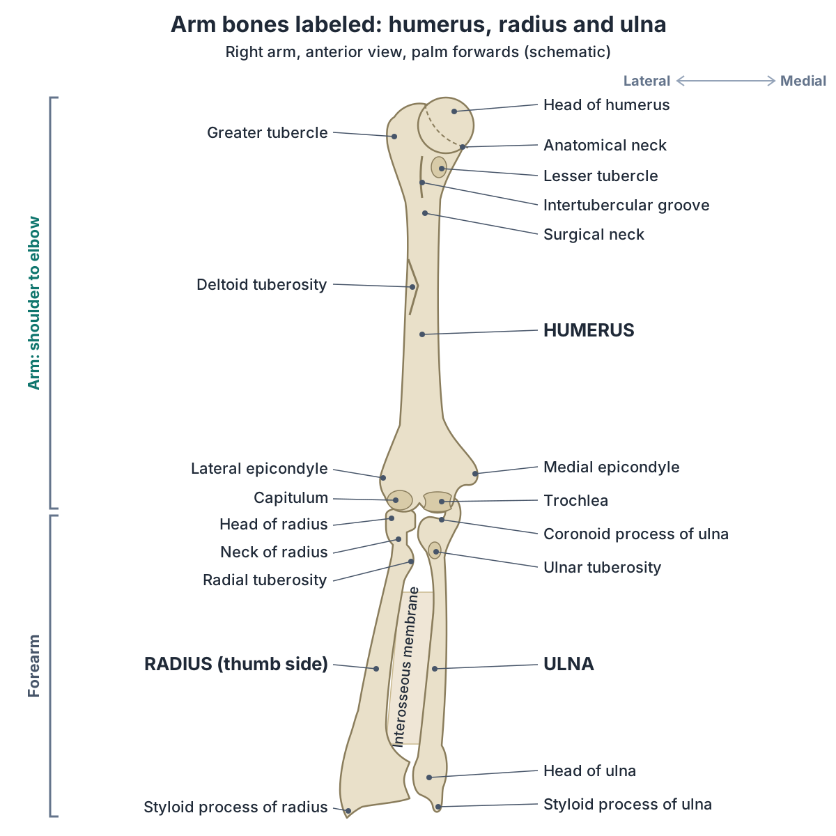
Arm bone diagrams are drawn in the anatomical position, with the palm facing forwards. In that position the radius and ulna are parallel to each other. When the palm is turned to face backwards, the radius crosses over the ulna and the two bones form an X, so always check which way the palm faces before you label the forearm. (OpenStax Anatomy and Physiology 2e, 9.5: Types of Body Movements.)
Start at the shoulder with the head of the humerus, which articulates with the glenoid cavity of the scapula, and the greater tubercle on the lateral side of the upper end. At the elbow the humerus widens into the medial and lateral epicondyles. Between them, the knob-like capitulum on the lateral side meets the head of the radius, and the pulley-shaped trochlea on the medial side fits into the trochlear notch of the ulna. (OpenStax Anatomy and Physiology 2e, 8.2: Bones of the Upper Limb.)
The diagram on this page shows the right arm from the front, so its lateral side, with the radius and the greater tubercle, is on the viewer's left. A left arm seen from the front is its mirror image. In any front view with the palm forwards, the radius is the forearm bone on the thumb side. (OpenStax Anatomy and Physiology 2e, 8.2: Bones of the Upper Limb.)
Arm bones at a glance
| Bone | Location | Key landmarks | Articulations |
|---|---|---|---|
| Humerus | Upper arm, from shoulder to elbow | Head, anatomical and surgical necks, greater and lesser tubercles, intertubercular groove, deltoid tuberosity, radial groove, medial and lateral epicondyles, capitulum, trochlea, coronoid, radial and olecranon fossae | Scapula (shoulder); ulna and radius (elbow) |
| Ulna | Medial (little-finger) side of the forearm | Olecranon, trochlear notch, coronoid process, radial notch, ulnar tuberosity, interosseous border, head, styloid process | Humerus (elbow); radius (proximal and distal radioulnar joints); no direct joint with the carpal bones |
| Radius | Lateral (thumb) side of the forearm | Head, neck, radial tuberosity, ulnar notch, styloid process, facets for the scaphoid and lunate | Humerus (elbow); ulna (proximal and distal radioulnar joints); scaphoid and lunate (wrist) |
Sources: OpenStax Anatomy and Physiology 2e, 8.2 and 9.6; TeachMeAnatomy; Kenhub.
Humerus: the upper arm bone
The humerus is the longest and largest bone of the upper limb. It articulates with the scapula at the shoulder (glenohumeral) joint and with the radius and ulna at the elbow. (Kenhub: The humerus.)
Its upper end carries a smooth, rounded head, and the margin of the head is the anatomical neck. The greater tubercle lies on the lateral side and the smaller lesser tubercle on the front, separated by the intertubercular (bicipital) groove; both tubercles are attachment sites for muscles that act across the shoulder joint. Below them, the narrower surgical neck is a common site of arm fractures. (OpenStax Anatomy and Physiology 2e, 8.2: Bones of the Upper Limb.)
Halfway down the lateral side of the shaft is the roughened, V-shaped deltoid tuberosity, the attachment site of the deltoid muscle. At the lower end are the medial and lateral epicondyles, the capitulum and the trochlea. Three hollows above them receive the forearm bones. At the front, the coronoid fossa above the trochlea and the radial fossa above the capitulum take the coronoid process and the radial head when the elbow bends. At the back, the larger olecranon fossa takes the olecranon when the forearm is fully extended. (OpenStax Anatomy and Physiology 2e, 8.2: Bones of the Upper Limb.)
Across the back of the shaft runs a shallow groove, the radial (spiral) groove, which carries the radial nerve and the profunda brachii (deep brachial) artery. (TeachMeAnatomy: The Humerus.)
Many of these landmarks are where muscles attach. The rotator cuff tendons insert on the greater tubercle (supraspinatus, infraspinatus and teres minor) and on the lesser tubercle (subscapularis). The medial epicondyle is the common origin of the superficial forearm flexors, and the lateral epicondyle the common origin of the superficial forearm extensors. (Kenhub: The humerus.)
At the elbow, triceps brachii inserts on the olecranon, brachialis on the ulnar tuberosity and biceps brachii on the radial tuberosity. (Kenhub: The radius and the ulna.)
For every landmark of the upper arm bone, its muscle attachments and the nerves at risk in fractures, see the humerus guide.
Forearm bones: radius vs ulna
The forearm has two bones, the radius and the ulna, which run parallel between the elbow and the wrist. In the anatomical position the radius is in the lateral (thumb side) forearm and the ulna in the medial (little-finger side) forearm. The radius is shorter than the ulna, which is the longer of the two. (Kenhub: The radius and the ulna.)
The two bones are shaped in opposite ways. The ulna has a larger upper end and tapers to a smaller lower end, while the radius does the opposite. (Radiopaedia: Ulna.)
So the ulna dominates at the elbow and the radius at the wrist. At the elbow, the trochlear notch of the ulna articulates with the trochlea of the humerus. At the wrist, the scaphoid and lunate articulate directly with the end of the radius, while the triquetrum rests on a pad of fibrocartilage that spans the radius and the styloid process of the ulna. The end of the ulna does not articulate directly with any carpal bone. (OpenStax Anatomy and Physiology 2e, 8.2: Bones of the Upper Limb.)
Turning the palm to face backwards (pronation) is produced by rotation of the radius at the proximal radioulnar joint, and its lower end crosses over the lower ulna at the distal radioulnar joint. Supination, the opposite movement, rotates the radius back so that the two bones are parallel again. (OpenStax Anatomy and Physiology 2e, 9.5: Types of Body Movements.)
TeachMeAnatomy describes the ulna as the stabilising bone of the forearm, with the radius pivoting around it to produce movement. (TeachMeAnatomy: The Ulna.)
For the muscles that turn the forearm and move the wrist and fingers, see the forearm muscles guide.
Radius vs ulna
| Feature | Radius | Ulna |
|---|---|---|
| Side of the forearm | Lateral, thumb side | Medial, little-finger side |
| Length | Shorter | Longer |
| Larger end | Lower end, at the wrist | Upper end, at the elbow |
| At the elbow | Head meets the capitulum of the humerus | Trochlear notch meets the trochlea of the humerus |
| At the wrist | Articulates with the scaphoid and lunate | No direct joint with the carpal bones |
| Turning the palm (pronation and supination) | Rotates and crosses over the ulna | Stays as the stable bone |
| Styloid process | On the lateral side of the lower end | On the posteromedial side of the lower end (little-finger side of the wrist) |
Sources: Kenhub, The radius and the ulna; Radiopaedia, Ulna; OpenStax Anatomy and Physiology 2e, 8.2 and 9.5; TeachMeAnatomy, The Ulna.
Ulna: the inner forearm bone
The upper end of the ulna looks like a crescent wrench. Its olecranon forms the bony tip of the elbow, and the large C-shaped trochlear notch, between the olecranon and a lip of bone in front called the coronoid process, articulates with the trochlea of the humerus. Beside the coronoid process is the small radial notch for the head of the radius, and just below the coronoid process, on the front of the bone, is a roughened area, the ulnar tuberosity. (OpenStax Anatomy and Physiology 2e, 8.2: Bones of the Upper Limb.)
The shaft has an interosseous border for the attachment of the interosseous membrane, which spans the space between the two forearm bones. At the wrist the ulna ends in a small rounded head with a projection, the ulnar styloid process, and the head articulates with the ulnar notch of the radius at the distal radioulnar joint. (TeachMeAnatomy: The Ulna.)
For the olecranon, coronoid process, ulnar head and ulna fractures in detail, see the ulna guide.
Radius: the outer forearm bone
The upper end of the radius is a disc-shaped head. The small depression on its top articulates with the capitulum of the humerus at the elbow, and its smooth outer margin turns against the radial notch of the ulna at the proximal radioulnar joint. Below the head is the narrow neck and then, on the medial side, the radial tuberosity. (OpenStax Anatomy and Physiology 2e, 8.2: Bones of the Upper Limb.)
The radial tuberosity is the attachment site of the biceps brachii muscle. At the lower end, the lateral side projects distally as the styloid process, the medial side has a concavity, the ulnar notch, that articulates with the head of the ulna, and the lower surface has two facets for the scaphoid and lunate bones of the wrist. (TeachMeAnatomy: The Radius.)
For the head, tuberosity, Lister's tubercle and fractures of the radius, see the radius guide.
How the radius and ulna are joined: radioulnar joints and interosseous membrane
The radius and ulna are joined at both ends and along their shafts. At the elbow, the head of the radius rotates within the annular ligament against the radial notch of the ulna at the proximal radioulnar joint. At the wrist, the ulnar notch of the radius slides over the head of the ulna at the distal radioulnar joint, where a fibrocartilage articular disc binds the two bones together. TeachMeAnatomy classifies both joints as pivot joints. (TeachMeAnatomy: The Radioulnar Joints.)
Between the shafts, the interosseous membrane is a sheet of connective tissue that joins the radius and ulna. It holds them together while the forearm turns, transfers forces from the radius to the ulna, and gives attachment to muscles of the front and back of the forearm. (TeachMeAnatomy: The Radioulnar Joints.)
A 2024 study of the membrane gives the same functions: it is a site of attachment for forearm muscles, and it transfers forces from the radius to the ulna while connecting them without restricting pronation and supination. (Ono T et al. Morphological changes in the interosseous membrane of the forearm during forearm rotation. J Phys Ther Sci. 2024.)
Because the two bones are bound together, a break in one often comes with an injury to the other. In a Monteggia fracture the proximal ulna breaks and the head of the radius dislocates at the proximal radioulnar joint; in a Galeazzi fracture the lower third of the radial shaft breaks and the head of the ulna dislocates at the distal radioulnar joint. (Kenhub: The radius and the ulna.)
For the elbow joint, its collateral and annular ligaments and bursae, see elbow anatomy.
Joints formed by the arm bones: shoulder, elbow and wrist
The shoulder joint, or glenohumeral joint, is a ball-and-socket joint between the head of the humerus and the glenoid cavity of the scapula. The elbow is a hinge joint formed by the humeroulnar joint, between the trochlea of the humerus and the trochlear notch of the ulna; the humeroradial joint and the proximal radioulnar joint are also associated with the elbow. (OpenStax Anatomy and Physiology 2e, 9.6: Anatomy of Selected Synovial Joints.)
At the wrist, the radius is the forearm bone that meets the hand: its lower end articulates directly with the scaphoid and lunate bones, while the ulna is separated from the carpal bones by a pad of fibrocartilage. (OpenStax Anatomy and Physiology 2e, 8.2: Bones of the Upper Limb.)
For the shoulder joint, the rotator cuff and the shoulder girdle, see shoulder anatomy.
Nerves that lie on the humerus, and the funny bone
Four major nerves of the upper limb lie close to different parts of the humerus: the axillary, radial, median and ulnar nerves. This is why a fractured humerus can also damage a nerve. (OpenStax Anatomy and Physiology 2e, 8.2: Bones of the Upper Limb.)
Three of these nerves lie directly against the bone. The axillary nerve lies against the surgical neck and is at risk in fractures there. The radial nerve lies in the radial groove on the back of the shaft and is at risk in mid-shaft fractures. The ulnar nerve passes in a groove on the back of the medial epicondyle, where it can be felt. (TeachMeAnatomy: The Humerus.)
Knocking that spot on the inner side of the elbow gives the shock-like tingling or burning known as hitting your funny bone. The funny bone is not a bone at all: it is the ulnar nerve, which runs just under the skin near the elbow, in a groove behind the bony bump of the medial epicondyle. (Cleveland Clinic: Ulnar Nerve.)
For the brachial plexus and the full course of the axillary, radial and ulnar nerves, see the upper limb nerves guide.
Common arm bone fractures
Falls onto the hand or elbow, and direct blows to the arm, can break the humerus. The surgical neck is a common fracture site. (OpenStax Anatomy and Physiology 2e, 8.2: Bones of the Upper Limb.)
In children the lower end of the humerus often breaks just above both epicondyles (a supracondylar fracture), usually after a fall onto an outstretched hand with the elbow straight; only a few per cent follow a fall onto a bent elbow. The brachial artery and the median nerve pass across the front of the elbow close to the fracture and can be injured. (Chomba D et al. Pediatric Supracondylar Humerus Fractures: Evaluation and Management Approach in Resource-limited Settings. J Am Acad Orthop Surg Glob Res Rev. 2026.)
A distal radius fracture is a break of the radius close to the wrist, and its most common cause is a fall onto an outstretched arm. In the type called a Colles fracture, the broken piece of the radius tilts backwards, towards the back of the wrist. (AAOS OrthoInfo: Distal Radius Fractures (Broken Wrist).)
TeachMeAnatomy calls the Colles fracture the most common type of radial fracture. A Smith fracture is a break of the lower radius caused by falling onto the back of the hand. (TeachMeAnatomy: The Radius.)
The olecranon, the point of the elbow, can also break, usually when someone falls onto a flexed elbow. These notes explain the anatomy only; anyone with an arm injury should see a clinician. (TeachMeAnatomy: The Ulna.)
For the wrist joints and the injuries around them, see wrist anatomy.
Hand bones in brief
Beyond the forearm, each wrist and hand has 27 bones: 8 carpal bones in the wrist, 5 metacarpal bones in the palm and 14 phalanges in the fingers and thumb. The thumb has two phalanges, a proximal and a distal phalanx, and each of the other four digits has three. (OpenStax Anatomy and Physiology 2e, 8.2: Bones of the Upper Limb.)
For all 27 bones of the wrist, palm and fingers with a labelled diagram, see the hand bones guide.
How to use the 3D model of the arm bones
The 3D model uses BodyParts3D, built from an adult male body. It highlights the humerus, radius and ulna, with the key labels on the left arm in the anatomical position, so the radius is on the viewer's right, on the thumb side, and the ulna on the inner side. The Female option uses the same arm bones. The model shows the outer shape of each bone only, and it does not include the nerves of the upper limb, so the axillary, radial and ulnar nerves described above are not shown.
Compare the model with your own elbow: both epicondyles of the humerus can be felt at the elbow, the medial one being the larger and reaching further down, and the ulnar nerve can be felt in its groove behind the medial epicondyle. (TeachMeAnatomy: The Humerus.)
Arm bones in the 3D model
Frequently asked questions
What are the 3 bones of the arm?
The three arm bones are the humerus, the radius and the ulna. The humerus is the single bone of the upper arm, from the shoulder to the elbow. The radius and ulna are the two bones of the forearm, from the elbow to the wrist: the radius on the thumb side and the ulna on the little-finger side. The wrist and hand add 27 more bones.
Which forearm bone is on the thumb side?
The radius. In the anatomical position, with the palm facing forwards, the radius lies on the lateral (thumb) side of the forearm and the ulna on the medial (little-finger) side. The radius ends at the wrist in a pointed styloid process, and it is the forearm bone that articulates directly with the wrist bones, the scaphoid and lunate.
Which is longer, the radius or the ulna?
The ulna is the longer of the two forearm bones; the radius is shorter. The ulna is largest at its upper end, where the olecranon forms the bony tip of the elbow, and it tapers towards the wrist. The radius is shaped the opposite way, with its larger end at the wrist, where it articulates with the scaphoid and lunate.
What is the upper arm bone called?
The upper arm bone is the humerus, the longest and largest bone of the upper limb. Its rounded head articulates with the glenoid cavity of the scapula at the shoulder, and its lower end meets the radius and ulna at the elbow. In anatomy the word arm strictly means only this segment, between the shoulder and the elbow.
How many bones are in the arm?
In everyday terms the arm has three long bones: the humerus, radius and ulna. In strict anatomy the arm, between the shoulder and the elbow, has one bone, the humerus. Counting the whole upper limb, each side has 30 bones: the humerus, radius, ulna, 8 carpals, 5 metacarpals and 14 phalanges. The clavicle and scapula of the shoulder girdle are counted separately.
Is the funny bone a bone?
No. The funny bone is the ulnar nerve. It passes in a groove on the back of the medial epicondyle of the humerus, on the inner side of the elbow, just under the skin. Knocking it there causes a sudden, shock-like tingling or burning feeling, which is what people mean by hitting the funny bone. The bony bump you feel beside it is the medial epicondyle.
Which forearm bone moves when you turn your palm over?
The radius. When you turn your palm to face backwards (pronation), the radius rotates at the proximal radioulnar joint and its lower end crosses over the ulna, so the two bones form an X. Turning the palm forwards again (supination) brings them back to parallel. The ulna acts as the stable bone that the radius pivots around.
References
- OpenStax. Anatomy and Physiology 2e, §8.2 Bones of the Upper Limb. Rice University, 2022.
- OpenStax. Anatomy and Physiology 2e, §8.1 The Pectoral Girdle. Rice University, 2022.
- OpenStax. Anatomy and Physiology 2e, §9.5 Types of Body Movements. Rice University, 2022.
- OpenStax. Anatomy and Physiology 2e, §9.6 Anatomy of Selected Synovial Joints. Rice University, 2022.
- OpenStax. Anatomy and Physiology 2e, §1.6 Anatomical Terminology. Rice University, 2022.
- TeachMeAnatomy. The Humerus.
- TeachMeAnatomy. The Radius.
- TeachMeAnatomy. The Ulna.
- TeachMeAnatomy. The Radioulnar Joints.
- TeachMeAnatomy. The Clavicle.
- Kenhub. The radius and the ulna.
- Kenhub. The humerus.
- Kenhub. Upper extremity anatomy.
- Radiopaedia. Radius.
- Radiopaedia. Ulna.
- Ono T, Iwamoto K, Monma M, Takahashi M, Tomita K. Morphological changes in the interosseous membrane of the forearm during forearm rotation. J Phys Ther Sci. 2024;36(11):739–744.
- American Academy of Orthopaedic Surgeons. Distal Radius Fractures (Broken Wrist). OrthoInfo.
- Cleveland Clinic. Ulnar Nerve.
- Chomba D, Mavrommatis S, Krishna SV, Challa ST, Simister SK, Dooregekant A, Mengesha MG, Agarwal-Harding KJ. Pediatric supracondylar humerus fractures: evaluation and management approach in resource-limited settings. J Am Acad Orthop Surg Glob Res Rev. 2026;10(1).
- Mitsuhashi N. et al. (2009). BodyParts3D: 3D structure database for anatomical concepts. Nucleic Acids Research 37, D782–D785. Data © The Database Center for Life Science, CC BY 4.0.
Related anatomy
- Arm anatomyArm anatomy explained: upper limb regions, its 30 bones, shoulder, elbow and wrist joints, muscle compartments…
- HumerusHumerus bone labeled: head, anatomical and surgical neck, tubercles, deltoid tuberosity, radial groove, epicondyles…
- RadiusRadius labeled: head of radius, neck, radial tuberosity, shaft, Lister's tubercle, ulnar notch and styloid process…
- UlnaUlna labeled: olecranon, trochlear notch, coronoid process, radial notch, shaft, head and styloid process, how the…
- Elbow jointElbow anatomy with labelled diagrams and a 3D model: humerus, radius and ulna, the three joints, collateral and…
- Hand bonesHow many bones are in the hand? A labeled hand bones diagram and 3D model name all 27: the 8 carpals, 5 metacarpals…
Explore the whole body in the interactive 3D human anatomy model, or browse the A–Z list of human structures.