

What is the head made of?
The framework of the head is the skull. Kenhub divides it into the neurocranium, which encloses and protects the brain, and the viscerocranium or facial skeleton, which mainly supports the facial muscles. Its head and neck chapter then covers the nose and nasal cavity, the eye, the ear, the mouth (oral cavity), the teeth and the neck, with the arteries and nerves of each. (Kenhub: Head and neck anatomy.)
The head carries the organs of the special senses. The bony orbits protect the eyes; the inner ear contains the cochlea for hearing and the vestibule for balance; olfactory receptor neurons for smell lie in a small area of the upper nasal cavity; and taste buds sit in raised bumps (papillae) on the tongue. (OpenStax Anatomy and Physiology 2e, 14.1: Sensory Perception.)
AMBOSS lists the organs of the head as the eye and orbit, the ear, the nose and sinuses, the skull and brain, and the oral cavity with the palate, tonsils, teeth and salivary glands, while the larynx, trachea, pharynx, oesophagus and the thyroid and parathyroid glands belong to the neck. (AMBOSS: Head and neck region.)
Regions of the head
To describe where something lies, anatomists divide the head into 14 surface regions. Six cover the cranium: frontal, parietal, occipital, temporal, auricular and mastoid. The face is split into eight subregions: orbital, infraorbital, nasal, zygomatic, buccal, parotid, oral and mental (chin). The neck has four regions of its own: the sternocleidomastoid, posterior cervical, lateral cervical and anterior cervical regions. (AMBOSS: Head and neck region.)
The temporal region, or temple, is a good example of how a region maps onto deeper anatomy. It lies on the side of the head between the temporal lines above, the lateral margin of the orbit in front and the zygomatic arch below, and it overlies the temporal fossa, where the frontal, parietal, sphenoid and temporal bones meet at the pterion. (IMAIOS e-Anatomy: Temporal region.)
The temple region, pterion, temporalis and temporal arteries. Temple anatomy.
Regions of the head
| Region | Where it is |
|---|---|
| Frontal | Forehead |
| Parietal | Top and upper sides of the head |
| Occipital | Back of the head |
| Temporal | Temple, on the side of the head above the zygomatic arch |
| Auricular | Around the ear |
| Mastoid | Behind the ear, over the mastoid process |
| Facial: orbital, infraorbital, nasal, zygomatic, buccal, parotid, oral, mental | Around the eye, below the eye, nose, cheekbone, cheek, in front of the ear over the parotid gland, mouth and chin |
AMBOSS divides the head into 14 regions: six regions of the cranium and eight facial subregions.
Bones of the head: the skull
The adult skull consists of 22 bones, 21 of which are locked together into one immobile unit. The brain case is made of eight bones and the facial skeleton of 14, six of them paired. The 22nd bone, the mandible (lower jaw), is the only movable bone of the skull. (OpenStax Anatomy and Physiology 2e, 7.2: The Skull.)
Several spaces of the head are built from skull bones. Each orbit, which holds an eye, has walls formed by seven bones, and on the side of the skull the temporal fossa lies above the zygomatic arch and the infratemporal fossa below it, both containing chewing muscles. (OpenStax Anatomy and Physiology 2e, 7.2: The Skull.)
The 8 cranial and 14 facial bones, sutures and fontanelles. Skull bones.
The scalp
The scalp covering the skull has five layers, remembered as SCALP: skin, dense connective tissue, the epicranial aponeurosis, loose areolar connective tissue and the pericranium (periosteum). The first three move together as one unit. The loose areolar layer is called the danger area because its valveless emissary veins connect the scalp veins with the venous sinuses inside the skull, a possible route for infection. (TeachMeAnatomy: The Scalp.)
The scalp is supplied by the superficial temporal, posterior auricular and occipital branches of the external carotid artery and, at the front, by the supraorbital and supratrochlear branches of the ophthalmic artery. Deep scalp wounds bleed heavily, because the vessels are held open by the dense connective tissue and richly connected with each other. (TeachMeAnatomy: The Scalp.)
The five layers of the scalp, its blood supply and nerves. Scalp.
The brain and its coverings
The adult brain is described in four major regions: the cerebrum, the diencephalon, the brainstem and the cerebellum. The cerebral cortex is divided into lobes by landmarks such as the lateral sulcus, which separates the temporal lobe, associated with hearing and memory. The midbrain, pons and medulla form the brainstem, which connects the brain to the spinal cord. (OpenStax Anatomy and Physiology 2e, 13.2: The Central Nervous System.)
Three membranes, the meninges, cover the brain: the tough dura mater against the skull, the thin arachnoid mater and the delicate pia mater on the brain surface. Cerebrospinal fluid made by the choroid plexuses in the ventricles flows into the subarachnoid space and acts as a liquid cushion for the brain and spinal cord. (OpenStax Anatomy and Physiology 2e, 13.3: Circulation and the Central Nervous System.)
The parts of the brain in 3D, with sagittal and coronal sections. Brain anatomy.
Nerves of the head: the 12 cranial nerves
Most nerves of the head are cranial nerves, which leave the brain directly. There are twelve, numbered CN I to CN XII: olfactory, optic, oculomotor, trochlear, trigeminal, abducens, facial, vestibulocochlear, glossopharyngeal, vagus, spinal accessory and hypoglossal. Three are purely sensory, five are purely motor and four are mixed. (OpenStax Anatomy and Physiology 2e, 13.4: The Peripheral Nervous System.)
Two cranial nerves do most of the work in the face. The trigeminal nerve carries skin sensation from the face and controls the muscles of mastication, while the facial nerve controls the muscles of facial expression and also contributes to taste and to the production of saliva. (OpenStax Anatomy and Physiology 2e, 13.4: The Peripheral Nervous System.)
All 12 cranial nerves, their functions, foramina and tests. Cranial nerves.
The face and muscles of facial expression
The muscles of facial expression insert into the skin rather than into bone, so when they contract they move the skin to create expressions. (OpenStax Anatomy and Physiology 2e, 11.3: Axial Muscles of the Head, Neck, and Back.)
AMBOSS groups them by area: cranial (occipitofrontalis and temporoparietalis), auricular, eye (orbicularis oculi and corrugator supercilii), nose (nasalis, depressor septi nasi and procerus) and mouth (orbicularis oris, buccinator and the upper and lower lip groups). All of them are supplied by the facial nerve (CN VII). (AMBOSS: Head and neck region.)
Every muscle of facial expression, its action and nerve. Facial muscles.
The jaw joint and chewing muscles
The mandible moves at the two temporomandibular joints, just in front of the ears, where its condyles meet the mandibular fossae and articular tubercles of the temporal bones. An articular disc divides each joint into an upper part, which allows the jaw to move forwards and backwards, and a lower part, which allows it to open and close. (TeachMeAnatomy: The Temporomandibular Joint.)
Four paired muscles of mastication move the jaw when chewing: masseter, temporalis, medial pterygoid and lateral pterygoid. OpenStax describes masseter as the main chewing muscle, with the pterygoids helping to move food within the mouth. (OpenStax Anatomy and Physiology 2e, 11.3: Axial Muscles of the Head, Neck, and Back.)
TMJ anatomy: condyle, disc, ligaments and how the jaw hinges and glides. Temporomandibular joint (TMJ).
Eyes and orbits
The bony orbits surround the eyeballs, protecting them and anchoring the soft tissues of the eye. Six extraocular muscles move each eye within its orbit, the eyelids protect it, and the lacrimal gland, just inside the orbit above and lateral to the eyeball, produces tears. (OpenStax Anatomy and Physiology 2e, 14.1: Sensory Perception.)
Each adult orbit holds about 30 mL, and the eyeball takes up only about 6.5 mL of it. The rest is filled by the extraocular muscles, cranial nerves II to VI, the ophthalmic artery and veins, orbital fat and the lacrimal gland. (Radiopaedia: Orbit.)
The parts of the eyeball, its layers and a labelled 3D eye model. Eye anatomy.
The ears
Each ear has three parts. The external ear contains the auricle, the ear canal and the tympanic membrane (eardrum). The middle ear contains the three ossicles, the malleus, incus and stapes, and is connected to the pharynx by the Eustachian tube. The inner ear contains the cochlea for hearing and the vestibule for balance. (OpenStax Anatomy and Physiology 2e, 14.1: Sensory Perception.)
The outer, middle and inner ear, ossicles and cochlea. Ear anatomy.
ENT anatomy: how the ear, nose and throat connect
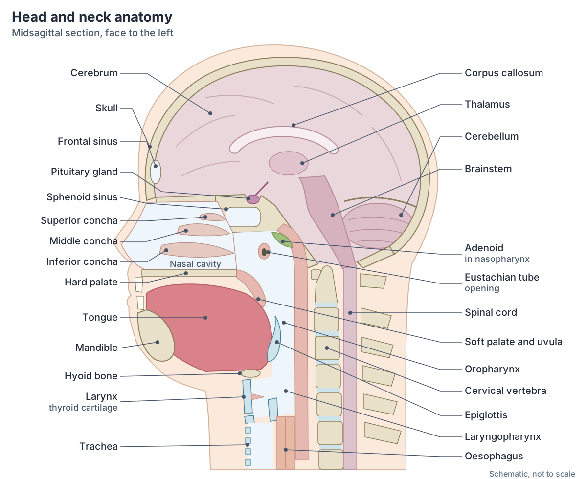
Ear, nose and throat are taught together because they form one connected system of air spaces. The Eustachian (pharyngotympanic) tube links the middle ear cavity with the nasopharynx. Its third nearest the ear is bony and its two thirds nearest the pharynx are cartilaginous. It ventilates the middle ear, keeping the air pressure equal on both sides of the eardrum, and drains middle ear secretions into the nasopharynx. (TeachMeAnatomy: The Eustachian Tube.)
The nose drains several neighbouring spaces. The frontal, maxillary and anterior ethmoidal sinuses open into the middle meatus, the posterior ethmoidal sinuses into the superior meatus, and the nasolacrimal duct, carrying tears from the eye, into the inferior meatus. Behind the nasal cavity, the Eustachian tube opens into the nasopharynx at the level of the inferior meatus. (TeachMeAnatomy: The Nasal Cavity.)
The pharynx (throat) has three regions. The nasopharynx, behind the nasal cavity, is an airway only; the oropharynx, behind the mouth, carries both air and food; and the laryngopharynx lies behind the larynx. The pharyngeal tonsil (adenoid) sits in the nasopharynx, the palatine tonsils at the sides of the oropharynx and the lingual tonsil at the base of the tongue. During swallowing the soft palate and uvula swing up to close off the nasopharynx. (OpenStax Anatomy and Physiology 2e, 22.1: Organs and Structures of the Respiratory System.)
Below the oropharynx the passages for air and food separate. The inferior border of the laryngopharynx connects to the oesophagus (esophagus), while the airway continues forwards into the larynx. (OpenStax Anatomy and Physiology 2e, 23.3: The Mouth, Pharynx, and Esophagus.)
The larynx is built around three large cartilages, the thyroid cartilage in front, the epiglottis above and the cricoid cartilage below, with three smaller paired cartilages. The epiglottis is a very flexible flap of elastic cartilage that covers the opening of the trachea, and the true vocal cords vibrate to produce sound. (OpenStax Anatomy and Physiology 2e, 22.1: Organs and Structures of the Respiratory System.)
The pharynx, its three regions, the tonsils and the larynx. Throat anatomy.
Nose and paranasal sinuses
The paired nasal bones lie under the root and bridge of the nose, and flexible septal cartilage continues from them to form the dorsum of the nose. The nasal septum is made of septal cartilage in front and the perpendicular plate of the ethmoid and the vomer behind. Each lateral wall of the nasal cavity carries three bony projections, the superior, middle and inferior nasal conchae. (OpenStax Anatomy and Physiology 2e, 22.1: Organs and Structures of the Respiratory System.)
Four pairs of paranasal sinuses, the frontal, maxillary, sphenoidal and ethmoidal sinuses, surround the nasal cavity. They warm and humidify incoming air, produce mucus and lighten the skull. (OpenStax Anatomy and Physiology 2e, 22.1: Organs and Structures of the Respiratory System.)
Smell is detected by olfactory receptor neurons in a small region of the upper nasal cavity. Their fibres reach the brain through the olfactory foramina in the cribriform plate of the ethmoid bone. (OpenStax Anatomy and Physiology 2e, 14.1: Sensory Perception.)
The external nose, nasal cavity, conchae and blood supply. Nose anatomy.
Mouth, teeth, tongue and salivary glands
The cheeks, tongue and palate frame the mouth (oral cavity), and the uvula hangs from the back of the soft palate. The vestibule is the pocket between the teeth and gums on one side and the cheeks and lips on the other. The tongue is anchored to the mandible, the styloid processes of the temporal bones and the hyoid bone. (OpenStax Anatomy and Physiology 2e, 23.3: The Mouth, Pharynx, and Esophagus.)
Three pairs of major salivary glands open into the mouth: the parotid glands between the skin and masseter near the ears, the submandibular glands in the floor of the mouth and the sublingual glands below the tongue. Twenty deciduous (baby) teeth begin to appear at about 6 months and are replaced between about 6 and 12 years by 32 permanent teeth. (OpenStax Anatomy and Physiology 2e, 23.3: The Mouth, Pharynx, and Esophagus.)
The hypoglossal nerve (CN XII) supplies all the intrinsic and extrinsic muscles of the tongue except palatoglossus, which is supplied by the vagus nerve. (TeachMeAnatomy: The Hypoglossal Nerve (CN XII).)
Lips, palate, teeth, tongue, gums and salivary glands. Mouth anatomy.
Muscles of the head and neck
Apart from the facial and chewing muscles, the head has six extraocular muscles that move each eyeball and a set of tongue muscles; the extrinsic tongue muscles all include the root glossus (genioglossus, styloglossus, palatoglossus and hyoglossus). In the front of the neck, the suprahyoid muscles lie above the hyoid bone and the infrahyoid muscles below it. (OpenStax Anatomy and Physiology 2e, 11.3: Axial Muscles of the Head, Neck, and Back.)
The sternocleidomastoid is the major muscle that flexes the head to the side and rotates it. Acting on one side, the muscles that move the head rotate it; acting on both sides together, they flex or extend it. They include sternocleidomastoid, semispinalis capitis, splenius capitis and longissimus capitis. (OpenStax Anatomy and Physiology 2e, 11.3: Axial Muscles of the Head, Neck, and Back.)
AMBOSS adds the nerve supply of the neck muscles: sternocleidomastoid is supplied by the accessory nerve with sensory fibres from C2 and C3, platysma by the cervical branch of the facial nerve, and the infrahyoid muscles by the ansa cervicalis, except thyrohyoid, which is supplied by a branch of C1. (AMBOSS: Head and neck region.)
Sternocleidomastoid, scalenes, suprahyoid and infrahyoid muscles. Neck muscles.
Muscles of the head and neck: main groups
| Group | Examples | Nerve | Main job |
|---|---|---|---|
| Muscles of facial expression | Occipitofrontalis, orbicularis oculi, orbicularis oris, buccinator, zygomaticus major, platysma | Facial nerve (VII) | Move the skin of the face, eyelids and lips |
| Muscles of mastication | Masseter, temporalis, medial and lateral pterygoid | Mandibular nerve (V3) of the trigeminal | Chewing: close, protrude and grind the jaw |
| Extraocular muscles | Superior, inferior, medial and lateral rectus; superior and inferior oblique | Oculomotor (III), trochlear (IV), abducens (VI) | Move the eyeball |
| Tongue muscles | Genioglossus, hyoglossus, styloglossus, palatoglossus and intrinsic muscles | Hypoglossal (XII); palatoglossus by the vagus (X) | Shape and move the tongue |
| Suprahyoid and infrahyoid muscles | Digastric, mylohyoid, geniohyoid, stylohyoid; sternohyoid, omohyoid, sternothyroid, thyrohyoid | Various; infrahyoids by the ansa cervicalis except thyrohyoid | Raise and lower the hyoid and larynx in swallowing |
| Muscles that move the head | Sternocleidomastoid, splenius capitis, semispinalis capitis, longissimus capitis | Sternocleidomastoid: accessory nerve (XI) | Flex, extend and rotate the head |
Sources: OpenStax 11.3 and 13.4, AMBOSS and TeachMeAnatomy. Each group has its own detailed guide.
The neck: where the head joins the body
The neck is supported by seven cervical vertebrae. The first, the atlas (C1), supports the skull and joins the occipital condyles; the second, the axis (C2), is the pivot for turning the head left and right. Each cervical transverse process has a transverse foramen, through which an important artery to the brain ascends. (OpenStax Anatomy and Physiology 2e, 7.3: The Vertebral Column.)
Kenhub describes the hyoid bone as the anchor point of the neck, with the suprahyoid and infrahyoid muscle groups attached to it. The neck also contains the thyroid and parathyroid glands, the pharynx and larynx, the common, external and internal carotid arteries and the cervical plexus of nerves. (Kenhub: Head and neck anatomy.)
Cervical spine, neck muscles and triangles, thyroid, vessels and nerves. Neck anatomy.
Blood supply of the head
Each common carotid artery divides into an internal and an external carotid artery. The external carotid supplies the face, lower jaw, neck, oesophagus and larynx through branches including the lingual, facial, occipital, maxillary and superficial temporal arteries. The internal carotid arteries and the vertebral arteries are the two main suppliers of the brain; the vertebral arteries climb through the cervical vertebrae and enter the skull through the foramen magnum. (OpenStax Anatomy and Physiology 2e, 20.5: Circulatory Pathways.)
The vertebral arteries join to form the basilar artery, and together with the internal carotids they form the circle of Willis at the base of the brain, which can keep blood flowing if one vessel is blocked. Blood leaves the brain through the dural venous sinuses, which drain into the internal jugular veins. (OpenStax Anatomy and Physiology 2e, 13.3: Circulation and the Central Nervous System.)
Blood from the brain and the superficial facial vein flows into each internal jugular vein, while the external jugular vein drains the more superficial parts of the head and scalp and empties into the subclavian vein. (OpenStax Anatomy and Physiology 2e, 20.5: Circulatory Pathways.)
The common, internal and external carotids, vertebral arteries and jugular veins. Carotid arteries.
Lymph nodes of the head and neck
Superficial lymph nodes form a ring where the head meets the neck: occipital, mastoid, pre-auricular, parotid, submandibular and submental nodes, with facial and superficial cervical nodes. All of them drain into the deep cervical nodes, a vertical chain alongside the internal jugular vein inside the carotid sheath. (TeachMeAnatomy: Lymphatic Drainage of the Head and Neck.)
Lymph from the deep cervical nodes collects in the jugular lymphatic trunks. On the left the trunk joins the thoracic duct and on the right it forms the right lymphatic duct; both empty into the veins at the root of the neck. Enlarged jugulodigastric (tonsillar) nodes are typical of tonsillitis. (TeachMeAnatomy: Lymphatic Drainage of the Head and Neck.)
Lymph node groups, neck levels I to VI and Waldeyer's ring. Lymph nodes of the head and neck.
Head anatomy in the 3D model
The 3D viewer on this page highlights the cerebral cortex, cerebellum and brainstem, the pituitary gland, the cornea, sclera and optic nerve of each eye, the lacrimal glands, the external ears and the internal carotid arteries, vertebral arteries and internal jugular veins, inside a see-through skeleton. The model is BodyParts3D, built from an adult male. Switch off Highlight topic to see the whole skull, or open the linked guides to see the skull bones, eye muscles, larynx, teeth and tongue highlighted on their own. The model has no scalp layers, facial or chewing muscles, cranial nerve branches of the face, or lymph nodes, so those are covered by diagrams on their own pages. (Mitsuhashi N et al. BodyParts3D: 3D structure database for anatomical concepts. Nucleic Acids Res 2009.)
Diagrams of the node groups the 3D model does not include. Lymph nodes of the head and neck.
Parts of the head and neck at a glance
| Part | Main structures | Detailed guide |
|---|---|---|
| Skull | 8 cranial bones, 14 facial bones, sutures | Skull bones |
| Scalp | Skin, dense connective tissue, aponeurosis, loose areolar tissue, pericranium | Scalp |
| Brain | Cerebrum, diencephalon, brainstem, cerebellum, meninges | Brain anatomy |
| Face and jaw | Facial muscles, masseter and temporalis, temporomandibular joint | Facial muscles; Temporomandibular joint |
| Eyes | Eyeball, extraocular muscles, lacrimal gland, bony orbit | Eye anatomy; Orbit |
| Ears | Outer, middle and inner ear, Eustachian tube | Ear anatomy |
| Nose | External nose, nasal cavity, conchae, paranasal sinuses | Nose anatomy; Paranasal sinuses |
| Mouth | Lips, palate, teeth, tongue, salivary glands | Mouth anatomy |
| Throat | Nasopharynx, oropharynx, laryngopharynx, larynx | Throat anatomy |
| Neck | Cervical vertebrae, neck muscles, thyroid, carotid arteries, jugular veins | Neck anatomy |
Head anatomy in the 3D model
- Angular gyrus
- The angular gyrus is a cortical region of the inferior parietal lobule that arches over the end of the superior temporal sulcus.
- Anterior part of superior temporal gyrus
- This is the anterior portion of the superior temporal gyrus, the uppermost gyrus on the lateral surface of the temporal lobe.
- Cerebellum
- The cerebellum is the large structure at the back of the brain that coordinates movement, posture and balance and takes part in motor learning.
- Cornea
- The cornea is the transparent front part of the outer coat of the eye and is the eye's major refractive surface.
- External ear
- The external ear is the part of the ear outside the tympanic membrane that collects sound.
- Inferior frontal gyrus
- The inferior frontal gyrus is the lowest of the three frontal gyri on the lateral frontal lobe, and contains Broca's area on the dominant side.
- Inferior temporal gyrus
- The inferior temporal gyrus is the lowest gyrus on the lateral surface of the temporal lobe.
- Internal carotid artery
- The internal carotid artery supplies the brain and the eye.
- Internal jugular vein
- The internal jugular vein is the main vein draining the head and neck.
- Lacrimal gland
- The lacrimal gland is the main tear-secreting gland of the eye.
- Medulla oblongata
- The medulla oblongata is the lowest part of the brainstem, continuous with the spinal cord.
- Midbrain
- The midbrain is the smallest part of the brainstem and links the pons with the diencephalon.
- Middle frontal gyrus
- The middle frontal gyrus is the central of the three frontal gyri on the lateral surface of the frontal lobe.
- Middle temporal gyrus
- The middle temporal gyrus is the central gyrus on the lateral surface of the temporal lobe.
- Occipital lobe
- The occipital lobe is the posterior lobe of the cerebral hemisphere, and the primary visual centre.
- Optic nerve
- The optic nerve (CN II) carries visual information from the retina to the brain.
- Pituitary gland
- The pituitary gland (hypophysis) is a pea-sized endocrine gland that regulates many other glands.
- Pons
- The pons is the part of the brainstem between the midbrain and medulla that relays signals between the cerebrum and cerebellum.
- Postcentral gyrus
- The postcentral gyrus is the primary somatosensory cortex of the parietal lobe.
- Posterior part of superior temporal gyrus
- This is the posterior portion of the superior temporal gyrus, important for hearing and language.
- Precentral gyrus
- The precentral gyrus is the primary motor cortex of the frontal lobe.
- Sclera
- The sclera is the tough, white outer coat of the eyeball.
- Superior frontal gyrus
- The superior frontal gyrus is the uppermost gyrus of the frontal lobe, extending onto its medial surface.
- Superior parietal lobule
- The superior parietal lobule is a parietal association area above the intraparietal sulcus.
- Supramarginal gyrus
- The supramarginal gyrus is part of the inferior parietal lobule that arches over the end of the lateral sulcus.
- Vertebral artery
- A branch of the subclavian artery supplying the brain and spinal cord.
Frequently asked questions
What are the main parts of the head?
The head is made of the skull and scalp, the brain and its coverings (the meninges), the face with its muscles of facial expression, and the sense organs: the eyes in the orbits, the ears, the nose and the tongue. It also contains the mouth, teeth and salivary glands, the jaw joints and chewing muscles, and the upper parts of the airway and food passage.
How many bones are in the head?
The adult skull has 22 bones: 8 cranial bones that form the brain case and 14 facial bones. All except the mandible are joined into one rigid unit. The tiny ear ossicles inside the temporal bones and the hyoid bone in the neck are counted separately.
What is head and neck anatomy?
Head and neck anatomy is the study of everything above the shoulders: the skull, brain, face, eyes, ears, nose, mouth and throat, together with the neck's cervical vertebrae, muscles, larynx, thyroid gland, blood vessels, nerves and lymph nodes. The two areas are taught together because the vessels, nerves, airway and food passage of the head all pass through the neck.
What are the 14 regions of the head?
The head is divided into six cranial regions, frontal, parietal, occipital, temporal, auricular and mastoid, and eight facial subregions: orbital, infraorbital, nasal, zygomatic, buccal (cheek), parotid, oral and mental (chin). These names help describe exactly where a structure, swelling or injury lies.
How are the ear, nose and throat connected?
The Eustachian tube connects the middle ear with the nasopharynx, the upper part of the throat behind the nose. The nasal cavity opens backwards into the nasopharynx, the paranasal sinuses and the tear duct drain into the nasal cavity, and the pharynx continues down into the larynx and oesophagus. This is why ear, nose and throat (ENT) are treated as one specialty.
Which arteries supply the head?
The head is supplied by the two common carotid arteries and the two vertebral arteries. Each common carotid divides into an external carotid, which supplies the face, scalp, jaws and neck, and an internal carotid, which with the vertebral arteries supplies the brain through the circle of Willis. Blood returns mainly through the internal and external jugular veins.
What muscles are in the head and neck?
The main groups are the muscles of facial expression (facial nerve), the four muscles of mastication (mandibular nerve), the six extraocular muscles of each eye, the tongue muscles (hypoglossal nerve), the suprahyoid and infrahyoid muscles of the front of the neck, and the muscles that move the head, led by the sternocleidomastoid.
References
- Betts JG, et al. Anatomy and Physiology 2e, §7.2 The Skull. OpenStax, Rice University, 2022.
- Betts JG, et al. Anatomy and Physiology 2e, §7.3 The Vertebral Column. OpenStax, Rice University, 2022.
- Betts JG, et al. Anatomy and Physiology 2e, §11.3 Axial Muscles of the Head, Neck, and Back. OpenStax, Rice University, 2022.
- Betts JG, et al. Anatomy and Physiology 2e, §13.2 The Central Nervous System. OpenStax, Rice University, 2022.
- Betts JG, et al. Anatomy and Physiology 2e, §13.3 Circulation and the Central Nervous System. OpenStax, Rice University, 2022.
- Betts JG, et al. Anatomy and Physiology 2e, §13.4 The Peripheral Nervous System. OpenStax, Rice University, 2022.
- Betts JG, et al. Anatomy and Physiology 2e, §14.1 Sensory Perception. OpenStax, Rice University, 2022.
- Betts JG, et al. Anatomy and Physiology 2e, §20.5 Circulatory Pathways. OpenStax, Rice University, 2022.
- Betts JG, et al. Anatomy and Physiology 2e, §22.1 Organs and Structures of the Respiratory System. OpenStax, Rice University, 2022.
- Betts JG, et al. Anatomy and Physiology 2e, §23.3 The Mouth, Pharynx, and Esophagus. OpenStax, Rice University, 2022.
- Kenhub. Head and neck anatomy. Accessed 2026-10-24.
- AMBOSS. Head and neck region. Accessed 2026-10-24.
- IMAIOS e-Anatomy. Temporal region. Accessed 2026-10-24.
- TeachMeAnatomy. The Scalp. Accessed 2026-10-24.
- TeachMeAnatomy. The Nasal Cavity. Accessed 2026-10-24.
- TeachMeAnatomy. The Eustachian Tube. Accessed 2026-10-24.
- TeachMeAnatomy. Lymphatic Drainage of the Head and Neck. Accessed 2026-10-24.
- TeachMeAnatomy. The Hypoglossal Nerve (CN XII). Accessed 2026-10-24.
- TeachMeAnatomy. The Temporomandibular Joint. Accessed 2026-10-24.
- Radiopaedia. Orbit. Accessed 2026-10-24.
- Mitsuhashi N, et al. BodyParts3D: 3D structure database for anatomical concepts. Nucleic Acids Res. 2009;37:D782-D785.
- Mitsuhashi N. et al. (2009). BodyParts3D: 3D structure database for anatomical concepts. Nucleic Acids Research 37, D782–D785. Data © The Database Center for Life Science, CC BY 4.0.
Related anatomy
- Skull bonesSkull anatomy with labelled diagrams and a 3D model: the 8 cranial and 14 facial bones, sutures, fontanelles, the…
- Brain anatomyBrain anatomy explained: cerebrum, lobes, gyri, insula, corpus callosum, thalamus, hippocampus, brainstem and…
- Neck anatomyNeck anatomy with labelled diagrams and a 3D model: landmarks, triangles, fascia, airway, thyroid, carotid sheath…
- OrbitOrbital anatomy: the 7 bones and 4 walls of the eye orbit, the optic canal and orbital fissures and what passes…
- Muscles of masticationThe muscles of mastication explained: masseter, temporalis, medial and lateral pterygoid, with labelled diagrams, the…
- Paranasal sinusesParanasal sinuses anatomy: the maxillary, frontal, ethmoid and sphenoid sinuses, where each drains, the nasal conchae…
Explore the whole body in the interactive 3D human anatomy model, or browse the A–Z list of human structures.