

What is the scalp and where are its borders?
The scalp is the layered skin and subcutaneous tissue that covers the bones of the cranial vault, the part of the skull around the brain. It is made of five layers, and the first three of them are tightly bound together and move as one unit over the skull. (TeachMeAnatomy: The Scalp.)
Radiopaedia gives its borders as the supraorbital margins of the frontal bone in front, the superior nuchal lines of the occipital bone behind and the zygomas (cheekbones) at the sides. The skin of the scalp is usually covered in hair, is thicker over the occiput at the back of the head, and has a rich arterial supply and venous and lymphatic drainage. (Radiopaedia: Scalp.)
The skin of the scalp is the thickest in the body, at about 3 to 8 mm. A review of scalp reconstruction adds that the scalp as a whole is inelastic, and that the parietal regions, on the upper sides of the head, are its most mobile parts. (Lin SJ, Hanasono MM, Skoracki RJ. Scalp and Calvarial Reconstruction. Semin Plast Surg 2008.)
A second review gives the same thickness of roughly 3 to 8 mm and notes that the thick galea and the curve of the skull make the scalp difficult to close after tissue is lost. At the temporal line the galea is attached to the pericranium, which reduces mobility there. (Sokoya M, Inman J, Ducic Y. Scalp and Forehead Reconstruction. Semin Plast Surg 2018.)
An overview of the head: skull, scalp, brain, face, sense organs, vessels and nerves. Head anatomy.
The 5 layers of the scalp (SCALP)
The five layers of the scalp, from the surface inwards, are skin, dense connective tissue, the epicranial aponeurosis, loose areolar connective tissue and the periosteum of the skull, called the pericranium. Their initials spell SCALP. The skin, connective tissue and aponeurosis are bound together and move as one unit. (TeachMeAnatomy: The Scalp.)
TeachMeAnatomy describes the skin as containing many hair follicles and sebaceous glands, which is why sebaceous cysts are common on the scalp. The dense connective tissue joins the skin to the aponeurosis and is richly supplied with vessels and nerves. The epicranial aponeurosis is a thin, tendon-like sheet joining the occipitalis and frontalis muscles. The loose areolar tissue separates the aponeurosis from the pericranium and contains many vessels, including emissary veins, and the pericranium is continuous with the endosteum, the lining of the bone, at the sutures. (TeachMeAnatomy: The Scalp.)
In Kenhub's histology account, the connective tissue layer is a fibrofatty superficial fascia, the galea aponeurotica is a thin but tough fibrous layer into which occipitofrontalis inserts, and the loose areolar tissue loosely connects the galea to the pericranium so that the upper layers can slide. The pericranium has an outer fibrous layer and an inner cambium layer containing progenitor cells that can become osteoblasts, the cells that form bone. (Kenhub: Scalp and hair histology.)
Radiopaedia calls the loose areolar layer sponge-like: it lets the superficial layers move freely, and because it is so loose it is a potential space in which fluid and blood can collect. The pericranium is dense fibrous tissue that forms the outer periosteum of the skull, is firmly attached to it and extends into the sutures. (Radiopaedia: Scalp.)
At the sides of the head the galea merges with the temporoparietal fascia of the temple, and the pericranium fuses with the deep temporal fascia. In front the galea continues into the frontal belly of occipitofrontalis and behind into its occipital belly. The galea is poorly elastic, and the pericranium hardly stretches at all. (Lin SJ, Hanasono MM, Skoracki RJ. Scalp and Calvarial Reconstruction. Semin Plast Surg 2008.)
How the scalp layers continue into the temple, with the pterion and temporalis. Temple anatomy.
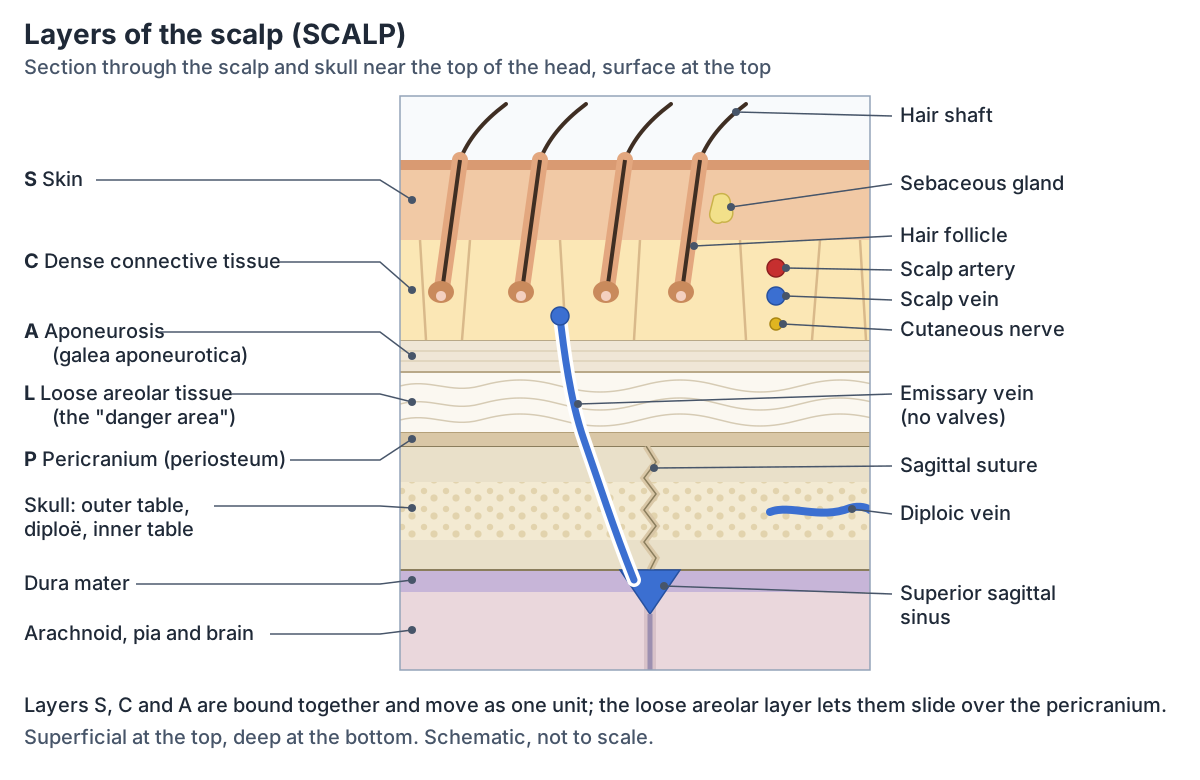
The five layers of the scalp
| Letter | Layer | What it is | Why it matters |
|---|---|---|---|
| S | Skin | Hair-bearing skin, the thickest in the body (about 3 to 8 mm), rich in hair follicles and sebaceous glands | Sebaceous cysts are common on the scalp |
| C | Dense connective tissue | Fibrofatty layer joining the skin to the galea; carries the arteries, veins and nerves | Its vessels cannot constrict when cut, so scalp wounds bleed freely |
| A | Aponeurosis (galea aponeurotica) | Tough fibrous sheet joining the frontal and occipital bellies of occipitofrontalis | Moves with the skin as one unit; a cut across it gapes |
| L | Loose areolar tissue | Loose layer that lets the top three layers slide; crossed by emissary veins | The danger area: infection and blood can spread in it |
| P | Pericranium | Periosteum on the outer surface of the skull, attached into the sutures | Bleeding beneath it (cephalohaematoma) stops at the sutures |
Compiled from TeachMeAnatomy, Kenhub, Radiopaedia and two scalp reconstruction reviews (Lin 2008; Sokoya 2018).
The scalp muscle: occipitofrontalis
Occipitofrontalis is a long, wide muscle of the scalp that runs from the eyebrows to the superior nuchal lines. Its frontal belly arises from the skin of the eyebrows and blends with procerus, orbicularis oculi and corrugator supercilii, and inserts into the epicranial aponeurosis in front of the coronal suture. Its occipital belly arises from the lateral two-thirds of the superior nuchal line and inserts into the aponeurosis behind the lambdoid suture. (Kenhub: Occipitofrontalis muscle.)
The frontal belly raises the eyebrows and the skin of the forehead, as in surprise, and its two halves can act separately. The occipital belly pulls the scalp backwards, and together the two bellies move the whole scalp. The frontal belly is supplied by temporal branches of the facial nerve and the occipital belly by the posterior auricular nerve, another branch of the facial nerve. (Kenhub: Occipitofrontalis muscle.)
Radiopaedia lists three muscles anchored to the galea: occipitofrontalis, temporoparietalis and the superior auricular muscle. All three are counted as muscles of facial expression and all are supplied by the facial nerve. (Radiopaedia: Scalp.)
The muscles of facial expression, including occipitofrontalis, and the facial nerve. Facial muscles.
Blood supply of the scalp: five pairs of arteries
Five paired arteries supply the scalp: the supratrochlear, supraorbital, superficial temporal, posterior auricular and occipital arteries. The supratrochlear and supraorbital arteries arise from the ophthalmic artery, the first branch of the internal carotid artery. The other three arise from the external carotid artery. (Lin SJ, Hanasono MM, Skoracki RJ. Scalp and Calvarial Reconstruction. Semin Plast Surg 2008.)
TeachMeAnatomy maps their territories. The superficial temporal artery supplies the frontal and temporal regions, the posterior auricular artery the area above and behind the ear, and the occipital artery the back of the scalp. The supraorbital and supratrochlear arteries supply the front and top of the scalp and run with the nerves of the same names. (TeachMeAnatomy: The Scalp.)
The superficial temporal artery is one of the two terminal branches of the external carotid artery. It arises in the parotid gland, crosses the zygomatic arch in front of the ear and ends by dividing into a frontal (anterior) branch and a parietal (posterior) branch. (Radiopaedia: Superficial temporal artery.)
Because both carotid systems feed the scalp and their terminal branches form extensive networks, the blood supply is redundant. The supraorbital artery is the lateral and the supratrochlear artery the medial artery of the forehead; the supratrochlear artery runs under the skin about 1.7 to 2.2 cm from the midline. Below the superior nuchal line, vessels that perforate trapezius and splenius capitis also reach the skin of the back of the head. (Sokoya M, Inman J, Ducic Y. Scalp and Forehead Reconstruction. Semin Plast Surg 2018.)
The common, internal and external carotid arteries and their branches. Carotid arteries.
Arteries of the scalp
| Artery | Arises from | Part of the scalp supplied |
|---|---|---|
| Supratrochlear artery | Ophthalmic artery (internal carotid system) | Front and top of the scalp, near the midline of the forehead |
| Supraorbital artery | Ophthalmic artery (internal carotid system) | Front and top of the scalp, lateral to the supratrochlear artery |
| Superficial temporal artery | External carotid artery (a terminal branch) | Frontal and temporal regions, through its frontal and parietal branches |
| Posterior auricular artery | External carotid artery | Above and behind the ear |
| Occipital artery | External carotid artery | Back of the scalp |
Each artery is paired (one on each side), and all five anastomose with each other. Compiled from TeachMeAnatomy, Lin 2008, Sokoya 2018 and Radiopaedia.
Why do scalp wounds bleed so much?
Deep cuts of the scalp bleed heavily for three reasons. The vessels are bound to the dense connective tissue, so they cannot constrict normally; the arteries of the scalp are joined by many anastomoses; and the pull of occipitofrontalis holds the wound, and the vessels in it, open. (TeachMeAnatomy: The Scalp.)
Kenhub gives the same explanation for the connective tissue layer: its blood vessels are attached to the surrounding tissue, so they cannot go into spasm (vasospasm) when they are cut, which can cause heavy bleeding after a scalp injury. (Kenhub: Scalp and hair histology.)
The same rich supply has an advantage in surgery. The subcutaneous layer is richly vascularised, and its network of small connecting vessels allows relatively long local flaps of scalp to survive when they are moved to close a defect. (Lin SJ, Hanasono MM, Skoracki RJ. Scalp and Calvarial Reconstruction. Semin Plast Surg 2008.)
Veins of the scalp and the danger area
The superficial veins of the scalp follow the arteries: the superficial temporal, occipital, posterior auricular, supraorbital and supratrochlear veins. The deep temporal region drains to the pterygoid venous plexus, which lies between temporalis and the lateral pterygoid muscle and empties into the maxillary vein. (TeachMeAnatomy: The Scalp.)
The loose areolar layer is called the danger area of the scalp. It contains emissary veins, which have no valves and connect the veins outside the skull with the dural venous sinuses inside it, so infection in this layer could spread into the cranial cavity. Emissary veins also connect the scalp veins with the diploic veins in the spongy bone of the skull. (TeachMeAnatomy: The Scalp.)
Radiopaedia describes emissary veins as thin-walled, valveless veins that pass through openings in the skull. The parietal emissary vein passes through the parietal foramen to join the superior sagittal sinus with scalp veins, the mastoid emissary vein joins the sigmoid sinus with the occipital or posterior auricular veins, and the frontal emissary vein passes through the foramen caecum. As well as being a route for infection, they act as collateral channels if a sinus is blocked and may help cool the brain with blood from the scalp. (Radiopaedia: Emissary veins.)
The superior sagittal and other sinuses that the emissary veins join. Dural venous sinuses.
Nerves of the scalp: in front of and behind the ear
Sensation from the scalp in front of the ear is carried by branches of the trigeminal nerve: the supratrochlear and supraorbital nerves of the ophthalmic division (V1), the zygomaticotemporal nerve of the maxillary division (V2) and the auriculotemporal nerve of the mandibular division (V3). Behind the ear it is carried by cervical nerves: the great auricular, lesser occipital, greater occipital and third occipital nerves. (TeachMeAnatomy: The Scalp.)
In TeachMeAnatomy's account, the supratrochlear nerve supplies the front of the forehead near the midline and the supraorbital nerve most of the scalp from the forehead to the vertex. The zygomaticotemporal nerve supplies the temple and the auriculotemporal nerve the skin above and in front of the ear. The greater occipital nerve (posterior ramus of C2) supplies the back of the head, the third occipital nerve (posterior ramus of C3) the lower occipital region, the lesser occipital nerve (anterior ramus of C2) the skin behind the ear, and the great auricular nerve (anterior rami of C2 and C3) the skin behind the ear and over the angle of the jaw. (TeachMeAnatomy: The Scalp.)
Sources differ in detail. Radiopaedia gives the lesser occipital nerve as C2 to C3, places the greater occipital (C2) and third occipital (C3) nerves at the back of the scalp, and also lists the lacrimal and zygomaticofacial nerves among the cutaneous nerves at the edges of the scalp. (Radiopaedia: Scalp.)
A surgical review lists six main sensory nerves of the scalp: supratrochlear (V1), supraorbital (V1), zygomaticotemporal (V2), auriculotemporal (V3), lesser occipital (C2 to C3) and greater occipital (C2). (Lin SJ, Hanasono MM, Skoracki RJ. Scalp and Calvarial Reconstruction. Semin Plast Surg 2008.)
The trigeminal and facial nerves and the other ten cranial nerves. Cranial nerves.
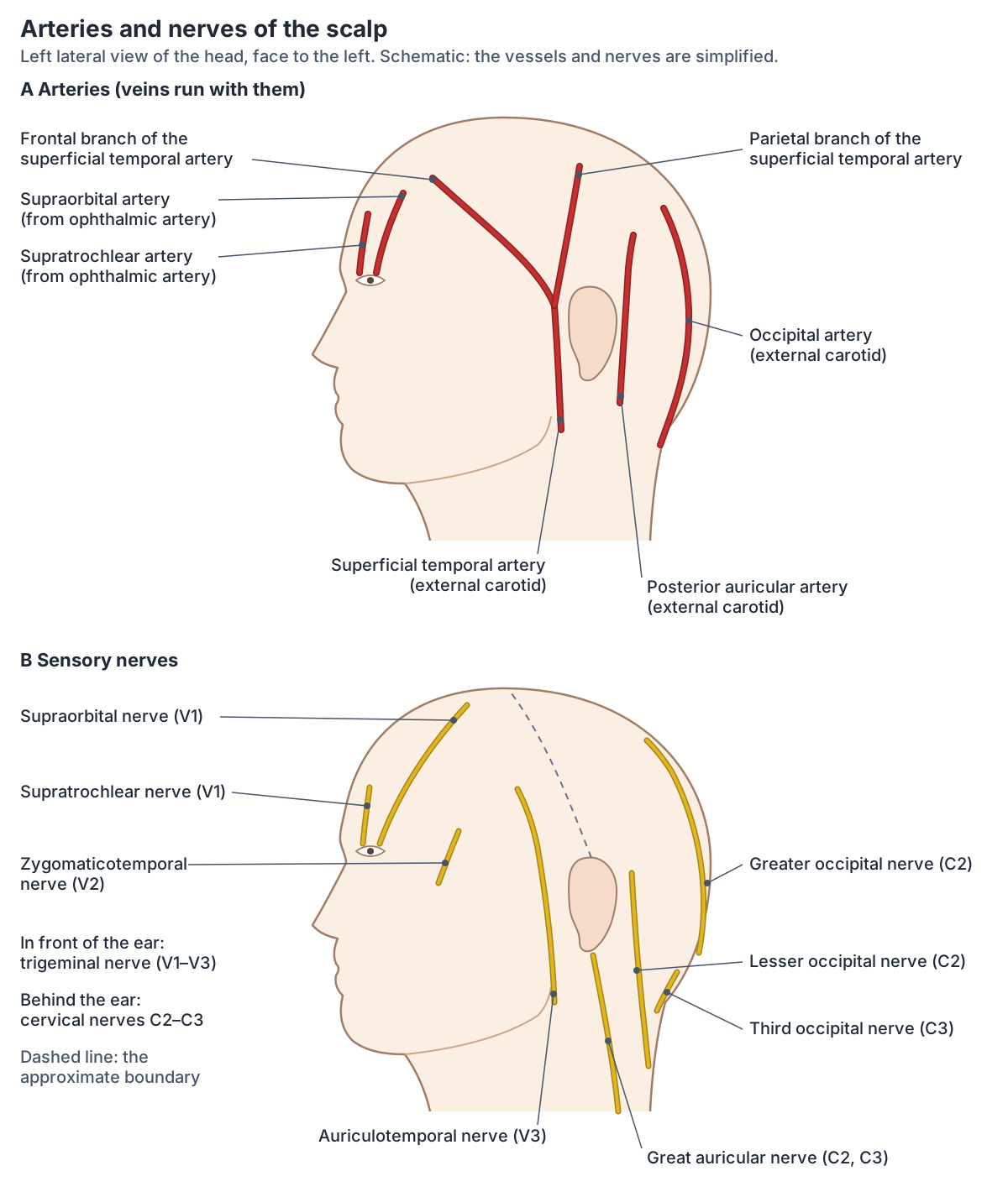
Sensory nerves of the scalp
| Nerve | Origin | Area of skin |
|---|---|---|
| Supratrochlear nerve | Ophthalmic division of the trigeminal nerve (V1) | Front of the forehead near the midline |
| Supraorbital nerve | Ophthalmic division (V1) | Most of the scalp from the forehead back to the vertex |
| Zygomaticotemporal nerve | Maxillary division (V2) | Temple |
| Auriculotemporal nerve | Mandibular division (V3) | Skin above and in front of the ear |
| Great auricular nerve | Anterior rami of C2 and C3 | Skin behind the ear and over the angle of the jaw |
| Lesser occipital nerve | Anterior ramus of C2 (C2 and C3 in some sources) | Skin behind the ear |
| Greater occipital nerve | Posterior ramus of C2 | Back of the head (occipital region) |
| Third occipital nerve | Posterior ramus of C3 | Lower occipital region |
Nerves in front of the ear come from the trigeminal nerve; nerves behind the ear come from the second and third cervical nerves. Based on TeachMeAnatomy, with Radiopaedia and Lin 2008.
Lymphatic drainage of the scalp
Like the face, the scalp has no lymph nodes of its own. Its lymph drains through small lymphatic channels to the submandibular, parotid, mastoid and occipital nodes, which form a collar of nodes at the junction of the head and neck. (Radiopaedia: Scalp.)
TeachMeAnatomy describes one to three occipital nodes at the back of the head, at the lateral border of trapezius, that collect lymph from the occipital area of the scalp, and one to three pre-auricular nodes in front of the ear that drain the superficial face and the temporal region. These superficial nodes drain on to the deep cervical nodes, which receive all of the lymph from the head and neck. (TeachMeAnatomy: Lymphatic Drainage of the Head and Neck.)
The ring of superficial nodes and the deep cervical chain. Lymph nodes of the head and neck.
Hair follicles of the scalp
Most people have about 100,000 hairs on the scalp. Each follicle cycles on its own, so the number of hairs stays fairly stable. On the scalp the growth phase (anagen) lasts about two to eight years, and about 9% of scalp hairs are in the resting phase (telogen) at any time, compared with 40 to 50% of hairs on the trunk. (Natarelli N, Gahoonia N, Sivamani RK. Integrative and Mechanistic Approach to the Hair Growth Cycle and Hair Loss. J Clin Med 2023.)
Hair follicles begin in the epidermis and can extend down through the dermis into the subcutaneous tissue. During the growth phase a hair typically grows about 0.3 mm a day, and each hair root is attached to a small smooth muscle, the arrector pili. (OpenStax Anatomy and Physiology 2e, 5.2: Accessory Structures of the Skin.)
Kenhub notes that the skin of the scalp has many blood vessels and Pacinian corpuscles, receptors for deep pressure, and that sebaceous glands are densely scattered across it. (Kenhub: Scalp and hair histology.)
The hair shaft, the layers of the follicle and the growth cycle. Hair anatomy.
Cephalohaematoma vs caput succedaneum vs subgaleal haematoma
Three swellings of a newborn's scalp are told apart by the layer they lie in. Caput succedaneum is a collection of blood-stained (serosanguineous) fluid in the subcutaneous tissue, outside the galea. It results from pressure of the presenting part of the skull against the dilating cervix during birth, extends across the midline and over suture lines, and usually resolves over the first few days. (Radiopaedia: Caput succedaneum.)
A subgaleal haematoma is bleeding into the potential space between the galea and the periosteum, the loose areolar layer. Because it lies above the periosteum it can cross suture lines, surround the whole skull and extend into the neck. Moderate to severe cases occur in about 1.5 per 10,000 live births, most often after vacuum-assisted or forceps delivery; it is rare but can be life-threatening. (Radiopaedia: Subgaleal haematoma.)
A cephalohaematoma (cephalohematoma) is bleeding under the periosteum, between it and the bone, usually from birth injury. The periosteum confines it, so it is bounded by the suture lines and stays over one bone. It occurs in about 1 to 2% of spontaneous vaginal births and 3 to 4% of forceps or vacuum-assisted births. Most resolve within weeks, though some take three to four months, and the edge may calcify after two to three weeks. (Radiopaedia: Cephalohaematoma.)
This section explains anatomy only. A swelling of a baby's head is assessed by a midwife or doctor, who uses these features together with the history and examination.
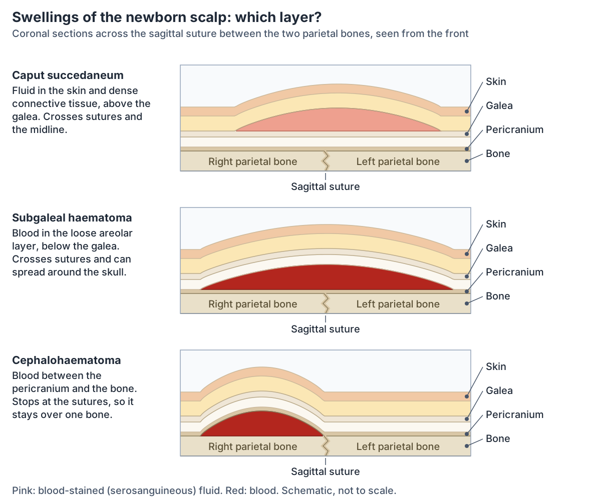
Caput succedaneum vs subgaleal haematoma vs cephalohaematoma
| Swelling | Layer | Crosses sutures? | Usual course |
|---|---|---|---|
| Caput succedaneum | Subcutaneous tissue, above the galea (blood-stained fluid) | Yes, and the midline | Usually resolves over the first few days |
| Subgaleal haematoma | Loose areolar layer, between the galea and the periosteum (blood) | Yes; can spread around the skull and into the neck | Rare but can be life-threatening |
| Cephalohaematoma | Under the periosteum, against the bone (blood) | No, it stays over one bone | Most resolve within weeks, some take 3 to 4 months; may calcify |
From the Radiopaedia articles on each condition. Educational summary only.
The scalp in the 3D model
The 3D viewer highlights the hair of the head and the supraorbital and supratrochlear nerves, over a ghosted skeleton seen from the left. The model is BodyParts3D, built from MRI of an adult male, and it represents the skin as a single surface: it has no separate scalp layers, no galea or occipitofrontalis, and no scalp arteries or occipital nerves. The hair belongs to the skin system, so raise the Skin slider to see it. Use the diagrams on this page for the layers, vessels and nerves. (Mitsuhashi N et al. BodyParts3D: 3D structure database for anatomical concepts. Nucleic Acids Res 2009.)
The frontal, parietal, occipital and other bones under the scalp, with the sutures. Skull bones.
Scalp in the 3D model
- Hair of head
- The hair of the head is made of keratinised hair shafts growing from follicles in the scalp.
- Supra-orbital nerve
- The supra-orbital nerve is the larger terminal branch of the frontal nerve supplying the forehead and scalp.
- Supratrochlear nerve
- The supratrochlear nerve is the smaller terminal branch of the frontal nerve.
Frequently asked questions
What are the 5 layers of the scalp?
From the surface inwards the five layers are skin, dense connective tissue, the epicranial aponeurosis (galea aponeurotica), loose areolar tissue and the pericranium, the periosteum of the skull. Their initials spell SCALP. The first three layers are bound together and move as one unit over the loose areolar layer.
Why do head wounds bleed so much?
The scalp has a very rich blood supply from five pairs of arteries that join each other. The vessels sit in dense connective tissue that holds them open, so they cannot constrict when cut, and the pull of the occipitofrontalis muscle on the galea can hold a deep wound open. Together these make even small scalp cuts bleed freely.
What is the danger area of the scalp?
The danger area is the fourth layer, the loose areolar tissue between the galea and the pericranium. It contains valveless emissary veins that connect the veins of the scalp with the diploic veins and the dural venous sinuses inside the skull, so an infection in this layer could spread into the cranial cavity.
Which arteries supply the scalp?
Five pairs of arteries: the supratrochlear and supraorbital arteries, which come from the ophthalmic branch of the internal carotid artery, and the superficial temporal, posterior auricular and occipital arteries, which come from the external carotid artery. They join each other freely, so the scalp has a rich, overlapping blood supply.
What nerves supply the scalp?
In front of the ear, branches of the trigeminal nerve: the supratrochlear and supraorbital nerves (V1), the zygomaticotemporal nerve (V2) and the auriculotemporal nerve (V3). Behind the ear, cervical nerves: the great auricular, lesser occipital, greater occipital and third occipital nerves from C2 and C3. The scalp muscles are supplied by the facial nerve.
What is the difference between a cephalohematoma and caput succedaneum?
Caput succedaneum is fluid in the subcutaneous tissue above the galea, caused by pressure during birth; it crosses sutures and the midline and usually settles within days. A cephalohaematoma is blood under the periosteum of one skull bone; it cannot cross a suture and can take weeks to months to resolve.
How thick is the scalp?
The skin of the scalp is the thickest skin in the body, about 3 to 8 mm according to two plastic surgery reviews, and it is thicker over the back of the head. Beneath the skin lie the connective tissue, galea, loose areolar tissue and pericranium, so the full scalp is thicker than the skin alone.
References
- TeachMeAnatomy. The Scalp. Accessed 2026-10-24.
- TeachMeAnatomy. Lymphatic Drainage of the Head and Neck. Accessed 2026-10-24.
- Radiopaedia. Scalp. Accessed 2026-10-24.
- Radiopaedia. Emissary veins. Accessed 2026-10-24.
- Radiopaedia. Superficial temporal artery. Accessed 2026-10-24.
- Radiopaedia. Subgaleal haematoma. Accessed 2026-10-24.
- Radiopaedia. Cephalohaematoma. Accessed 2026-10-24.
- Radiopaedia. Caput succedaneum. Accessed 2026-10-24.
- Kenhub. Scalp and hair histology. Accessed 2026-10-24.
- Kenhub. Occipitofrontalis muscle. Accessed 2026-10-24.
- Lin SJ, Hanasono MM, Skoracki RJ. Scalp and Calvarial Reconstruction. Semin Plast Surg. 2008;22(4):281-293.
- Sokoya M, Inman J, Ducic Y. Scalp and Forehead Reconstruction. Semin Plast Surg. 2018;32(2):90-94.
- Natarelli N, Gahoonia N, Sivamani RK. Integrative and Mechanistic Approach to the Hair Growth Cycle and Hair Loss. J Clin Med. 2023;12(3):893.
- Betts JG, et al. Anatomy and Physiology 2e, §5.2 Accessory Structures of the Skin. OpenStax, Rice University, 2022.
- Mitsuhashi N, et al. BodyParts3D: 3D structure database for anatomical concepts. Nucleic Acids Res. 2009;37:D782-D785.
- Mitsuhashi N. et al. (2009). BodyParts3D: 3D structure database for anatomical concepts. Nucleic Acids Research 37, D782–D785. Data © The Database Center for Life Science, CC BY 4.0.
Related anatomy
- Head anatomyHead anatomy guide: skull, brain, face, sense organs and ENT structures, head and neck muscles, vessels, nerves and…
- Skull bonesSkull anatomy with labelled diagrams and a 3D model: the 8 cranial and 14 facial bones, sutures, fontanelles, the…
- TempleTemple anatomy: where the temple is, the four bones that meet at the pterion, temporalis, the temporal arteries and…
- Hair anatomyHair anatomy with labelled diagrams: shaft layers, follicle root sheaths, bulb and dermal papilla, arrector pili, hair…
- Facial musclesFace muscles labeled: labelled diagrams and a table of every muscle of facial expression, from procerus and…
- Dural venous sinusesDural venous sinuses explained: superior sagittal, straight, transverse, sigmoid, occipital and cavernous sinus…
Explore the whole body in the interactive 3D human anatomy model, or browse the A–Z list of human structures.