

Where is the liver located?
The liver lies in the right upper quadrant of the abdomen, mainly in the right hypochondrium and epigastric region, and extends into the left hypochondrium. It is the largest gland in the body and the largest organ in the abdominal cavity. (TeachMeAnatomy: The Liver.)
It sits just below the diaphragm and is protected by the surrounding ribs. OpenStax gives its adult weight as about three pounds. (OpenStax Anatomy and Physiology 2e, 23.6: Accessory Organs in Digestion: The Liver, Pancreas, and Gallbladder.)
The liver is wedge-shaped and largely covered by the costal cartilages. On imaging, a normal liver measures about 10 to 12.5 cm from top to bottom (15 to 16 cm is the upper limit of normal) and 20 to 23 cm from side to side. (Radiopaedia: Liver.)
Kenhub gives the adult weight as approximately 1.5 kg. (Kenhub: Liver.)
Cleveland Clinic gives a wider range of 3 to 5 pounds and compares the liver's size and shape to a football. (Cleveland Clinic: Liver.)
Quadrants, regions and every organ of the abdomen around the liver. Abdominal anatomy.
Surfaces of the liver and the bare area
The liver has two surfaces. The diaphragmatic surface faces up and forwards; it is smooth and convex and fits under the diaphragm, and its back part has no peritoneum, forming the bare area where the liver touches the diaphragm directly. The visceral surface faces down and back; it is covered by peritoneum except at the gallbladder fossa and porta hepatis, and is moulded by the organs beneath it. (TeachMeAnatomy: The Liver.)
Neighbouring organs leave impressions on the visceral surface: the stomach and oesophagus on the left; the gallbladder, right kidney, right suprarenal gland and hepatic flexure of the colon on the right; and the pancreas and duodenum in the midline. (Radiopaedia: Liver.)
Around the liver are peritoneal recesses where pus or fluid can collect. The subphrenic spaces lie between the liver and the diaphragm, divided into right and left by the falciform ligament. The subhepatic space lies below the liver above the transverse colon, and the hepatorenal recess (Morison's pouch) lies between the liver and the right kidney. (TeachMeAnatomy: The Liver.)
Morison's pouch is bounded in front by the right lobe of the liver and the gallbladder and behind by the right kidney, right adrenal gland, duodenum and hepatic flexure. It is one of the lowest spaces in the peritoneal cavity, so ascites and blood tend to collect there. (Radiopaedia: Hepatorenal pouch.)
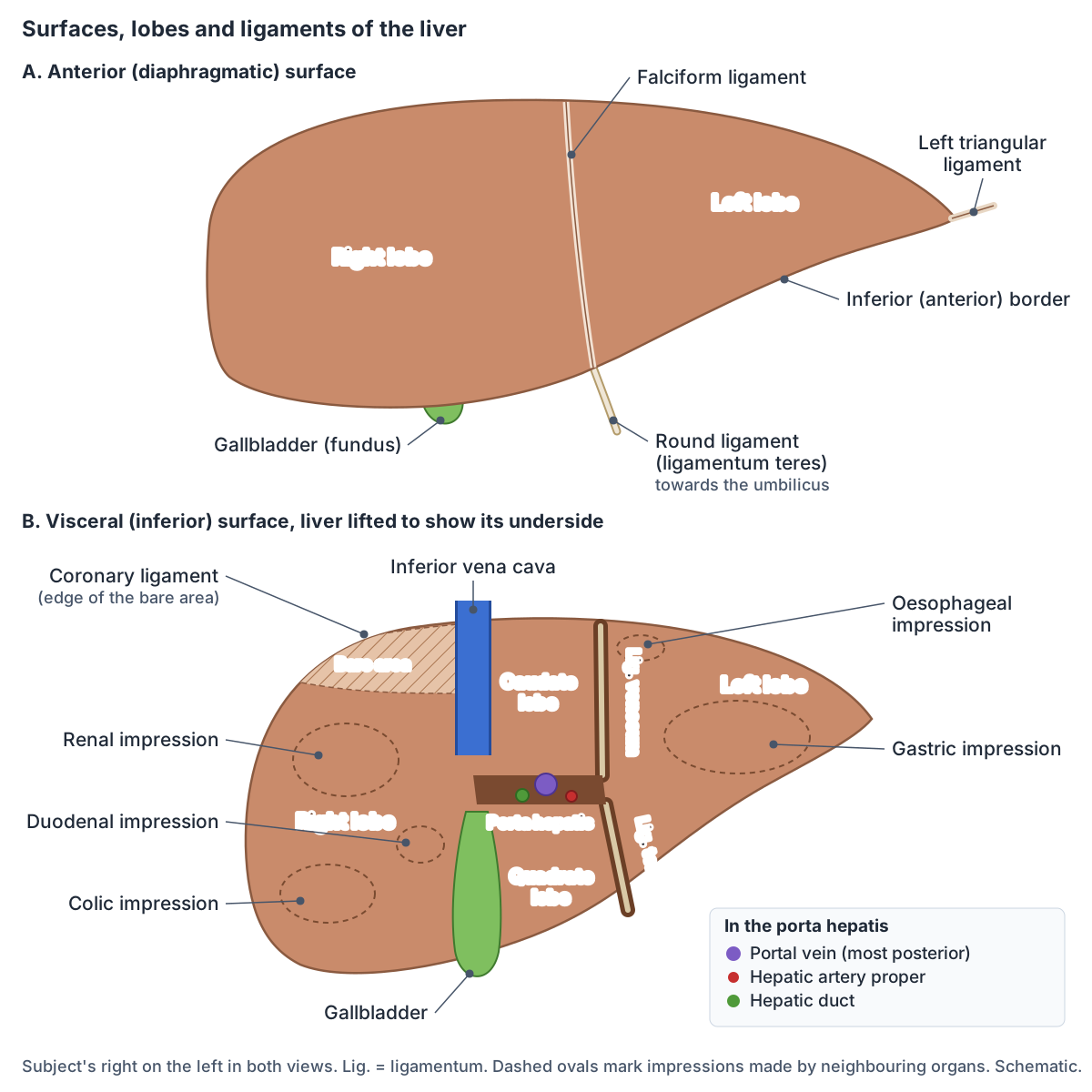
Lobes of the liver: right, left, caudate and quadrate
Anatomically the liver has four lobes. The right lobe is the largest; the left lobe is flatter and smaller. On the visceral surface, the caudate lobe lies between the fissure for the ligamentum venosum and the inferior vena cava, and the quadrate lobe lies between the gallbladder and the fissure for the ligamentum teres. (Kenhub: Liver.)
On the front, the falciform ligament marks the traditional boundary between the right and left lobes. This description by external shape has now been largely replaced by the Couinaud classification of functional segments. (Radiopaedia: Liver.)
The fissures on the visceral surface form an H. The left limb is the fissure for the ligamentum venosum above and the fissure for the ligamentum teres below; the right limb is the groove for the inferior vena cava above and the gallbladder fossa below; the crossbar is the porta hepatis. The caudate lobe lies behind the porta and the quadrate lobe in front of it. (Radiopaedia: Porta hepatis.)
The caudate and quadrate lobes belong to the anatomical right lobe, but in Kenhub's functional division they are part of the functional left lobe. (Kenhub: Functional division of the liver.)
Anatomical lobes and Couinaud segments
| Anatomical lobe | Where it is | Couinaud segments |
|---|---|---|
| Right lobe | Right of the falciform ligament on the front; right of the IVC groove and gallbladder fossa underneath | V, VI, VII, VIII and the upper part of IV (IVa) |
| Left lobe | Left of the falciform ligament and of the fissures for the ligamenta teres and venosum | II and III |
| Quadrate lobe | Underside, between the gallbladder fossa and the fissure for the ligamentum teres, in front of the porta hepatis | IVb |
| Caudate lobe | Back and underside, between the IVC groove and the fissure for the ligamentum venosum, behind the porta hepatis | I |
Compiled from Kenhub, Radiopaedia and TeachMeAnatomy (see references). The caudate and quadrate lobes count as part of the anatomical right lobe but the functional left liver.
Ligaments of the liver
The ligaments of the liver are double folds of peritoneum. The sickle-shaped falciform ligament attaches the front of the liver to the anterior abdominal wall, and its free edge carries the ligamentum teres, the remnant of the umbilical vein. The anterior and posterior folds of the coronary ligament attach the top of the liver to the diaphragm and border the bare area; at each side they meet as the right and left triangular ligaments. The lesser omentum (hepatogastric and hepatoduodenal ligaments) joins the liver to the stomach and duodenum. (TeachMeAnatomy: The Liver.)
The ligamentum teres runs from the inferior border into the porta hepatis and attaches to the left portal vein. The ligamentum venosum continues from it into the upper part of the porta hepatis. (Radiopaedia: Liver.)
The ligamentum venosum is the remnant of the ductus venosus, which before birth shunted blood from the umbilical vein to the inferior vena cava, bypassing the liver. (Kenhub: Liver.)
Ligaments of the liver
| Ligament | Connects | Notes |
|---|---|---|
| Falciform ligament | Front of the liver to the anterior abdominal wall | Traditional border of the right and left lobes |
| Ligamentum teres (round ligament) | Umbilicus to the left portal vein, in the free edge of the falciform ligament | Remnant of the umbilical vein |
| Ligamentum venosum | Runs in its fissure from the ligamentum teres into the upper porta hepatis | Remnant of the ductus venosus |
| Coronary ligament | Top of the liver to the diaphragm | Anterior and posterior folds border the bare area |
| Right and left triangular ligaments | Right and left lobes to the diaphragm | Where the coronary folds meet at each side |
| Lesser omentum | Liver to the lesser curvature of the stomach and the duodenum | Hepatogastric and hepatoduodenal ligaments; the latter carries the portal triad |
Compiled from TeachMeAnatomy, Kenhub and Radiopaedia (see references).
Porta hepatis: where vessels enter the liver
The porta hepatis (transverse hepatic fissure) is a deep fissure on the visceral surface through which all the vessels, nerves and hepatic ducts enter or leave the liver, except the hepatic veins. It lies between the caudate lobe behind and the quadrate lobe in front, within the hepatoduodenal ligament, and gives attachment to the lesser omentum. (Radiopaedia: Porta hepatis.)
From front to back the order is duct, artery, vein: the common hepatic duct lies in front and to the right, the hepatic artery proper in front and to the left, and the portal vein behind both. The hepatic plexus of autonomic nerves also passes through. (Radiopaedia: Porta hepatis.)
Kenhub lists the structures passing through the porta hepatis (also called the hilum of the liver) as the portal vein, hepatic arteries, hepatic ducts, hepatic nerve plexus and lymphatic vessels. (Kenhub: Liver.)
Hepatic ducts, cystic duct, common bile duct and Calot's triangle. Gallbladder and biliary tree.
Liver segments: the Couinaud classification
The Couinaud classification is the most widely used system for the functional anatomy of the liver. It divides the liver into eight independent segments, each with its own inflow from the hepatic artery and portal vein and its own biliary and lymphatic drainage. Each segment is roughly wedge-shaped, with its apex at the porta hepatis. Segments 2 to 8 are numbered clockwise starting from the top of the left lobe, and Arabic numerals (1 to 8) are now preferred to Roman numerals. (Radiopaedia: Couinaud classification of hepatic segments.)
Three hepatic veins run in vertical planes from the inferior vena cava. The right hepatic vein divides the right lobe into a posterior (lateral) section, segments 6 and 7, and an anterior (medial) section, segments 5 and 8. The middle hepatic vein runs in the main fissure along Cantlie's line, from the IVC to the gallbladder fossa, separating the right and left hemilivers. The left hepatic vein separates segment 4 from segments 2 and 3. A horizontal portal plane at the bifurcation of the portal vein then divides each section into an upper segment (2, 4a, 8, 7) and a lower one (3, 4b, 5, 6). (Radiopaedia: Couinaud classification of hepatic segments.)
Kenhub describes the numbering as running anticlockwise when the liver is viewed with its lower border lifted towards the diaphragm, which is the same sequence seen from the other side. It also calls the right anterior and posterior sections the right medial and lateral sectors. (Kenhub: Functional division of the liver.)
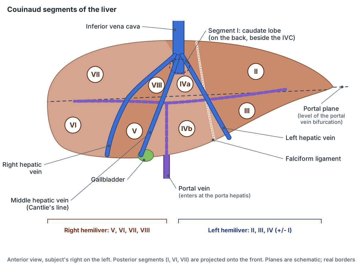
The Couinaud segments
| Segment | Section | Position | Main draining vein |
|---|---|---|---|
| I | Caudate lobe | Back of the liver beside the IVC, behind the porta hepatis | Small veins straight into the IVC |
| II | Left lateral | Upper, left of the left hepatic vein | Left hepatic vein |
| III | Left lateral | Lower, left of the left hepatic vein | Left hepatic vein |
| IVa | Left medial | Upper, between the middle and left hepatic veins | Middle and left hepatic veins |
| IVb | Left medial (quadrate lobe) | Lower, between the middle and left hepatic veins | Middle and left hepatic veins |
| V | Right anterior | Lower, between the middle and right hepatic veins | Middle and right hepatic veins |
| VI | Right posterior | Lower, right of the right hepatic vein | Right hepatic vein |
| VII | Right posterior | Upper, right of the right hepatic vein | Right hepatic vein |
| VIII | Right anterior | Upper, between the middle and right hepatic veins | Middle and right hepatic veins |
Based on Radiopaedia's Couinaud classification and hepatic veins articles (see references). Upper and lower are relative to the portal plane.
The caudate lobe (segment I)
Segment 1, the caudate lobe, is bounded behind and to the side by the groove for the inferior vena cava, in front by the ligamentum venosum and below by the porta hepatis. Its lower part has a lateral caudate process and a medial papillary process. It may receive blood from both the right and left portal veins, and it drains straight into the IVC through small hepatic veins. (Radiopaedia: Couinaud classification of hepatic segments.)
Because it has its own supply and drainage, Kenhub calls segment I the autonomous zone of the liver. (Kenhub: Functional division of the liver.)
In advanced cirrhosis the caudate lobe and the lateral segments of the left lobe typically enlarge while the posterior right segments (6 and 7) shrink, so radiologists compare the size of the caudate lobe with the right lobe. (Radiopaedia: Cirrhosis.)
Functional right and left liver
Surgeons divide the liver into a right hemiliver (segments 5 to 8) and a left hemiliver (segments 2 to 4, with or without segment 1). Removing one is a right or left hepatectomy. Smaller resections follow the sections and segments: for example, a left lateral sectionectomy removes segments 2 and 3, and a segmentectomy removes a single segment. These names follow the Brisbane 2000 terminology. (Radiopaedia: Couinaud classification of hepatic segments.)
Segmental anatomy makes living-donor transplantation possible. An adult usually receives a right hemiliver cut along a plane to the right of the middle hepatic vein, and a child most often receives the left lateral segments 2 and 3 cut just right of the falciform ligament. The donor's remaining liver regenerates to replace the removed volume. (Radiopaedia: Liver transplantation.)
Blood supply: hepatic artery and portal vein
The liver has a dual blood supply. The hepatic portal vein brings about 75% of its blood: nutrient-rich, partly deoxygenated blood from the gut, and it is the main supply to the liver tissue. The hepatic artery proper, from the coeliac trunk, brings about 25% and supplies the non-parenchymal structures. (TeachMeAnatomy: The Liver.)
Although the portal vein carries about three-quarters of the blood volume, roughly half of the liver's oxygen comes from the portal vein and half from the hepatic arteries. The hepatic arteries also supply the biliary system. (Radiopaedia: Liver.)
Kenhub gives slightly different figures, about 70% of liver blood flow from the portal vein and 30% from the hepatic artery. (Kenhub: Functional division of the liver.)
The common hepatic artery is a terminal branch of the coeliac trunk. It gives off the right gastric artery and ends as the proper hepatic artery and the gastroduodenal artery. The textbook pattern, in which the proper hepatic artery divides into right and left hepatic arteries that supply the whole liver, is found in only 55 to 60% of people; the rest have variant arteries. (Radiopaedia: Common hepatic artery.)
The portal vein forms behind the neck of the pancreas, where the splenic vein joins the superior mesenteric vein. Where portal and systemic veins meet, at the oesophagus and rectum for example, blocked portal flow can open up varices that may bleed. (TeachMeAnatomy: Venous Drainage of the Abdomen.)
The hepatic portal system, its tributaries and portal hypertension. Portal vein.
Venous drainage: the hepatic veins
Three large hepatic veins, right, middle and left, drain the liver into the inferior vena cava. The right hepatic vein drains segments 5 to 8, the middle hepatic vein segments 4a, 4b, 5 and 8, and the left hepatic vein segments 2, 3, 4a and 4b. Small separate veins from the caudate lobe drain straight into the IVC. The commonest variant is an accessory right inferior hepatic vein. (Radiopaedia: Hepatic veins.)
The hepatic veins have no valves and help to hold the liver in place. (Radiopaedia: Liver.)
Within the liver, central veins join to form the hepatic veins, which open into the inferior vena cava just before it passes through the diaphragm. (Kenhub: Liver.)
The large vein that receives the hepatic veins just below the diaphragm. Inferior vena cava.
How bile leaves the liver
Hepatocytes make about 1 litre of bile a day, an alkaline (pH 7.6 to 8.6) yellow-brown or yellow-green fluid of water, bile salts, bile pigments, phospholipids, cholesterol and electrolytes. Bile salts emulsify fats so enzymes can digest them, and most are reabsorbed in the ileum and returned to the liver (the enterohepatic circulation). The main pigment, bilirubin, comes from old red blood cells. (OpenStax Anatomy and Physiology 2e, 23.6: Accessory Organs in Digestion: The Liver, Pancreas, and Gallbladder.)
Bile passes from tiny bile canaliculi between the hepatocytes into bile ductules and bile ducts, then into the right and left hepatic ducts, which merge to form the common hepatic duct. Between meals bile is diverted into the gallbladder for storage. (OpenStax Anatomy and Physiology 2e, 23.6: Accessory Organs in Digestion: The Liver, Pancreas, and Gallbladder.)
Liver lobules and histology
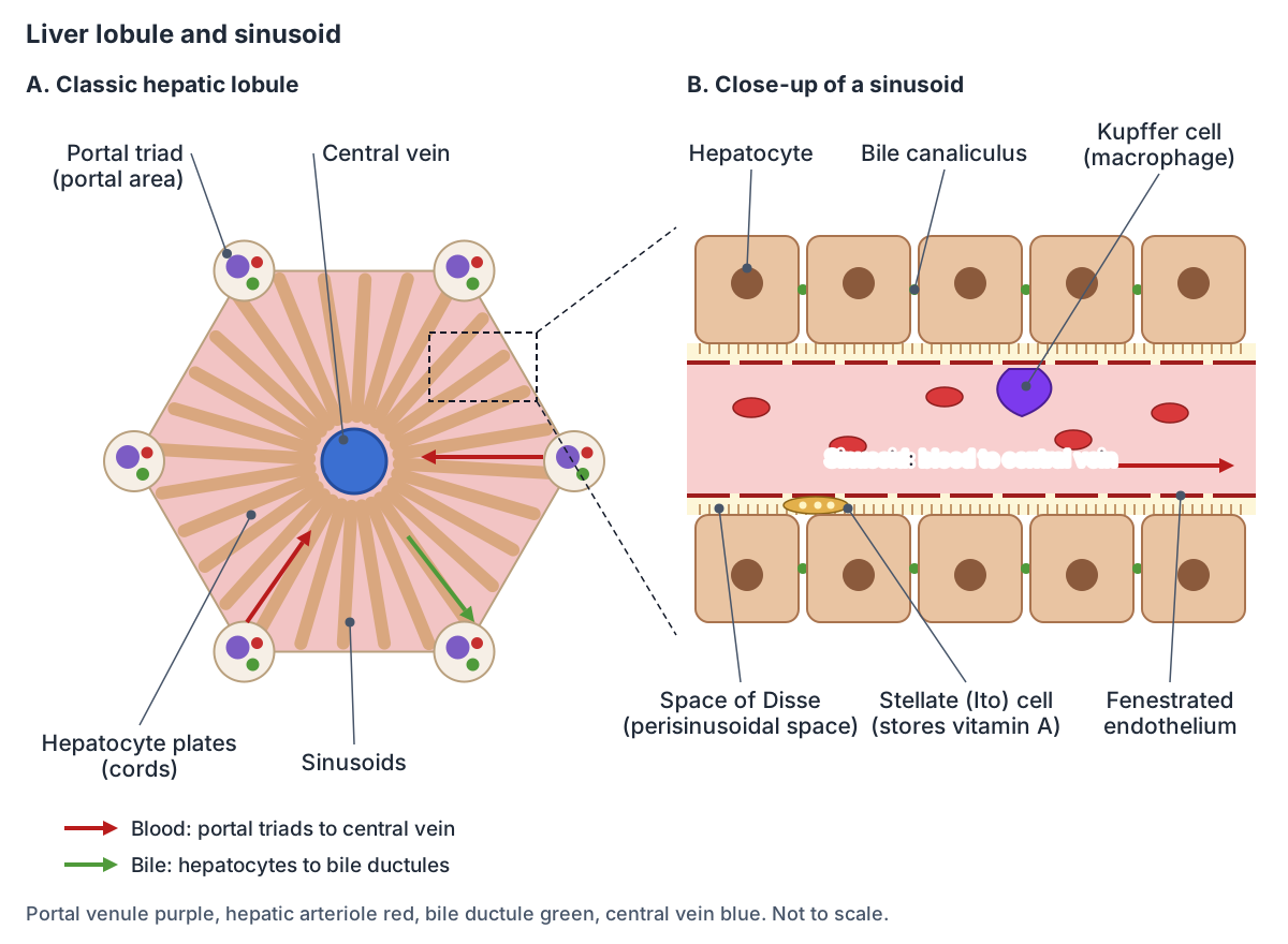
Under the microscope the liver is built of hexagonal classic lobules. Plates of hepatocytes radiate from a central vein like the spokes of a wheel, with sinusoids running between them, and a portal triad (a branch of the portal vein, a branch of the hepatic artery and a bile duct) sits at each of the six corners. (Kenhub: Liver histology.)
Hepatocytes make up about 80% of the liver's volume. The sinusoids are leaky, fenestrated capillaries, and star-shaped Kupffer cells in their walls remove old blood cells, bacteria and debris. Blood flows through the sinusoids towards the central vein, while bile flows the opposite way, towards the portal triads. (OpenStax Anatomy and Physiology 2e, 23.6: Accessory Organs in Digestion: The Liver, Pancreas, and Gallbladder.)
Between the hepatocytes and the sinusoid lining is the space of Disse (perisinusoidal space). Stellate (Ito) cells there store vitamin A and lay down collagen after injury, which drives fibrosis. Bile canaliculi, about 1 to 2 micrometres wide, run between neighbouring hepatocytes and join the canals of Hering, which contain liver stem cells. (Kenhub: Liver histology.)
Two other units describe the same tissue. The triangular portal lobule groups the tissue that drains bile into one bile duct. The oval liver acinus is divided into zones by oxygen supply: zone 1 lies closest to the portal triad and gets the most oxygen, and zone 3 lies next to the central vein and gets the least. (Kenhub: Liver histology.)
Nerves and lymph drainage
The hepatic plexus carries sympathetic fibres from the coeliac plexus and parasympathetic fibres from the vagus nerves into the porta hepatis, along the hepatic artery and portal vein. Glisson's capsule, the fibrous covering of the liver, is supplied by the lower intercostal nerves, so stretching it causes sharp, well-localised pain. (TeachMeAnatomy: The Liver.)
Irritation of the diaphragm by an inflamed liver or gallbladder can cause pain felt at the tip of the shoulder. (TeachMeAnatomy: The Peritoneum.)
Deep lymph vessels follow the portal vein branches to hepatic nodes at the porta hepatis, then to coeliac nodes, the cisterna chyli and the thoracic duct. Some leave with the hepatic veins through the bare area to posterior mediastinal nodes. Superficial vessels under the capsule drain to hepatic, lumbar, parasternal or posterior mediastinal nodes depending on the surface. (Kenhub: Liver.)
What does the liver do?
The liver makes bile, stores and metabolises fat-soluble vitamins, breaks down drugs and other chemicals, processes bilirubin, takes part in thyroid hormone metabolism and makes plasma proteins such as albumin and clotting factors. (Kenhub: Liver.)
Because all the blood from the digestive tract passes through the liver, it processes absorbed nutrients and toxins. For the same reason the liver is a common site for cancers of the digestive tract to spread to. (OpenStax Anatomy and Physiology 2e, 23.6: Accessory Organs in Digestion: The Liver, Pancreas, and Gallbladder.)
Cleveland Clinic notes that the liver has more than 500 functions, is one of the few organs that can regenerate itself, and is affected by more than 100 types of disease. (Cleveland Clinic: Liver.)
Variants and clinical anatomy
A Riedel lobe is a tongue-like downward projection of the right lobe, a variant of normal shape rather than a true extra lobe. It is reported in about 17.5% of people (3.3 to 31% across studies), is about three times more common in women, and has been mistaken for an abdominal mass. (Radiopaedia: Riedel lobe.)
Rarer variants include agenesis (absence) of the right or left lobe, accessory lobes or islands of liver tissue elsewhere, a beaver tail liver and liver tissue above the diaphragm. (Radiopaedia: Liver.)
Cirrhosis is the end stage of many chronic liver diseases, with fibrosis, regenerating nodules and distorted architecture. Common causes are chronic hepatitis B and C, alcohol-related liver disease and metabolic dysfunction-associated steatotic liver disease (MASLD); complications include portal hypertension, ascites, liver failure and hepatocellular carcinoma. (Radiopaedia: Cirrhosis.)
Portal hypertension is a portal venous pressure persistently above 12 mmHg. Blood then diverts through the five portacaval anastomoses (oesophageal, periumbilical, retroperitoneal, at the bare area of the liver and anal), causing oesophageal varices and dilated veins around the umbilicus (caput medusae). (Kenhub: Functional division of the liver.)
In a percutaneous liver biopsy, a needle is passed into the liver under ultrasound guidance, usually from below the ribs, along a path free of vessels while the patient holds their breath. If clotting is abnormal, a sample can instead be taken through the hepatic veins from the internal jugular vein. (TeachMeAnatomy: The Liver.)
The liver in the 3D model
The model highlights the liver as Couinaud segments plus the caudate lobe, together with the gallbladder, the portal vein, hepatic artery proper, hepatic and common hepatic ducts, the hepatic veins and the inferior vena cava. Click a segment to see its name, turn the body to look at the visceral surface from below, or switch off the highlight to see how much of the liver hides behind the lower right ribs.
Because each segment has its own blood supply and bile drainage, a surgeon can remove a single segment (segmentectomy) or a pair of segments, and the model shows those units in place. (Radiopaedia: Couinaud classification of hepatic segments.)
The segment meshes come from the BodyParts3D dataset and are approximate. Two of its labels were wrong: its "segment VII" duplicated segment VI, and segment VII itself was labelled VIII and IX. This site corrected them using the portal vein branches that supply each segment, but the borders between meshes are still rough, so use the Couinaud diagram above for exact borders.
Liver in the 3D model
- Caudate lobe of liver
- The caudate lobe (Couinaud segment I) is a small liver lobe on the posterior surface between the inferior vena cava and the fissure for the ligamentum venosum.
- Common hepatic duct
- The common hepatic duct is formed by the union of the right and left hepatic ducts and carries bile out of the liver.
- Gallbladder
- The gallbladder is a pear-shaped sac that stores and concentrates bile.
- Hepatic artery proper
- The proper hepatic artery is the continuation of the common hepatic artery and supplies the liver and gallbladder.
- Hepatic duct
- The right and left hepatic ducts drain bile from the functional right and left halves of the liver and unite to form the common hepatic duct.
- Hepatic portal vein
- The hepatic portal vein carries nutrient-rich blood from the gut and spleen to the liver.
- Hepatic vein
- The hepatic veins carry blood from the liver into the inferior vena cava.
- Hepatovenous segment II
- Couinaud segment II is the superior part of the left lateral section of the liver.
- Hepatovenous segment III
- Couinaud segment III is the inferior part of the left lateral section of the liver.
- Hepatovenous segment IV
- Couinaud segment IV is the left medial section of the liver, also called the quadrate lobe in its inferior part.
- Hepatovenous segment V
- Couinaud segment V is the inferior part of the right anterior section of the liver.
- Hepatovenous segment VI
- Couinaud segment VI is the inferior part of the right posterior section of the liver.
- Hepatovenous segment VII
- Couinaud segment VII is the superior part of the right posterior section of the liver.
- Hepatovenous segment VIII
- Couinaud segment VIII is the superior part of the right anterior section of the liver.
- Inferior vena cava
- The inferior vena cava is the largest vein in the body and returns blood from the lower body to the right atrium.
- Middle hepatic vein
- One of three main hepatic veins, draining the central part of the liver.
Frequently asked questions
How many lobes does the liver have?
Anatomically the liver has four lobes: a large right lobe, a smaller left lobe and, on the underside, the caudate and quadrate lobes. Functionally it is divided differently, into right and left hemilivers along Cantlie's line and then into eight Couinaud segments, each with its own blood supply and bile drainage.
What are the 8 segments of the liver?
The Couinaud segments are I (caudate lobe), II and III (left lateral section), IV (left medial section, split into IVa and IVb), V and VIII (right anterior section) and VI and VII (right posterior section). The hepatic veins separate the sections and the portal plane separates the upper segments from the lower ones.
Where is the liver located in the body?
The liver sits in the right upper part of the abdomen, just under the diaphragm, mostly behind the lower right ribs. It fills the right hypochondrium, crosses the epigastric region and reaches into the left hypochondrium. Its underside rests on the stomach, duodenum, gallbladder, right kidney and the hepatic flexure of the colon.
What is the porta hepatis?
The porta hepatis is the deep transverse fissure on the underside of the liver, between the caudate and quadrate lobes, where the portal vein, hepatic artery proper, hepatic ducts, nerves and lymph vessels enter or leave. From front to back the order is duct, artery, vein. The hepatic veins are the only vessels that do not pass through it.
What are the two blood supplies to the liver?
The liver receives blood from the hepatic portal vein, about 70 to 75% of the flow, carrying nutrient-rich blood from the gut, and from the hepatic artery proper, about 25 to 30%, carrying oxygenated blood from the coeliac trunk. Each supplies roughly half of the liver's oxygen. Blood leaves through the hepatic veins into the inferior vena cava.
What is the caudate lobe?
The caudate lobe is Couinaud segment I, a small lobe on the back of the liver between the inferior vena cava and the fissure for the ligamentum venosum, above the porta hepatis. It gets blood from both right and left vessels and drains straight into the vena cava, which is why it often enlarges in cirrhosis.
Can the liver regenerate?
Yes. The liver is one of the few organs that can regenerate. After part of it is removed, for example when a living donor gives segments for a transplant, the remaining liver grows back to replace the lost volume. Long-term damage, as in cirrhosis, instead produces scarring and regenerating nodules that distort its structure.
How much does the liver weigh?
An adult liver weighs about 1.5 kg according to Kenhub, or about three pounds according to OpenStax; Cleveland Clinic gives a range of 3 to 5 pounds. On imaging it normally measures about 10 to 12.5 cm from top to bottom and 20 to 23 cm across.
References
- OpenStax. Anatomy and Physiology 2e, §23.6 Accessory Organs in Digestion: The Liver, Pancreas, and Gallbladder. Rice University, 2022.
- Jones O. The Liver. TeachMeAnatomy.
- Jones O. Venous Drainage of the Abdomen. TeachMeAnatomy.
- Jones O. The Peritoneum. TeachMeAnatomy.
- Liver. Kenhub anatomy library.
- Functional division of the liver. Kenhub anatomy library.
- Liver histology. Kenhub anatomy library.
- Liver. Radiopaedia reference article.
- Couinaud classification of hepatic segments. Radiopaedia reference article.
- Porta hepatis. Radiopaedia reference article.
- Hepatic veins. Radiopaedia reference article.
- Common hepatic artery. Radiopaedia reference article.
- Riedel lobe. Radiopaedia reference article.
- Cirrhosis. Radiopaedia reference article.
- Liver transplantation. Radiopaedia reference article.
- Hepatorenal pouch. Radiopaedia reference article.
- Liver: Anatomy and Functions. Cleveland Clinic health library.
- Mitsuhashi N. et al. (2009). BodyParts3D: 3D structure database for anatomical concepts. Nucleic Acids Research 37, D782–D785. Data © The Database Center for Life Science, CC BY 4.0.
Related anatomy
- Portal veinHepatic portal vein explained: how it forms, its tributaries, the porta hepatis, portal vs hepatic veins…
- GallbladderWhere is the gallbladder situated? Under the liver in the right upper abdomen. Parts, cystic duct, biliary tree…
- AbdomenAbdominal anatomy with labelled quadrant and nine-region diagrams and a 3D model: organs on each side, abdominal wall…
- Inferior vena cavaInferior vena cava anatomy: course, relations, tributaries by vertebral level, embryology, variants and IVC filters…
- Digestive systemGI tract anatomy: each digestive system organ from mouth to anus in order, the accessory organs, wall layers and blood…
- Caudate lobe of liverCaudate lobe of liver: The caudate lobe (Couinaud segment I) is a small liver lobe on the posterior surface between…
Explore the whole body in the interactive 3D human anatomy model, or browse the A–Z list of human structures.