

What is the abdomen?
The abdomen is the upper part of the abdominopelvic cavity, which the diaphragm separates from the thoracic cavity above. The abdominal division houses the digestive organs and the pelvic division the reproductive organs. No membrane divides the abdomen from the pelvis, so the border between them is a convention. (OpenStax Anatomy and Physiology 2e, 1.6: Anatomical Terminology.)
The anterolateral abdominal wall is bounded above by the xiphoid process and the costal margins (the lower edges of the rib cage) and below by the pelvic bones and the inguinal ligament. Behind, the abdomen is bounded by the vertebral column. (Kenhub: Anterior abdominal wall.)
The posterior abdominal wall is formed by the lumbar vertebrae, the pelvic girdle, the posterior abdominal muscles (psoas major, psoas minor, iliacus and quadratus lumborum) with their fascia, and the back part of the diaphragm. Large vessels, nerves and organs lie on its inner surface. (TeachMeAnatomy: Muscles of the Posterior Abdominal Wall.)
In everyday speech the whole region is often called the stomach or belly. Cleveland Clinic describes the belly region as the area between the ribs and the pelvis, home to the stomach, liver, gallbladder, bile ducts, pancreas, spleen, kidneys, intestines, appendix and bladder. (Cleveland Clinic: Abdominal pain.)
Every major organ of the torso by system, with its location. Organs of the body.
The four abdominal quadrants
The four abdominal quadrants are made by two lines that cross at the umbilicus. A vertical line runs down the midline over the linea alba, from the xiphoid process to the pubic symphysis, and a horizontal line passes through the umbilicus. The quadrants are the right upper (RUQ), left upper (LUQ), right lower (RLQ) and left lower (LLQ). (Radiopaedia: Abdominal surface anatomy.)
OpenStax notes that this simpler four-quadrant approach is the one more commonly used in medicine, while the nine regions give finer detail. (OpenStax Anatomy and Physiology 2e, 1.6: Anatomical Terminology.)
Large organs cross the lines, so each quadrant holds parts of several organs (see the table). Parts of the bladder and uterus lie in both lower quadrants. Each lower quadrant also contains an ovary and a uterine tube in females and a ductus deferens in males. (Kenhub: Regions of the abdomen.)
Clinicians use the quadrants to say where pain or a mass is. Cleveland Clinic lists liver and gallbladder problems among causes of right upper quadrant pain, stomach, pancreas and spleen problems in the left upper quadrant, appendicitis in the right lower quadrant and diverticulitis in the left lower quadrant. (Cleveland Clinic: Abdominal pain.)
Right and left always mean the patient's own right and left. On a diagram or a person facing you, the right upper quadrant is on your left.
Regional terms for every part of the body, from cephalic to pedal. Body regions.
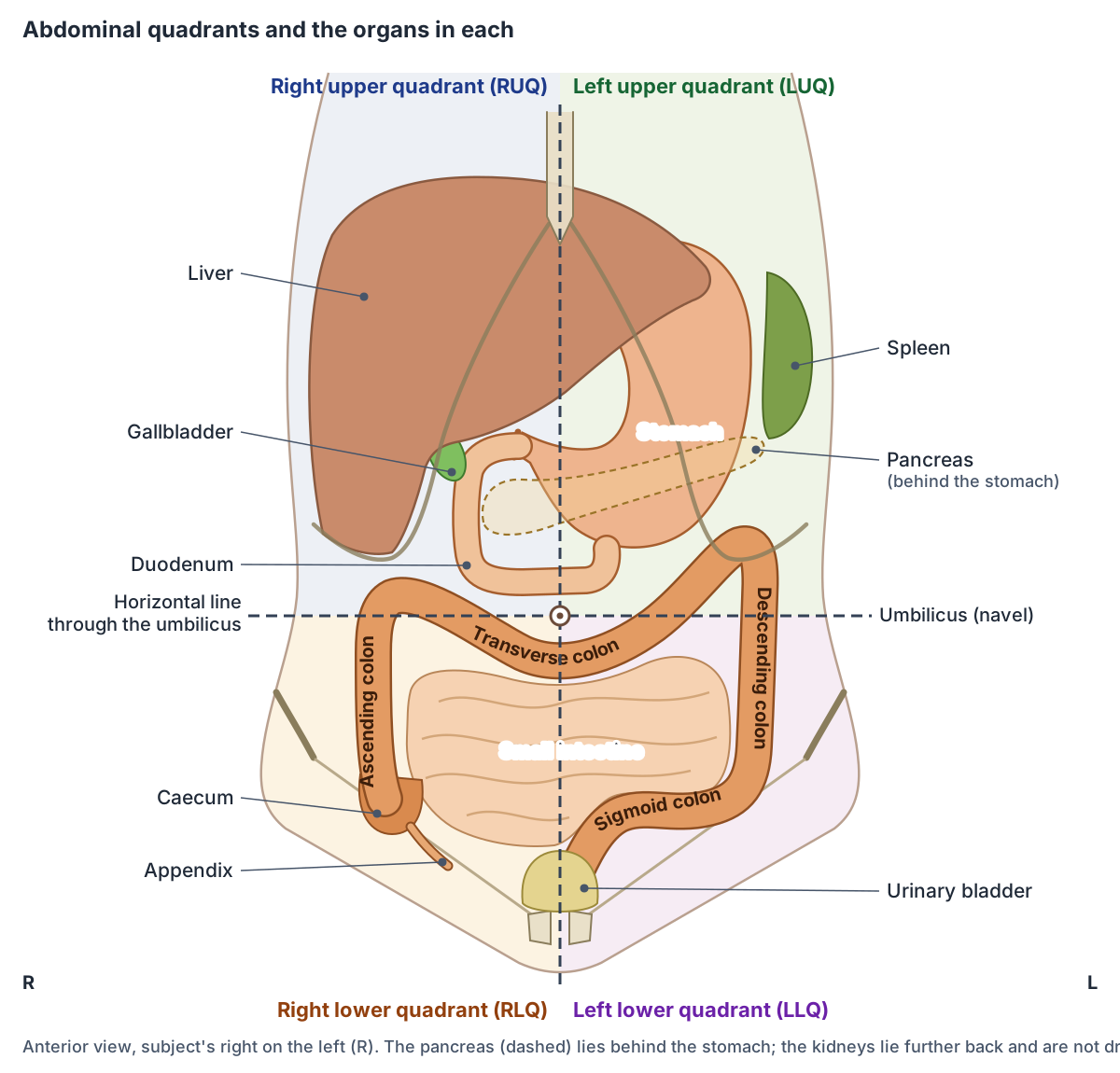
What is in each abdominal quadrant?
| Quadrant | Main contents |
|---|---|
| Right upper (RUQ) | Right lobe of the liver, gallbladder, pylorus, first three parts of the duodenum, head of the pancreas, right kidney and adrenal gland, upper ascending colon, hepatic flexure, right half of the transverse colon |
| Left upper (LUQ) | Left lobe of the liver, stomach, spleen, jejunum and upper ileum, body and tail of the pancreas, left kidney and adrenal gland, left half of the transverse colon, splenic flexure, upper descending colon |
| Right lower (RLQ) | Most of the ileum, caecum, appendix, lower ascending colon, right ureter; right ovary and uterine tube in females |
| Left lower (LLQ) | Lower descending colon, sigmoid colon, left ureter; left ovary and uterine tube in females |
| Both lower quadrants | Parts of the bladder (when full) and uterus; ductus deferens in males |
Based on Kenhub's regions of the abdomen (see references). Organs cross the lines and move with posture and breathing.
The nine regions of the abdomen
The nine regions of the abdomen are made by two vertical and two horizontal lines. Each vertical line runs from the midclavicular point to the mid-inguinal point. The upper horizontal line is the subcostal plane, through the lowest point of the rib cage (usually the 10th costal cartilage, at the body of L3). The lower one is the transtubercular plane, joining the tubercles of the iliac crests at the upper border of L5. (Radiopaedia: Abdominal surface anatomy.)
The regions form three rows. The upper row is the right hypochondrium, the epigastric region (epigastrium) and the left hypochondrium. The middle row is the right lumbar region (right flank), the umbilical region and the left lumbar region (left flank). The lower row is the right iliac fossa, the suprapubic (hypogastric) region and the left iliac fossa. (Radiopaedia: Abdominal surface anatomy.)
Some textbooks draw the upper line at the transpyloric plane instead of the subcostal plane, and call the lower line the intertubercular plane. The transpyloric plane lies midway between the suprasternal (jugular) notch and the upper surface of the pubic symphysis. (Radiopaedia: General topography of the abdomen.)
The transpyloric plane (Addison's plane) lies at about the level of L1. The pylorus, the first part of the duodenum, the neck of the pancreas, the fundus of the gallbladder, the hila of the kidneys and the origin of the superior mesenteric artery are traditionally placed on it, although positions vary between people. (Radiopaedia: Transpyloric plane.)
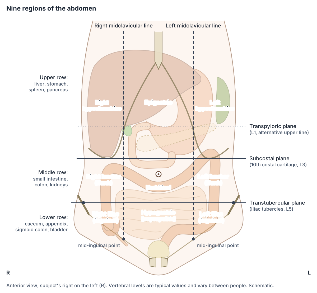
What is in each of the nine abdominal regions?
| Region | Where | Main contents |
|---|---|---|
| Right hypochondriac | Upper right, under the ribs | Right lobe of the liver, gallbladder, upper part of the right kidney, hepatic flexure of the colon |
| Epigastric | Upper centre, below the xiphoid | Much of the stomach, parts of the liver, pancreas, duodenum, adrenal glands |
| Left hypochondriac | Upper left, under the ribs | Stomach, spleen, tail of the pancreas, upper part of the left kidney, splenic flexure |
| Right lumbar (flank) | Middle right | Ascending colon, lower part of the right kidney, small intestine |
| Umbilical | Centre, around the navel | Loops of small intestine, transverse colon, lower parts of the stomach and pancreas; aorta and inferior vena cava behind |
| Left lumbar (flank) | Middle left | Descending colon, lower part of the left kidney, small intestine |
| Right iliac (inguinal) | Lower right | Caecum, appendix, end of the ileum; right ovary and uterine tube in females |
| Hypogastric (suprapubic) | Lower centre, above the pubis | Small intestine, sigmoid colon, rectum, urinary bladder; uterus in females |
| Left iliac (inguinal) | Lower left | Descending and sigmoid colon, small intestine; left ovary and uterine tube in females |
Compiled from Kenhub and Medmastery (see references). Sources differ in detail because large organs span several regions.
Abdominal organs: what is inside the abdomen?
The digestive organs fill most of the abdomen. They are of two kinds: parts of the alimentary canal, the continuous tube that includes the stomach, small intestine and large intestine, and accessory digestive organs, which in the abdomen are the liver, gallbladder and pancreas. (OpenStax Anatomy and Physiology 2e, 23.1: Overview of the Digestive System.)
Other organs lie behind the peritoneum, against the posterior abdominal wall: the adrenal (suprarenal) glands, the abdominal aorta and inferior vena cava, most of the duodenum, the pancreas except its tail, the ureters, the ascending and descending colon and the kidneys. (TeachMeAnatomy: The Peritoneum.)
The spleen sits in the left upper quadrant under the diaphragm and rib cage, against ribs 9 to 11. It is roughly the size of a clenched fist and is not normally palpable unless it is enlarged. (TeachMeAnatomy: The Spleen.)
The whole GI tract from mouth to anus, with the accessory organs. Digestive system anatomy.
Main abdominal organs at a glance
| Organ | Usual position | Key facts |
|---|---|---|
| Liver | Right hypochondrium and epigastrium, into the left hypochondrium | Largest gland in the body; under the diaphragm |
| Stomach | Epigastric and left hypochondriac regions | J-shaped; cardia at T11, pylorus at L1 |
| Gallbladder | Under the right lobe of the liver | 8 to 10 cm long; stores and concentrates bile |
| Pancreas | Across the posterior wall at L1 | Retroperitoneal except the tail; head in the duodenal curve |
| Spleen | Left upper quadrant, against ribs 9 to 11 | Size of a clenched fist; most often injured abdominal organ |
| Small intestine | Centre of the abdomen; jejunum upper left, ileum lower right | About 3 m long in life; duodenum, jejunum, ileum |
| Large intestine | Frames the small intestine | Caecum, appendix, colon, rectum; about 7.6 cm wide |
| Kidneys | Either side of the spine, T12 to L3 | Retroperitoneal; right kidney slightly lower |
| Abdominal aorta | In front of the vertebral column, T12 to L4 | Coeliac trunk, superior and inferior mesenteric arteries |
Compiled from TeachMeAnatomy and OpenStax (see references). Positions vary between people.
Upper abdomen: liver, stomach, gallbladder, pancreas and spleen
The liver is the largest gland in the body and the largest organ in the abdominal cavity. It lies mainly in the right hypochondrium and epigastric region and extends into the left hypochondrium, under the diaphragm. (TeachMeAnatomy: The Liver.)
The stomach is a J-shaped organ between the oesophagus (esophagus) and the duodenum, lying mainly in the epigastric and left hypochondriac regions. Its upper opening, the cardia, is at about T11 and its outlet, the pylorus, at the transpyloric plane (L1). Its position changes with body build, breathing and posture. (TeachMeAnatomy: The Stomach.)
The gallbladder, 8 to 10 cm long, sits in a shallow hollow on the back of the right lobe of the liver. Between meals it stores bile and concentrates it up to 10-fold, then pushes it into the duodenum through the common bile duct when stimulated. (OpenStax Anatomy and Physiology 2e, 23.6: Accessory Organs in Digestion: The Liver, Pancreas, and Gallbladder.)
The pancreas lies across the posterior abdominal wall at the transpyloric plane (L1). Its head sits in the C-shaped curve of the duodenum, the stomach lies in front of it with the lesser sac between them, and its tail is joined to the spleen by the splenorenal ligament. It is retroperitoneal except for the tail. (TeachMeAnatomy: The Pancreas.)
Lobes, Couinaud segments, ligaments, porta hepatis and blood supply. Liver anatomy.
The intestines: small and large bowel
The small intestine is about 3.05 m (10 ft) long in a living person, and roughly twice that after death when muscle tone is lost. Its three parts are the duodenum (about 25.4 cm), the jejunum (about 0.9 m) and the ileum (about 1.8 m), the longest part. (OpenStax Anatomy and Physiology 2e, 23.5: The Small and Large Intestines.)
The duodenum curves in a C-shape around the head of the pancreas. The jejunum begins at the duodenojejunal flexure and lies mainly in the left upper quadrant, and the ileum lies mainly in the right lower quadrant and ends at the ileocaecal junction. The jejunum and ileum are intraperitoneal, hung from the posterior abdominal wall by the mesentery. (TeachMeAnatomy: The Small Intestine.)
The large intestine is about half as long as the small intestine but more than twice as wide. It frames the small intestine: the caecum and appendix lie low on the right, the ascending colon runs up the right side, the transverse colon crosses to the left, the descending colon runs down the left side and the sigmoid colon curves to the midline. (OpenStax Anatomy and Physiology 2e, 23.5: The Small and Large Intestines.)
Duodenum, jejunum and ileum, villi, and jejunum vs ileum. Small intestine anatomy.
Organs on the left and right side of the abdomen
On the left side of the abdomen lie the stomach, the spleen, the jejunum, the body and tail of the pancreas, the left kidney and adrenal gland, the left lobe of the liver, the left half of the transverse colon with the splenic (left colic) flexure, and the descending and sigmoid colon with the left ureter. In females the left ovary and uterine tube lie low on the left. (Kenhub: Regions of the abdomen.)
On the right side lie most of the liver (its right lobe), the gallbladder, the pylorus and the first three parts of the duodenum, the head of the pancreas, the right kidney and adrenal gland, the hepatic flexure and ascending colon, most of the ileum, the caecum and the appendix. In females the right ovary and uterine tube lie low on the right. (Kenhub: Regions of the abdomen.)
The kidneys lie either side of the vertebral column, from about T12 to L3. The right kidney usually sits slightly lower than the left because of the liver above it. (TeachMeAnatomy: The Kidneys.)
Gut pain is often not felt on the organ's own side. Visceral pain is referred by embryological origin: foregut pain to the epigastric region, midgut pain to the umbilical region and hindgut pain to the pubic region. In appendicitis, pain starts around the umbilicus, then moves to the right lower quadrant. (TeachMeAnatomy: The Peritoneum.)
Pain from retroperitoneal organs such as the kidney or pancreas may be felt in the back, and irritation of the diaphragm, for example by inflammation of the liver, gallbladder or duodenum, can cause shoulder tip pain. (TeachMeAnatomy: The Peritoneum.)
Parts, curvatures, wall layers and blood supply of the stomach. Stomach anatomy.
Layers of the abdominal wall
From superficial to deep, the anterolateral abdominal wall has six layers: skin, superficial fascia, the muscles with their fasciae, transversalis fascia, extraperitoneal fat and the parietal peritoneum. (Kenhub: Anterior abdominal wall.)
Above the umbilicus the superficial fascia is a single sheet. Below it, the fascia has a fatty superficial layer (Camper's fascia) and a membranous deep layer (Scarpa's fascia), with superficial vessels and nerves running between the two. (TeachMeAnatomy: The Anterolateral Abdominal Wall.)
Five muscles form each side of the wall. Three flat muscles lie at the side (external oblique, internal oblique and transversus abdominis) and two vertical muscles at the front (rectus abdominis and the small pyramidalis). The aponeuroses of the flat muscles form the rectus sheath around rectus abdominis and meet in the midline at the linea alba, which runs from the xiphoid process to the pubic symphysis. (TeachMeAnatomy: The Anterolateral Abdominal Wall.)
The skin and parietal peritoneum of the anterolateral wall are supplied by spinal nerves T7 to L1. The muscles receive the lower intercostal nerves (T7 to T11), the subcostal nerve (T12) and the iliohypogastric and ilioinguinal nerves (L1). (Kenhub: Anterior abdominal wall.)
Obliques, transversus, rectus abdominis, rectus sheath and inguinal canal. Abdominal muscles.
The peritoneum and peritoneal cavity
The peritoneum is a continuous membrane of mesothelium. Its parietal layer lines the abdominopelvic wall and has the same somatic nerves as the wall, so pain from it is well localised. Its visceral layer covers most of the organs, has autonomic nerves and senses only stretch and chemical irritation, so its pain is poorly localised. (TeachMeAnatomy: The Peritoneum.)
The peritoneal cavity is the space between the two layers. It normally contains only a few millilitres of watery fluid that reduces friction as the organs move. (OpenStax Anatomy and Physiology 2e, 23.1: Overview of the Digestive System.)
The cavity has a large greater sac and a smaller lesser sac (omental bursa) behind the stomach, joined through the epiploic foramen (of Winslow). The transverse mesocolon divides the greater sac into a supracolic compartment (stomach, liver and spleen) and an infracolic compartment (small intestine and ascending and descending colon). Excess fluid in the cavity is called ascites, most often due to portal hypertension from cirrhosis. (TeachMeAnatomy: The Peritoneal Cavity.)
A mesentery is a double layer of peritoneum that attaches an organ to the body wall and carries its vessels and nerves. The greater omentum hangs like an apron from the greater curvature of the stomach and moves to sites of infection (the abdominal policeman). The lesser omentum joins the lesser curvature and first part of the duodenum to the liver. (TeachMeAnatomy: The Peritoneum.)
Peritonitis, inflammation of the peritoneum, can be chemical (for example after a stomach ulcer perforates), haemorrhagic or bacterial (for example from appendicitis). It is life-threatening, with a mortality that remains around 30 to 40%. (OpenStax Anatomy and Physiology 2e, 23.1: Overview of the Digestive System.)
Intraperitoneal vs retroperitoneal organs
Intraperitoneal organs are wrapped in visceral peritoneum in front and behind; examples are the stomach, liver and spleen. Retroperitoneal organs have no visceral peritoneum and are covered only on their front surface by parietal peritoneum. Primarily retroperitoneal organs, such as the oesophagus, kidneys and rectum, developed outside the peritoneum. Secondarily retroperitoneal organs, such as the ascending and descending colon, began on a mesentery and later fused to the posterior wall. (TeachMeAnatomy: The Peritoneum.)
A common memory aid for the retroperitoneal organs is SAD PUCKER: suprarenal (adrenal) glands, aorta and inferior vena cava, duodenum (except its first 2 cm), pancreas (except the tail), ureters, colon (ascending and descending parts), kidneys, (o)esophagus and rectum. (TeachMeAnatomy: The Peritoneum.)
A retroperitoneal gland: head, neck, body, tail, ducts and islets. Pancreas anatomy.
Intraperitoneal and retroperitoneal organs
| Group | Organs |
|---|---|
| Intraperitoneal | Stomach, liver, spleen, first 2 cm of the duodenum (duodenal cap), jejunum, ileum, tail of the pancreas |
| Retroperitoneal (SAD PUCKER) | Suprarenal glands, aorta and inferior vena cava, rest of the duodenum, pancreas except the tail, ureters, ascending and descending colon, kidneys, oesophagus, rectum |
| Primarily retroperitoneal (examples) | Oesophagus, kidneys, rectum |
| Secondarily retroperitoneal (examples) | Ascending and descending colon |
From TeachMeAnatomy's peritoneum article (see references).
Blood vessels of the abdomen
The abdominal aorta begins at T12, just below the aortic hiatus of the diaphragm, runs down in front of the vertebral column and ends at L4 by dividing into the right and left common iliac arteries. (TeachMeAnatomy: The Abdominal Aorta.)
Three unpaired arteries supply the gut (see the table): the coeliac (celiac) trunk at T12 for the foregut, the superior mesenteric artery at L1 for the midgut and the inferior mesenteric artery at L3 for the hindgut. Paired branches go to the diaphragm, adrenal glands, kidneys (renal arteries at L1 to L2) and gonads (L2). (TeachMeAnatomy: The Abdominal Aorta.)
Blood from the gut does not return straight to the heart. The splenic vein and superior mesenteric vein join behind the neck of the pancreas to form the hepatic portal vein, which carries it to the liver. The liver's blood then reaches the inferior vena cava through the hepatic veins. Where portal and systemic veins meet (portosystemic anastomoses), blocked portal flow can produce varices. (TeachMeAnatomy: Venous Drainage of the Abdomen.)
The inferior vena cava begins where the two common iliac veins join, on the transtubercular plane at the upper border of L5. (Radiopaedia: Abdominal surface anatomy.)
Thoracic and abdominal aorta and every major branch. Aorta anatomy.
The three arteries of the gut
| Artery | Leaves the aorta at | Supplies |
|---|---|---|
| Coeliac trunk | T12 | Foregut: liver, stomach, abdominal oesophagus, spleen, upper duodenum, upper pancreas |
| Superior mesenteric artery | L1 | Midgut: lower duodenum, jejunum, ileum, ascending colon, part of the transverse colon |
| Inferior mesenteric artery | L3 | Hindgut: large intestine from the splenic flexure to the upper rectum |
From TeachMeAnatomy's abdominal aorta article (see references). Levels are typical.
Female abdominal anatomy (and how the male abdomen differs)
Above the pelvis, female and male abdominal anatomy is the same: the same organs in the same quadrants. The differences are low down. Parts of the uterus and bladder reach into both lower quadrants, and each lower quadrant contains an ovary and a uterine tube in females, where males have a ductus deferens. (Kenhub: Regions of the abdomen.)
The uterus normally lies just behind and above the bladder and in front of the rectum, tilted forwards (anteverted and anteflexed). Its exact position changes with how full the bladder is. (TeachMeAnatomy: The Uterus.)
Outside pregnancy the uterus is about 7 cm long and 5 cm wide. The ovaries lie in the pelvic cavity, held by the mesovarium, and the broad ligament, a fold of peritoneum, attaches the sides of the uterus to the pelvic wall. (OpenStax Anatomy and Physiology 2e, 27.2: Anatomy and Physiology of the Ovarian Reproductive System.)
The peritoneal cavity differs between the sexes. In males it is completely closed, with a rectovesical pouch between the rectum and bladder. In females the uterine tubes open into it, and there are two pouches: the vesicouterine pouch and the rectouterine pouch of Douglas. (TeachMeAnatomy: The Peritoneal Cavity.)
Before pregnancy the uterus is about the size of a fist; in the second and third trimesters it grows dramatically to contain the fetus. Its upward pressure on the diaphragm reduces the volume of each breath, pressure on the stomach causes heartburn, and downward pressure on the bladder causes frequent urination. (OpenStax Anatomy and Physiology 2e, 28.4: Changes During Pregnancy, Labor, and Birth.)
Gallstones are found in about 10% of the population and are about twice as common in women as in men; female sex, middle age, obesity, a family history and rapid weight loss are risk factors for cholesterol stones. (Radiopaedia: Cholelithiasis.)
In the lower abdominal wall the inguinal canal, about 3 to 5 cm long in adults, carries the spermatic cord in males and the round ligament of the uterus in females. (Kenhub: Anterior abdominal wall.)
Because pelvic organs sit just below the abdomen, conditions such as endometriosis, ovarian cysts and pelvic inflammatory disease can cause pain felt in the lower abdomen. (Cleveland Clinic: Abdominal pain.)
Uterus, ovaries, uterine tubes, cervix and vagina. Female reproductive system.
Exploring the abdomen in the 3D model
The model highlights the digestive organs, spleen, kidneys, adrenal glands, ureters, abdominal aorta and inferior vena cava (all listed below). Rotate the body to see which organs lie in front and which lie against the posterior wall, and click any part to read about it.
The liver is shown as separate Couinaud segments, the functional units used in liver surgery. Each segment has its own branch of the portal vein and hepatic artery and its own bile drainage, so clicking the liver selects one segment at a time. (Radiopaedia: Couinaud classification of hepatic segments.)
The model is the BodyParts3D adult male. Its caecum and sigmoid colon are split from the BodyParts3D ascending and descending colon at the ileocaecal junction and the pelvic brim. It lacks the omenta and most abdominal wall muscles, so use the diagrams for those. Switch to the Female body to see the female pelvic organs.
Abdomen in the 3D model
- Abdominal aorta
- The abdominal aorta is the part of the aorta that runs through the abdomen and supplies the abdominal organs, pelvis and lower limbs.
- Adrenal gland
- The adrenal (suprarenal) gland is a small endocrine gland on top of each kidney.
- Appendix
- The vermiform appendix is a narrow blind-ended tube arising from the cecum.
- Ascending colon
- The ascending colon is the part of the large intestine rising from the cecum to the right colic (hepatic) flexure.
- Caudate lobe of liver
- The caudate lobe (Couinaud segment I) is a small liver lobe on the posterior surface between the inferior vena cava and the fissure for the ligamentum venosum.
- Cecum
- The caecum is the first part of the large intestine, a blind-ending pouch in the right lower abdomen below the point where the small intestine joins the colon.
- Descending colon
- The descending colon passes down from the left colic (splenic) flexure to the sigmoid colon.
- Distal part of ileum
- The distal part of the ileum is a segment of the lower small intestine.
- Distal part of jejunum
- The distal part of the jejunum is a segment of the middle small intestine.
- Duodenum
- The duodenum is the first and shortest part of the small intestine, about 20 to 25 cm long and C-shaped.
- Gallbladder
- The gallbladder is a pear-shaped sac that stores and concentrates bile.
- Hepatovenous segment II
- Couinaud segment II is the superior part of the left lateral section of the liver.
- Hepatovenous segment III
- Couinaud segment III is the inferior part of the left lateral section of the liver.
- Hepatovenous segment IV
- Couinaud segment IV is the left medial section of the liver, also called the quadrate lobe in its inferior part.
- Hepatovenous segment V
- Couinaud segment V is the inferior part of the right anterior section of the liver.
- Hepatovenous segment VI
- Couinaud segment VI is the inferior part of the right posterior section of the liver.
- Hepatovenous segment VII
- Couinaud segment VII is the superior part of the right posterior section of the liver.
- Hepatovenous segment VIII
- Couinaud segment VIII is the superior part of the right anterior section of the liver.
- Ileocecal junction
- The ileocecal junction is where the terminal ileum opens into the cecum of the large intestine.
- Inferior vena cava
- The inferior vena cava is the largest vein in the body and returns blood from the lower body to the right atrium.
- Kidney
- The kidney is a bean-shaped organ that filters blood to form urine and regulate fluid, electrolytes and blood pressure.
- Middle part of ileum
- The middle part of the ileum is a segment of the lower small intestine.
- Middle part of jejunum
- The middle part of the jejunum is a segment of the middle small intestine.
- Pancreas
- The pancreas is a soft, elongated gland with both exocrine and endocrine functions.
- Proximal part of ileum
- The proximal part of the ileum is a segment of the lower small intestine.
- Proximal part of jejunum
- The proximal part of the jejunum is a segment of the middle small intestine.
- Sigmoid colon
- The sigmoid colon is the S-shaped last part of the colon. It runs from the end of the descending colon at the pelvic brim to the rectum.
- Spleen
- The spleen is the largest lymphoid organ, filtering blood and supporting immune responses.
- Stomach
- The stomach is a J-shaped, expandable part of the digestive tract that mixes food with acid and enzymes.
- Transverse colon
- The transverse colon is the part of the large intestine that crosses the abdomen between the right and left colic flexures.
- Ureter
- The ureter is a muscular tube carrying urine from the renal pelvis to the urinary bladder.
Frequently asked questions
What organs are in the abdomen?
The abdomen holds the stomach, small intestine and most of the large intestine, the liver, gallbladder and pancreas, and the spleen. Behind the peritoneum, against the back wall, lie the kidneys, adrenal glands, ureters, abdominal aorta and inferior vena cava. The bladder and, in females, the uterus and ovaries sit just below in the pelvis and reach into the lower abdomen.
What are the four quadrants of the abdomen?
The four quadrants are the right upper, left upper, right lower and left lower quadrants (RUQ, LUQ, RLQ and LLQ). They are made by a vertical line down the midline from the xiphoid process to the pubic symphysis and a horizontal line through the umbilicus. Right and left are the patient's own sides.
What are the 9 regions of the abdomen?
The nine regions are the right hypochondriac, epigastric and left hypochondriac regions in the upper row; the right lumbar, umbilical and left lumbar regions in the middle; and the right iliac (inguinal), hypogastric (suprapubic) and left iliac (inguinal) regions below. They are drawn with the two midclavicular lines, the subcostal plane and the transtubercular plane.
What organs are on the left side of the abdomen?
The left side holds the stomach, spleen, body and tail of the pancreas, left kidney and adrenal gland, the left lobe of the liver, the jejunum, the splenic flexure and left half of the transverse colon, and lower down the descending and sigmoid colon and left ureter. In females the left ovary and uterine tube lie low on the left.
What organs are in the right upper quadrant?
The right upper quadrant contains the right lobe of the liver, the gallbladder, the pylorus of the stomach, the first three parts of the duodenum, the head of the pancreas, the right kidney and adrenal gland, and the upper ascending colon, hepatic flexure and right half of the transverse colon. Liver and gallbladder problems are common causes of pain here.
What is behind the belly button?
The belly button is the scar of the umbilical cord. Behind it, all the layers of the abdominal wall fuse at the umbilical ring in the linea alba, together with the remnants of fetal structures: the round ligament of the liver, the median umbilical ligament and the two medial umbilical ligaments. Deeper still lie loops of small intestine and the transverse colon.
Is female abdominal anatomy different from male?
Above the pelvis the organs are the same. The differences are low down: females have the uterus, ovaries and uterine tubes, which reach into the lower quadrants, and the uterine tubes open into the peritoneal cavity, which is closed in males. During pregnancy the growing uterus pushes the other abdominal organs upwards.
What are the layers of the abdominal wall?
From outside in, the anterior abdominal wall has skin, superficial fascia (Camper's and Scarpa's fascia below the umbilicus), the muscles with their fasciae (external oblique, internal oblique, transversus abdominis and rectus abdominis in its sheath), transversalis fascia, extraperitoneal fat and the parietal peritoneum.
References
- OpenStax. Anatomy and Physiology 2e, §1.6 Anatomical Terminology. Rice University, 2022.
- OpenStax. Anatomy and Physiology 2e, §23.1 Overview of the Digestive System. Rice University, 2022.
- OpenStax. Anatomy and Physiology 2e, §23.5 The Small and Large Intestines. Rice University, 2022.
- OpenStax. Anatomy and Physiology 2e, §23.6 Accessory Organs in Digestion: The Liver, Pancreas, and Gallbladder. Rice University, 2022.
- OpenStax. Anatomy and Physiology 2e, §27.2 Anatomy and Physiology of the Ovarian Reproductive System. Rice University, 2022.
- OpenStax. Anatomy and Physiology 2e, §28.4 Changes During Pregnancy, Labor, and Birth. Rice University, 2022.
- Abdominal surface anatomy. Radiopaedia reference article.
- General topography of the abdomen. Radiopaedia reference article.
- Transpyloric plane. Radiopaedia reference article.
- Umbilicus. Radiopaedia reference article.
- Umbilical hernia. Radiopaedia reference article.
- Cholelithiasis. Radiopaedia reference article.
- Riedel lobe. Radiopaedia reference article.
- Hepatorenal pouch. Radiopaedia reference article.
- Couinaud classification of hepatic segments. Radiopaedia reference article.
- Regions of the abdomen. Kenhub anatomy library.
- Anterior abdominal wall. Kenhub anatomy library.
- Umbilicus. Kenhub anatomy library.
- Dermatomes. Kenhub anatomy library.
- Jones O. The Peritoneum. TeachMeAnatomy.
- Jones O. The Peritoneal Cavity. TeachMeAnatomy.
- Jones O. The Anterolateral Abdominal Wall. TeachMeAnatomy.
- Jones O. Muscles of the Posterior Abdominal Wall. TeachMeAnatomy.
- Jones O. The Stomach. TeachMeAnatomy.
- Jones O. The Liver. TeachMeAnatomy.
- Jones O. The Pancreas. TeachMeAnatomy.
- Jones O. The Spleen. TeachMeAnatomy.
- Jones O. The Kidneys. TeachMeAnatomy.
- Jones O. The Small Intestine. TeachMeAnatomy.
- Jones O. The Abdominal Aorta. TeachMeAnatomy.
- Jones O. Venous Drainage of the Abdomen. TeachMeAnatomy.
- Jones O. The Uterus. TeachMeAnatomy.
- What are the anatomical regions and quadrants of the abdomen? Medmastery, Abdominal examination clinical guide.
- Abdominal pain. Cleveland Clinic health library.
- Mitsuhashi N. et al. (2009). BodyParts3D: 3D structure database for anatomical concepts. Nucleic Acids Research 37, D782–D785. Data © The Database Center for Life Science, CC BY 4.0.
Related anatomy
- StomachStomach anatomy with labelled diagrams and a 3D model: cardia, fundus, body, antrum and pylorus, curvatures, rugae…
- LiverLiver anatomy with labelled diagrams and a 3D model: surfaces, four lobes, eight Couinaud segments, ligaments, porta…
- Abdominal musclesAbdominal muscles explained: external and internal oblique, transversus abdominis, rectus abdominis, pyramidalis, the…
- Digestive systemGI tract anatomy: each digestive system organ from mouth to anus in order, the accessory organs, wall layers and blood…
- Organs of the bodyHuman anatomy organs explained with a labelled internal organs diagram: organs by body system and location, left vs…
- KidneyWhere are your kidneys? Kidney location on the back (T12 to L3), coverings, relations, cortex, medulla, calyces, renal…
Explore the whole body in the interactive 3D human anatomy model, or browse the A–Z list of human structures.