

Organs of the digestive system in order
In order from start to finish, the alimentary canal is the mouth, pharynx, oesophagus, stomach, small intestine and large intestine, ending at the anus. The accessory digestive organs sit outside this tube: the teeth and tongue begin mechanical digestion, the salivary glands begin chemical digestion, and the liver, gallbladder and pancreas release secretions such as bile and enzymes into the small intestine. (OpenStax Anatomy and Physiology 2e, 23.1: Overview of the Digestive System.)
Divided more finely, the canal runs: oral (buccal) cavity, pharynx, oesophagus, stomach, the three parts of the small intestine (duodenum, jejunum and ileum), the caecum (cecum) with the appendix, the ascending, transverse, descending and sigmoid colon, the rectum and the anal canal. It is a continuous tube divided into wider regions that are separated by sphincters. (Kenhub: Digestive system.)
The organs of the GI tract are hollow, and food moves through them by peristalsis: the muscle behind the food contracts to push it forward while the muscle ahead of it relaxes. (NIDDK: Your Digestive System & How It Works.)
Knowing this anatomy is the basis for working out the cause of abdominal pain, bowel obstruction, inflammatory disease and gastrointestinal bleeding, for reading scans, and for planning endoscopy and surgery. (TeachMeAnatomy: The Gastrointestinal Tract.)
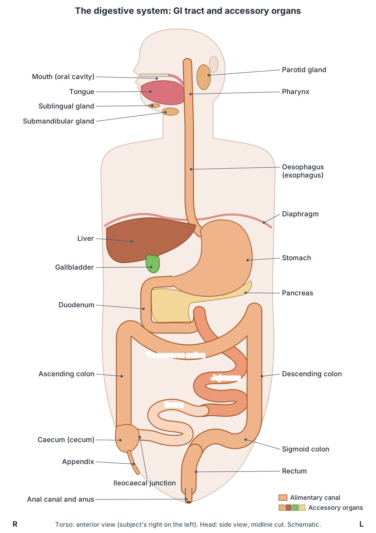
GI tract organs in order: position, size and main job
| Organ | Where it is | Size in an adult | Main job |
|---|---|---|---|
| Mouth (oral cavity) | Below the nasal cavity, behind the lips | 32 permanent teeth; 1 to 1.5 L of saliva a day | Chews food; saliva begins starch digestion |
| Pharynx | Behind the nose, mouth and larynx | Oropharynx and laryngopharynx carry food | Swallowing |
| Oesophagus (esophagus) | Neck and chest, behind the trachea | About 25 cm long | Carries food to the stomach by peristalsis |
| Stomach | Upper abdomen, mostly on the left | Stretches to hold up to about 4 L | Churns food into chyme; starts protein digestion |
| Small intestine | Central and lower abdomen | About 3 m in life; duodenum about 25 cm | Completes digestion; absorbs most nutrients and water |
| Large intestine | Frames the small intestine | About half the length of the small intestine; about 7.6 cm wide | Absorbs remaining water and salts; forms faeces |
| Anal canal and anus | Below the rectum, in the perineum | Anal canal 3.8 to 5 cm | Defecation, controlled by two sphincters |
Compiled from OpenStax Anatomy and Physiology 2e, sections 23.3 to 23.5, and Cleveland Clinic (see references). Lengths are for living adults.
Upper vs lower GI tract
Clinicians split the tract at the duodenojejunal flexure, where the suspensory ligament of the duodenum (ligament of Treitz) holds the end of the duodenum to the back wall of the abdomen. Bleeding above this ligament is classed as upper GI bleeding and bleeding below it as lower GI bleeding. (Kenhub: Ligaments of the gastrointestinal tract.)
The embryonic terms foregut, midgut and hindgut are still used for the blood supply. The coeliac trunk supplies the foregut organs, the superior mesenteric artery the midgut, and the inferior mesenteric artery the hindgut. (Kenhub: Digestive system.)
Mouth, pharynx and salivary glands
The mouth (oral cavity) is bounded in front by the lips, which cover the orbicularis oris muscle, and at the sides by the cheeks, which contain the buccinator muscles. Its roof is the bony hard palate in front and the muscular soft palate behind, from which the uvula hangs. The fauces is the opening from the mouth into the oropharynx. (OpenStax Anatomy and Physiology 2e, 23.3: The Mouth, Pharynx, and Esophagus.)
Three pairs of major salivary glands drain into the mouth: the parotid glands, between the skin and the masseter muscle, whose ducts open near the second upper molar; the submandibular glands in the floor of the mouth; and the sublingual glands below the tongue. With many minor glands, they make about 1 to 1.5 litres of saliva a day. Salivary amylase in saliva starts the breakdown of carbohydrates. (OpenStax Anatomy and Physiology 2e, 23.3: The Mouth, Pharynx, and Esophagus.)
Adults have 32 permanent teeth and children 20 deciduous teeth. Of the three parts of the pharynx, only the oropharynx and laryngopharynx carry food; the nasopharynx is used only for breathing and speech. Swallowing has a voluntary oral phase followed by involuntary pharyngeal and oesophageal phases, and takes about 4 to 8 seconds for solid food and about 1 second for liquids. (OpenStax Anatomy and Physiology 2e, 23.3: The Mouth, Pharynx, and Esophagus.)
Lips, palate, cheeks, floor of the mouth and the salivary ducts in detail. Mouth anatomy.
Oesophagus (esophagus)
The oesophagus is a muscular tube about 25 cm long that lies behind the trachea and runs through the mediastinum of the chest. It passes through the oesophageal hiatus of the diaphragm to reach the stomach. An upper oesophageal sphincter, continuous with the inferior pharyngeal constrictor, controls entry from the pharynx, and a lower oesophageal sphincter controls entry to the stomach and stops stomach contents flowing back up. (OpenStax Anatomy and Physiology 2e, 23.3: The Mouth, Pharynx, and Esophagus.)
It begins at the level of the C6 vertebra and pierces the diaphragm at T10; only about 2.5 cm of it lies in the abdomen. Peristaltic waves carry a swallowed bolus along it at about 4 cm per second. (Kenhub: Digestive system.)
Course, constrictions, sphincters and blood supply of the food pipe. Esophagus anatomy.
Stomach
The stomach links the oesophagus to the duodenum. It has four regions, the cardia, fundus, body and pylorus, and the pyloric sphincter controls emptying into the duodenum. Empty, it is about the size of a fist, but it can stretch to hold as much as 4 litres of food and fluid. When it is empty, its lining lies in large folds called rugae. (OpenStax Anatomy and Physiology 2e, 23.4: The Stomach.)
Its wall adds an inner oblique muscle layer to the usual circular and longitudinal layers, which lets it churn food into a liquid called chyme. Parietal cells secrete hydrochloric acid, giving a pH of about 1.5 to 3.5, and intrinsic factor, which is needed to absorb vitamin B12; chief cells secrete pepsinogen. The stomach usually empties within 2 to 4 hours, passing about 3 mL of chyme at a time; fatty meals can stay 6 hours or more. (OpenStax Anatomy and Physiology 2e, 23.4: The Stomach.)
Parts, curvatures, wall layers and blood supply of the stomach. Stomach anatomy.
Small intestine: duodenum, jejunum and ileum
The small intestine is the longest part of the tract: about 3 m in a living person and roughly twice that in a cadaver, once the muscle has lost its tone. Its parts are the duodenum (about 25 cm), which curves around the head of the pancreas, the jejunum (about 0.9 m in life) and the ileum (about 1.8 m), which joins the caecum at the ileocaecal valve. (OpenStax Anatomy and Physiology 2e, 23.5: The Small and Large Intestines.)
Almost all ingested food, about 80% of electrolytes and about 90% of water are absorbed in the small intestine. About 9 litres of fluid enter it each day. (OpenStax Anatomy and Physiology 2e, 23.7: Chemical Digestion and Absorption: A Closer Look.)
Circular folds, villi and microvilli multiply its absorptive surface more than 600-fold, and chyme takes about 3 to 5 hours to pass through it. (OpenStax Anatomy and Physiology 2e, 23.5: The Small and Large Intestines.)
Duodenum, jejunum vs ileum, villi and the mesentery, with diagrams. Small intestine anatomy.
Large intestine, rectum and anus
The large intestine is about half as long as the small intestine but more than twice as wide, about 7.6 cm. Its regions are the caecum (about 6 cm long) with the appendix attached, the ascending, transverse, descending and sigmoid colon, the rectum and the anal canal. The anal canal is 3.8 to 5 cm long and is closed by an involuntary internal sphincter of smooth muscle and a voluntary external sphincter of skeletal muscle. (OpenStax Anatomy and Physiology 2e, 23.5: The Small and Large Intestines.)
Its wall has no circular folds or villi. The outer longitudinal muscle is gathered into three bands, the teniae coli, whose tone pulls the wall into pouches called haustra. Food residue usually stays in the large intestine for 12 to 24 hours while water and salts are absorbed, and trillions of bacteria from more than 700 species ferment what is left; some make vitamins, mainly biotin, pantothenic acid and vitamin K. (OpenStax Anatomy and Physiology 2e, 23.5: The Small and Large Intestines.)
Caecum, appendix, colon, rectum and anal canal. Large intestine anatomy.
Liver
The liver is the largest gland in the body, weighing about 1.4 kg (3 lb) in an adult. It lies below the diaphragm in the right upper quadrant, protected by the ribs. Its cells (hepatocytes) make about 1 litre of bile a day. Bile salts and phospholipids in bile emulsify fats, breaking large droplets into tiny ones that enzymes can digest. (OpenStax Anatomy and Physiology 2e, 23.6: Accessory Organs in Digestion: The Liver, Pancreas, and Gallbladder.)
Blood leaving the intestines carries absorbed sugars, amino acids, glycerol and some vitamins and salts to the liver, which stores, processes and distributes them. (NIDDK: Your Digestive System & How It Works.)
Lobes, segments, blood supply and bile flow. Liver anatomy.
Gallbladder and bile ducts
The gallbladder is a muscular sac about 8 to 10 cm long nested on the underside of the right lobe of the liver. Between meals it stores bile and concentrates it up to 10-fold by absorbing water and ions. When stimulated, it ejects bile through the cystic duct into the common bile duct, which carries it to the duodenum. (OpenStax Anatomy and Physiology 2e, 23.6: Accessory Organs in Digestion: The Liver, Pancreas, and Gallbladder.)
Cholecystokinin (CCK), a hormone released by the duodenum, makes the gallbladder release bile and the pancreas release enzymes, while secretin from the duodenum triggers a watery, bicarbonate-rich pancreatic juice. (OpenStax Anatomy and Physiology 2e, 23.2: Digestive System Processes and Regulation.)
Where the gallbladder sits and how bile reaches the duodenum. Gallbladder and biliary tree.
Pancreas
The pancreas is a soft, oblong gland lying crosswise behind the stomach in the retroperitoneum. Its head sits in the C-shaped curve of the duodenum and its tail ends at the hilum of the spleen. Grape-like clusters of exocrine cells (acini) make more than a litre of pancreatic juice a day, with bicarbonate and enzymes: amylase for starch, lipase for fat, nucleases, and protein-digesting enzymes released in inactive forms such as trypsinogen. (OpenStax Anatomy and Physiology 2e, 23.6: Accessory Organs in Digestion: The Liver, Pancreas, and Gallbladder.)
Acidic chyme entering the duodenum triggers secretin, which releases bicarbonate-rich juice, while proteins and fats trigger CCK, which releases enzyme-rich juice. The main pancreatic duct joins the common bile duct at the hepatopancreatic ampulla. Scattered islets of Langerhans form the endocrine pancreas, which produces insulin, glucagon, somatostatin and pancreatic polypeptide. (OpenStax Anatomy and Physiology 2e, 23.6: Accessory Organs in Digestion: The Liver, Pancreas, and Gallbladder.)
Head, neck, body and tail, ducts, islets and blood supply. Pancreas anatomy.
Layers of the GI tract wall
From the oesophagus to the anus, the wall of the tract has the same four layers, from the lumen outwards: mucosa, submucosa, muscularis and serosa. The mucosa has three parts: a lining epithelium, a connective tissue lamina propria with blood and lymph vessels and mucosa-associated lymphoid tissue (MALT), and a thin layer of smooth muscle, the muscularis mucosae. (OpenStax Anatomy and Physiology 2e, 23.1: Overview of the Digestive System.)
The epithelium is non-keratinised stratified squamous in the mouth, pharynx, oesophagus and anal canal, and simple columnar in the stomach and intestines, where it contains goblet cells and hormone-secreting enteroendocrine cells. It is renewed every few days in the mouth and about every week in the gut. (OpenStax Anatomy and Physiology 2e, 23.1: Overview of the Digestive System.)
The submucosa is loose connective tissue with larger vessels, glands and the submucosal nerve plexus. The muscularis is usually an inner circular and an outer longitudinal layer of smooth muscle. It is skeletal muscle in the mouth, pharynx, upper oesophagus and external anal sphincter, which is why swallowing and defecation can be controlled voluntarily. The stomach adds an oblique layer, and the colon's longitudinal muscle forms the teniae coli. (OpenStax Anatomy and Physiology 2e, 23.1: Overview of the Digestive System.)
The outer layer is a serosa, made of visceral peritoneum, only inside the abdominal cavity. The mouth, pharynx and oesophagus have an adventitia of connective tissue instead. (OpenStax Anatomy and Physiology 2e, 23.1: Overview of the Digestive System.)
The four layers of the GI tract wall
| Layer (lumen outwards) | Made of | Notes |
|---|---|---|
| Mucosa | Epithelium, lamina propria, muscularis mucosae | Stratified squamous epithelium in the mouth, pharynx, oesophagus and anal canal; simple columnar in the stomach and intestines; contains lymphoid tissue (MALT) |
| Submucosa | Loose connective tissue | Larger blood and lymph vessels, glands, submucosal (Meissner) nerve plexus |
| Muscularis (externa) | Inner circular and outer longitudinal smooth muscle | Myenteric (Auerbach) plexus; extra oblique layer in the stomach; teniae coli in the colon; skeletal muscle in the mouth, pharynx, upper oesophagus and external anal sphincter |
| Serosa or adventitia | Visceral peritoneum (serosa) or connective tissue (adventitia) | Serosa only inside the abdominal cavity; adventitia in the mouth, pharynx and oesophagus |
Source: OpenStax Anatomy and Physiology 2e, section 23.1.
Peritoneum, mesentery and retroperitoneal organs
The peritoneum is a serous membrane. Its parietal layer lines the abdominal wall and its visceral layer covers the organs; between them is the peritoneal cavity, which holds only a few millilitres of lubricating fluid. Organs that lie fully or partly behind the peritoneum are called retroperitoneal: the duodenum, pancreas, ascending and descending colon, and rectum. (OpenStax Anatomy and Physiology 2e, 23.1: Overview of the Digestive System.)
Folds of peritoneum hold the organs in place and carry their vessels. The greater omentum hangs like an apron over the small intestine and transverse colon. The lesser omentum suspends the stomach from the liver. The mesentery anchors the small intestine, except the duodenum, to the back wall of the abdomen, the mesocolon attaches the transverse and sigmoid colon, and the falciform ligament attaches the liver to the front wall and diaphragm. (OpenStax Anatomy and Physiology 2e, 23.1: Overview of the Digestive System.)
Quadrants, regions and which organs lie in each. Abdomen anatomy.
Blood supply: coeliac trunk, superior and inferior mesenteric arteries
The first parts of the canal are supplied by branches of the aortic arch and thoracic aorta, and the parts below the diaphragm by the abdominal aorta. During rest and digestion, about a quarter of the blood pumped by each heartbeat enters the intestinal arteries. (OpenStax Anatomy and Physiology 2e, 23.1: Overview of the Digestive System.)
Three unpaired arteries leave the front of the abdominal aorta for the gut: the coeliac trunk at T12, the superior mesenteric artery (SMA) at L1 and the inferior mesenteric artery (IMA) at L3. (TeachMeAnatomy: The Abdominal Aorta.)
Each supplies one embryonic region of the gut. The coeliac trunk supplies the foregut: the abdominal oesophagus, stomach, upper duodenum, liver, spleen and pancreas. The SMA supplies the midgut, from the distal duodenum through the jejunum, ileum, caecum, appendix and ascending colon to the proximal transverse colon. The IMA supplies the hindgut, from the distal transverse colon through the descending and sigmoid colon to the rectum. (Kenhub: Digestive system.)
The foregut artery and its left gastric, splenic and common hepatic branches. Celiac trunk anatomy.
Arteries of the gut
| Artery | Leaves the aorta at | Gut region | Organs supplied |
|---|---|---|---|
| Coeliac (celiac) trunk | T12 | Foregut | Abdominal oesophagus, stomach, upper duodenum, liver, pancreas and spleen |
| Superior mesenteric artery | L1 | Midgut | Distal duodenum, jejunum, ileum, caecum, appendix, ascending colon and the proximal part of the transverse colon |
| Inferior mesenteric artery | L3 | Hindgut | Distal part of the transverse colon, descending and sigmoid colon, rectum |
Vertebral levels from TeachMeAnatomy; territories from Kenhub and TeachMeAnatomy. Levels vary between people.
Venous drainage: the hepatic portal system
Veins from the small intestine do not go straight back to the heart. They drain into the hepatic portal system, which carries absorbed nutrients to the liver first. Fats are the exception: after absorption they enter lymphatic capillaries called lacteals instead of the blood. (OpenStax Anatomy and Physiology 2e, 23.1: Overview of the Digestive System.)
The hepatic portal vein forms from the union of the superior mesenteric and splenic veins. After passing through the liver, blood leaves through the hepatic veins into the inferior vena cava. (Kenhub: Digestive system.)
How gut blood reaches the liver, and the portosystemic anastomoses. Portal vein anatomy.
Nerves: the enteric nervous system
The gut has its own nervous system, the enteric nervous system, which runs from the oesophagus to the anus and contains about 100 million neurons. Its myenteric (Auerbach) plexus in the muscularis controls motility; its submucosal (Meissner) plexus regulates secretion and responds to food. Parasympathetic activity increases secretion and motility, while sympathetic activity decreases them. (OpenStax Anatomy and Physiology 2e, 23.1: Overview of the Digestive System.)
Sympathetic fibres reach the gut through the thoracic and lumbar splanchnic nerves, which relay in the coeliac, aorticorenal, superior mesenteric and inferior mesenteric ganglia. Parasympathetic fibres come from the vagus nerve, as far as the transverse colon, and from the pelvic splanchnic nerves for the descending colon, sigmoid colon and rectum. (Kenhub: Digestive system.)
Short reflexes run entirely within the plexuses of the gut wall; for example, stretching of the stomach increases local secretion. Long reflexes involve the brain: the sight, smell or taste of food signals the medulla oblongata, which stimulates the stomach to secrete. (OpenStax Anatomy and Physiology 2e, 23.2: Digestive System Processes and Regulation.)
Sympathetic and parasympathetic control of the organs. Autonomic nervous system.
What does the digestive system do? Six digestive processes
Digestion is described as six processes. Ingestion is taking food into the mouth. Propulsion moves it along, first by voluntary swallowing and then by peristalsis, alternating waves of contraction and relaxation of smooth muscle. Mechanical digestion breaks food into smaller pieces without changing it chemically: chewing (mastication), churning in the stomach and segmentation in the small intestine. (OpenStax Anatomy and Physiology 2e, 23.2: Digestive System Processes and Regulation.)
Chemical digestion uses enzymes, acid and salts to split large molecules into their building blocks, such as proteins into amino acids; it starts in the mouth and is completed in the small intestine. Absorption moves nutrients through the lining into the blood or lymph, mainly in the small intestine. Defecation removes undigested material as faeces. (OpenStax Anatomy and Physiology 2e, 23.2: Digestive System Processes and Regulation.)
Bacteria help as well. Those in the small intestine make some of the enzymes needed to digest carbohydrates, and those in the large intestine break down remaining nutrients and make vitamin K. (NIDDK: Your Digestive System & How It Works.)
How long is the digestive tract?
The alimentary canal is about 7.6 m (25 ft) long in a living adult. After death, when its smooth muscle loses tone, it measures about 10.7 m (35 ft), which is why books quote such different lengths. (OpenStax Anatomy and Physiology 2e, 23.1: Overview of the Digestive System.)
Cleveland Clinic gives about 9 m (30 ft) for the tract stretched out, and points out that it takes up far less room in the body because the intestines are coiled. (Cleveland Clinic: Digestive System.)
Its inner surface is smaller than often claimed. Using microscopic measurements, Helander and Fändriks (2014) estimated the mucosal surface of the whole digestive tract at about 32 m², of which about 30 m² is in the small intestine and 1.9 m² in the large intestine. The mouth, oesophagus and stomach add only a fraction of a square metre. (BioNumbers 111126: Average surface area of parts of the gastrointestinal tract (Helander and Fändriks 2014).)
That is far smaller than the figure of about 200 m² that OpenStax gives for the small intestine. As for timing, chyme leaves the small intestine in about 3 to 5 hours, and residue then usually spends 12 to 24 hours in the large intestine. Mass movements push faeces towards the rectum three or four times a day, usually during or just after a meal. (OpenStax Anatomy and Physiology 2e, 23.5: The Small and Large Intestines.)
Digestive system in the 3D model
- Appendix
- The vermiform appendix is a narrow blind-ended tube arising from the cecum.
- Ascending colon
- The ascending colon is the part of the large intestine rising from the cecum to the right colic (hepatic) flexure.
- Caudate lobe of liver
- The caudate lobe (Couinaud segment I) is a small liver lobe on the posterior surface between the inferior vena cava and the fissure for the ligamentum venosum.
- Cecum
- The caecum is the first part of the large intestine, a blind-ending pouch in the right lower abdomen below the point where the small intestine joins the colon.
- Descending colon
- The descending colon passes down from the left colic (splenic) flexure to the sigmoid colon.
- Distal part of ileum
- The distal part of the ileum is a segment of the lower small intestine.
- Distal part of jejunum
- The distal part of the jejunum is a segment of the middle small intestine.
- Duodenum
- The duodenum is the first and shortest part of the small intestine, about 20 to 25 cm long and C-shaped.
- Esophagus
- The esophagus is a muscular tube about 22 to 25 cm long that carries swallowed food from the pharynx to the stomach.
- Gallbladder
- The gallbladder is a pear-shaped sac that stores and concentrates bile.
- Hepatovenous segment II
- Couinaud segment II is the superior part of the left lateral section of the liver.
- Hepatovenous segment III
- Couinaud segment III is the inferior part of the left lateral section of the liver.
- Hepatovenous segment IV
- Couinaud segment IV is the left medial section of the liver, also called the quadrate lobe in its inferior part.
- Hepatovenous segment V
- Couinaud segment V is the inferior part of the right anterior section of the liver.
- Hepatovenous segment VI
- Couinaud segment VI is the inferior part of the right posterior section of the liver.
- Hepatovenous segment VII
- Couinaud segment VII is the superior part of the right posterior section of the liver.
- Hepatovenous segment VIII
- Couinaud segment VIII is the superior part of the right anterior section of the liver.
- Ileocecal junction
- The ileocecal junction is where the terminal ileum opens into the cecum of the large intestine.
- Lip
- The lips are two muscular, mobile folds that surround the opening of the mouth.
- Middle part of ileum
- The middle part of the ileum is a segment of the lower small intestine.
- Middle part of jejunum
- The middle part of the jejunum is a segment of the middle small intestine.
- Pancreas
- The pancreas is a soft, elongated gland with both exocrine and endocrine functions.
- Parotid gland
- The parotid gland is the salivary gland in front of and below each ear. It makes serous saliva, a watery fluid rich in enzymes.
- Proximal part of ileum
- The proximal part of the ileum is a segment of the lower small intestine.
- Proximal part of jejunum
- The proximal part of the jejunum is a segment of the middle small intestine.
- Rectum
- The rectum is the final straight portion of the large intestine before the anal canal.
- Sigmoid colon
- The sigmoid colon is the S-shaped last part of the colon. It runs from the end of the descending colon at the pelvic brim to the rectum.
- Stomach
- The stomach is a J-shaped, expandable part of the digestive tract that mixes food with acid and enzymes.
- Sublingual gland
- The sublingual gland is the smallest of the three major salivary glands.
- Submandibular gland
- The submandibular gland is a major salivary gland located below the mandible.
- Tongue
- The tongue is a muscular organ of the mouth involved in taste, chewing, swallowing and speech.
- Transverse colon
- The transverse colon is the part of the large intestine that crosses the abdomen between the right and left colic flexures.
Frequently asked questions
What are the organs of the digestive system in order?
Food passes through the mouth, pharynx, oesophagus, stomach, small intestine (duodenum, jejunum and ileum) and large intestine (caecum, colon, rectum and anal canal) before leaving through the anus. The teeth, tongue, salivary glands, liver, gallbladder and pancreas are accessory organs: food does not pass through them, but they help digestion.
What are the accessory organs of the digestive system?
They are the teeth, tongue, salivary glands, liver, gallbladder and pancreas. The teeth and tongue break food up mechanically, the salivary glands start chemical digestion, the liver makes bile, the gallbladder stores and concentrates it, and the pancreas makes digestive enzymes and bicarbonate. Bile and pancreatic juice flow into the duodenum.
How long is the human GI tract?
In a living adult the tract from mouth to anus is about 7.6 m (25 ft) long. Measured after death, when its muscle has relaxed, it is about 10.7 m (35 ft). The small intestine makes up most of it: about 3 m in life and roughly twice that in a cadaver.
What are the four layers of the GI tract wall?
From the inside out: the mucosa (epithelium, lamina propria and muscularis mucosae), the submucosa (connective tissue with vessels, glands and the submucosal nerve plexus), the muscularis (usually inner circular and outer longitudinal smooth muscle, with the myenteric plexus between them) and the serosa, which is replaced by an adventitia in the oesophagus.
What is the difference between the upper and lower GI tract?
The usual dividing line is the ligament of Treitz at the duodenojejunal flexure, where the duodenum becomes the jejunum. The upper GI tract is everything above it: mouth, pharynx, oesophagus, stomach and duodenum. The lower GI tract is the jejunum, ileum and large intestine. Doctors use this line to class bleeding as upper or lower GI bleeding.
Where does most absorption take place?
In the small intestine, which absorbs almost all ingested food, about 80% of electrolytes and about 90% of water. Its circular folds, villi and microvilli give it a large absorptive surface. The large intestine absorbs much of the remaining water and salts, and the stomach absorbs very little apart from alcohol and some drugs such as aspirin.
How long does food take to pass through the digestive system?
It depends on the meal. The stomach usually empties in 2 to 4 hours, or 6 hours or more after a fatty meal. Chyme then takes about 3 to 5 hours to pass through the small intestine, and the residue usually stays 12 to 24 hours in the large intestine before mass movements push it into the rectum.
References
- OpenStax. Anatomy and Physiology 2e, §23.1 Overview of the Digestive System. Rice University, 2022.
- OpenStax. Anatomy and Physiology 2e, §23.2 Digestive System Processes and Regulation. Rice University, 2022.
- OpenStax. Anatomy and Physiology 2e, §23.3 The Mouth, Pharynx, and Esophagus. Rice University, 2022.
- OpenStax. Anatomy and Physiology 2e, §23.4 The Stomach. Rice University, 2022.
- OpenStax. Anatomy and Physiology 2e, §23.5 The Small and Large Intestines. Rice University, 2022.
- OpenStax. Anatomy and Physiology 2e, §23.6 Accessory Organs in Digestion: The Liver, Pancreas, and Gallbladder. Rice University, 2022.
- OpenStax. Anatomy and Physiology 2e, §23.7 Chemical Digestion and Absorption: A Closer Look. Rice University, 2022.
- National Institute of Diabetes and Digestive and Kidney Diseases. Your Digestive System & How It Works. NIH.
- Digestive system. Kenhub anatomy library.
- Ligaments of the gastrointestinal tract. Kenhub anatomy library.
- The Gastrointestinal Tract. TeachMeAnatomy.
- The Abdominal Aorta. TeachMeAnatomy.
- Digestive System. Cleveland Clinic health library.
- Helander HF, Fändriks L. Surface area of the digestive tract, revisited. Scand J Gastroenterol. 2014;49(6):681-9. Via BioNumbers (Harvard Medical School), entry 111126.
- Mitsuhashi N. et al. (2009). BodyParts3D: 3D structure database for anatomical concepts. Nucleic Acids Research 37, D782–D785. Data © The Database Center for Life Science, CC BY 4.0.
- Browne K., Schlehlein H. (2026). 3D Reference Organ Set for Male, v1.10 (Visible Human Male). Human Reference Atlas 3D Reference Object Library, HuBMAP / NIH. CC BY 4.0.
Related anatomy
- StomachStomach anatomy with labelled diagrams and a 3D model: cardia, fundus, body, antrum and pylorus, curvatures, rugae…
- Small intestineSmall intestine anatomy: duodenum parts, jejunum vs ileum, villi and circular folds, length and blood supply, with…
- Large intestineColon anatomy with labelled diagrams and a 3D model: caecum and appendix, each part of the colon, rectum, anal canal…
- LiverLiver anatomy with labelled diagrams and a 3D model: surfaces, four lobes, eight Couinaud segments, ligaments, porta…
- PancreasPancreas anatomy: head, uncinate process, neck, body and tail, ducts, acini and islets, blood supply and variants…
- GallbladderWhere is the gallbladder situated? Under the liver in the right upper abdomen. Parts, cystic duct, biliary tree…
Explore the whole body in the interactive 3D human anatomy model, or browse the A–Z list of human structures.