

Parts of the penis: root, body (shaft) and glans
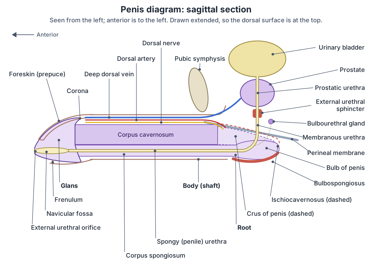
The penis has three parts: the root, the body and the glans. It lies in the urogenital triangle of the perineum. Its dorsal part is made of the paired corpora cavernosa, and its ventral part of the corpus spongiosum, which contains the urethra. Anatomical directions refer to the erect penis, so the dorsal surface is the one that faces the head when erect and the ventral surface the one that faces the feet. (Sam P, LaGrange CA. Anatomy, Abdomen and Pelvis, Penis. StatPearls, 2023.)
The root is the most proximal, fixed part. It contains three masses of erectile tissue, the two crura and the bulb, and two muscles. The body is made of three cylinders of erectile tissue: two corpora cavernosa and the corpus spongiosum. The glans is the cone-shaped end, formed by the expanded end of the corpus spongiosum, and carries the opening of the urethra, the external urethral orifice. (TeachMeAnatomy: The Penis.)
Unlike the root, the body of the penis has no muscles. The proximal rim of the glans is called the corona, and the neck just behind the corona separates the glans from the body. (Sam P, LaGrange CA. Anatomy, Abdomen and Pelvis, Penis. StatPearls, 2023.)
Cleveland Clinic lists three jobs of the penis: carrying urine out of the body through the urethra, carrying semen out of the body in reproduction, and sexual sensation, since the head (glans) has many sensitive nerve endings. (Cleveland Clinic: Penis.)
In the 3D model the corpora cavernosa, the corpus spongiosum and the glans come from BodyParts3D, an adult male data set, and hang in a resting (pendulous) position. The dorsal artery (red) and the deep dorsal vein (blue) run along the top near the root and then down the front (dorsal) surface of the hanging shaft, and the prostate is highlighted behind the root for orientation. The view is from the left; turn the model to see the corpus spongiosum underneath and how it widens into the glans.
The testes, ducts and glands that make and carry sperm, with a sagittal section of the male pelvis. Male reproductive system.
Parts of the penis
| Part | Where it is | Made of or contains |
|---|---|---|
| Root | Fixed part in the perineum (superficial perineal pouch) | Two crura and the bulb, with the ischiocavernosus and bulbospongiosus muscles |
| Crura (left and right) | Sides of the root, attached to the ischiopubic rami | The back ends of the corpora cavernosa |
| Bulb | Midline of the root, attached to the perineal membrane | The back end of the corpus spongiosum; the urethra passes through it |
| Body (shaft) | Free, hanging part | Two corpora cavernosa (dorsal) and the corpus spongiosum (ventral) with the spongy urethra |
| Glans | Cone-shaped end | Expanded end of the corpus spongiosum; corona at its base; external urethral orifice at its tip |
| Prepuce (foreskin) and frenulum | Fold of skin over the glans | The frenulum joins the prepuce to the underside of the glans |
From TeachMeAnatomy, Radiopaedia and StatPearls (see references).
Corpora cavernosa and corpus spongiosum: the erectile tissue
The two crura lie at the sides of the root, each attached to the ischial ramus on its own side, and continue forwards as the two corpora cavernosa, which are separated by the septum of the penis, often incompletely. The bulb lies in the midline of the root, is traversed by the urethra and continues forwards as the corpus spongiosum on the ventral side. (TeachMeAnatomy: The Penis.)
Radiopaedia describes the crura as attached to the pubic arch, the bulb as a midline structure between the crura attached to the perineal membrane, and the corpus spongiosum as lying in the groove on the underside of the corpora cavernosa. (Radiopaedia: Penis.)
OpenStax describes the corpora cavernosa as the two larger lateral chambers of erectile tissue running the length of the shaft, and the corpus spongiosum as a smaller chamber around the urethra that can be felt as a raised ridge on the erect penis. (OpenStax Anatomy and Physiology 2e, 27.1: Anatomy and Physiology of the Testicular Reproductive System.)
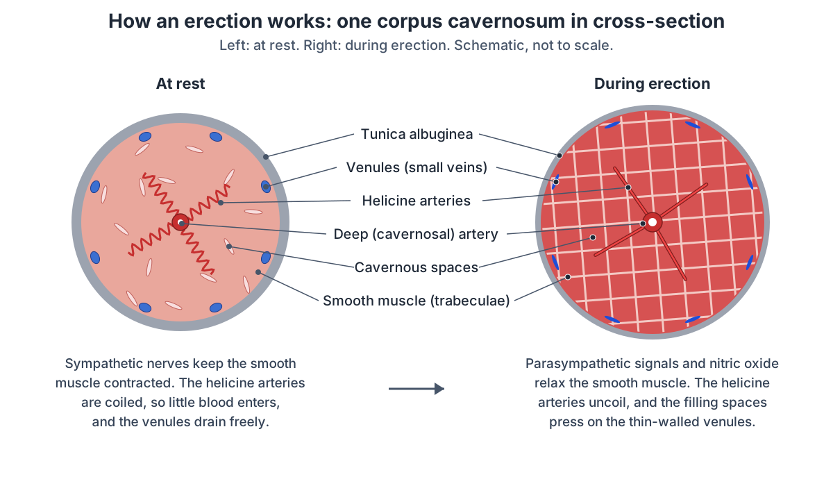
Erectile tissue is a sponge of blood-filled cavernous spaces (sinusoids) separated by trabeculae containing smooth muscle. A deep artery runs through the centre of each corpus cavernosum, and its branches, the helicine arteries, spread into the cavernous spaces. (Jung J, Jo HW, Kwon H, Jeong NY. Clinical neuroanatomy and neurotransmitter-mediated regulation of penile erection. Int Neurourol J. 2014.)
Cross-section of the penis: skin, fascia and tunica albuginea
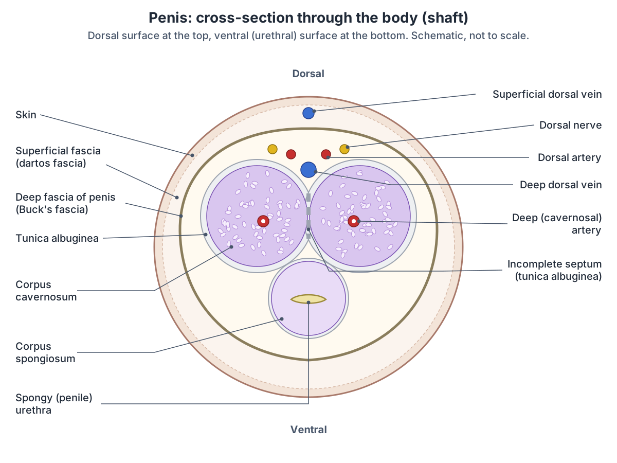
From the outside in, a section through the shaft shows: skin, more heavily pigmented than elsewhere on the body; the superficial (dartos) fascia of the penis immediately under the skin, a loose layer with smooth muscle that is continuous with Colles' fascia of the perineum; the deep fascia of the penis (Buck's fascia), which holds all three erectile bodies together; and the tunica albuginea, a capsule around each corpus cavernosum that fuses with its partner in the midline. (TeachMeAnatomy: The Penis.)
Buck's fascia is a continuation of the deep perineal fascia. It lies outside the tunica albuginea and covers the corpora cavernosa and corpus spongiosum together. (Sam P, LaGrange CA. Anatomy, Abdomen and Pelvis, Penis. StatPearls, 2023.)
Recent studies describe the tunica albuginea of the corpora cavernosa as two layers: a complete inner circular layer and an incomplete outer longitudinal layer. (Hsieh CH et al. Advances in understanding of mammalian penile evolution, human penile anatomy and human erection physiology. Med Sci Monit. 2012.)
On the dorsal surface, under the deep fascia and on the fibrous sheaths of the corpora cavernosa, runs a neurovascular bundle. The deep dorsal vein lies medial to each dorsal artery and the dorsal nerve lies lateral to it, so from the midline outwards the order is vein, artery, nerve. (Radiopaedia: Dorsal artery of the penis.)
Gray's classic description, reproduced by IMAIOS, gives the same order: on the penis the dorsal artery lies between the dorsal nerve and the deep dorsal vein, with the nerve on its lateral side. (IMAIOS e-Anatomy: Dorsal artery of penis.)
Hsieh and colleagues found more veins than the classic picture of one deep dorsal vein between two arteries: one deep dorsal vein, two cavernosal veins and two pairs of para-arterial veins run between Buck's fascia and the tunica albuginea. (Hsieh CH et al. Advances in understanding of mammalian penile evolution, human penile anatomy and human erection physiology. Med Sci Monit. 2012.)
Ligaments and muscles at the root of the penis
Two ligaments hold the penis to the front of the pelvis. The suspensory ligament, a condensation of deep fascia, connects the erectile bodies to the pubic symphysis. The fundiform ligament, a condensation of the subcutaneous tissue of the abdomen, runs down from the linea alba and surrounds the penis like a sling. (TeachMeAnatomy: The Penis.)
StatPearls gives the same two ligaments, describing the suspensory ligament as part of the deep fascia and the fundiform ligament as descending in the midline from the linea alba. (Sam P, LaGrange CA. Anatomy, Abdomen and Pelvis, Penis. StatPearls, 2023.)
The root lies in the superficial perineal pouch, which contains the erectile tissues and three muscles: ischiocavernosus, bulbospongiosus and superficial transverse perineal. The perineal membrane above it gives attachment to the muscles of the external genitalia. (TeachMeAnatomy: The Perineum.)
The ischiocavernosus contracts to force blood from the cavernous spaces of the crura into the corpora cavernosa. The bulbospongiosus contracts to empty the spongy urethra of any remaining semen and urine. Both are supplied by the pudendal nerve. (TeachMeAnatomy: The Penis.)
During erection both muscles also press on the veins of the corpora cavernosa, which restricts the outflow of blood. (Jung J, Jo HW, Kwon H, Jeong NY. Clinical neuroanatomy and neurotransmitter-mediated regulation of penile erection. Int Neurourol J. 2014.)
Glans, corona, foreskin and frenulum
The glans has a high concentration of nerve endings. The skin of the shaft extends over the glans as a collar called the prepuce (foreskin). Circumcision, often performed for religious or social reasons, removes the prepuce. (OpenStax Anatomy and Physiology 2e, 27.1: Anatomy and Physiology of the Testicular Reproductive System.)
The prepuce is a second layer of skin that covers the glans, and the frenulum of the prepuce connects it to the urethral (under) surface of the glans. The corona is the proximal rim of the glans, and the opening of the urethra, the meatus, lies at the tip. (Sam P, LaGrange CA. Anatomy, Abdomen and Pelvis, Penis. StatPearls, 2023.)
Radiopaedia describes the skin folding back on itself at the corona to form the prepuce, and the frenulum as a median fold of skin on the urethral side of the glans. (Radiopaedia: Penis.)
Phimosis is a prepuce that fits tightly over the glans and cannot be pulled back. Paraphimosis is a tight prepuce that has been pulled back and left trapped behind the glans. (TeachMeAnatomy: The Penis.)
The urethra in the penis
The male urethra is about 15 to 20 cm long. It has three parts: the prostatic part, which runs through the prostate and is the widest; the membranous part, which passes through the pelvic floor and deep perineal pouch, is surrounded by the external urethral sphincter and is the narrowest part; and the penile (spongy) part, which runs through the bulb and corpus spongiosum and receives the bulbourethral glands. In the glans the urethra widens into the navicular fossa and ends at the external urethral orifice. (TeachMeAnatomy: The Urethra.)
Radiopaedia gives an average length of 18 to 20 cm and divides the urethra into a posterior part (prostatic and membranous) and an anterior part (bulbar and penile). It calls the membranous urethra the shortest and narrowest portion, describes the navicular fossa as a small widening at the end of the penile urethra, and names the external meatus as the narrowest point. (Radiopaedia: Male urethra.)
The urethra carries both urine and semen out of the body through its opening, the meatus, at the tip of the glans. (Cleveland Clinic: Penis.)
The gland around the first part of the urethra, with its zones and the ejaculatory ducts. Prostate.
Blood supply of the penis: arteries, veins and lymph
The erectile tissue and glans are supplied by branches of the internal pudendal artery. There are three kinds: the dorsal arteries, the deep (cavernosal) arteries and the arteries of the bulb (bulbourethral arteries). (TeachMeAnatomy: The Penis.)
The skin of the shaft and foreskin also gets blood from the superficial and deep external pudendal arteries, which are branches of the femoral artery. (Fawzy M, Hadidi AT. Buried penis; what buried the penis? Front Pediatr. 2025.)
The deep arteries run in the centre of each corpus cavernosum and give off helicine arteries into the cavernous spaces. The dorsal arteries run along each side of the deep dorsal vein, and the arteries of the bulb supply the bulb of the corpus spongiosum. (Jung J, Jo HW, Kwon H, Jeong NY. Clinical neuroanatomy and neurotransmitter-mediated regulation of penile erection. Int Neurourol J. 2014.)
Each dorsal artery runs forwards along the dorsum of the penis to the glans. It supplies the glans and penile skin and, through connections with the artery of the bulb, part of the corpus spongiosum and urethra. (Radiopaedia: Dorsal artery of the penis.)
The cavernous spaces are drained by the deep dorsal vein, which empties into the prostatic venous plexus. The superficial dorsal veins drain the skin and other superficial tissues. (TeachMeAnatomy: The Penis.)
Lymph from the skin of the penis drains to the superficial inguinal nodes, lymph from the glans to the deep inguinal and external iliac nodes, and lymph from the erectile tissue and penile urethra to the internal iliac nodes. (Radiopaedia: Penis.)
Blood supply, nerves and lymph of the penis
| Supply | Details |
|---|---|
| Arteries | Branches of the internal pudendal artery: dorsal arteries, deep (cavernosal) arteries with helicine branches, arteries of the bulb; the skin of the shaft and foreskin also from the superficial and deep external pudendal arteries (from the femoral artery) |
| Veins | Deep dorsal vein to the prostatic venous plexus; superficial dorsal veins from the skin; also cavernosal and para-arterial veins (Hsieh 2012) |
| Sensory nerves | Dorsal nerve of the penis, a terminal branch of the pudendal nerve (S2 to S4), for the glans and most of the skin of the shaft; ilioinguinal nerve (L1) for the skin at the root |
| Autonomic nerves | Cavernous nerves from the pelvic (inferior hypogastric) plexus: parasympathetic (S2 to S4) for erection, sympathetic for ejaculation and detumescence |
| Lymph | Skin: superficial inguinal nodes; glans: deep inguinal and external iliac nodes; erectile tissue and penile urethra: internal iliac nodes |
From TeachMeAnatomy, Radiopaedia, StatPearls, Kenhub, Jung et al. 2014 and Fawzy and Hadidi 2025.
Nerves of the penis
Sensation comes through the dorsal nerve of the penis, a smaller terminal branch of the pudendal nerve, which arises from the S2, S3 and S4 nerve roots. The dorsal nerve runs along the dorsum of the penis and ends in the glans, carrying sensation from the penile skin and the glans. (Weech D, Ameer MA, Ashurst JV. Anatomy, Abdomen and Pelvis, Penis Dorsal Nerve. StatPearls, 2023.)
The erectile tissue is supplied by the cavernous nerves, which arise from the pelvic (inferior hypogastric) plexus. Their parasympathetic fibres (S2 to S4) are responsible for erection, while sympathetic fibres are involved in ejaculation and in the return to the resting state (detumescence). (TeachMeAnatomy: The Penis.)
Jung and colleagues trace the route: pelvic splanchnic nerves from S2 to S4 join the hypogastric nerves to form the pelvic plexus, branches of the pelvic plexus form the prostatic plexus, and the cavernous nerves arise from it to reach the erectile tissue. (Jung J, Jo HW, Kwon H, Jeong NY. Clinical neuroanatomy and neurotransmitter-mediated regulation of penile erection. Int Neurourol J. 2014.)
How an erection works: the physiology
An erection is a change in blood flow. At rest, sympathetic nerves keep the smooth muscle of the erectile tissue contracted, the helicine arteries are coiled and little blood enters the cavernous spaces. Parasympathetic signals through the cavernous nerves relax this smooth muscle and let the helicine arteries uncoil, so blood flows into the empty spaces, and the corpora cavernosa expand more than the corpus spongiosum. (Jung J, Jo HW, Kwon H, Jeong NY. Clinical neuroanatomy and neurotransmitter-mediated regulation of penile erection. Int Neurourol J. 2014.)
A key chemical signal is nitric oxide, released by nerve endings near the blood vessels and by the cells lining them. It relaxes the smooth muscle around the penile arteries so they widen. As the chambers fill, they press on the thin-walled veins and block the outflow of blood, which keeps the penis firm. (OpenStax Anatomy and Physiology 2e, 27.1: Anatomy and Physiology of the Testicular Reproductive System.)
The ischiocavernosus and bulbospongiosus muscles add to this by compressing the veins. When the nerve signals stop, the smooth muscle contracts again and the trapped blood flows out, returning the penis to its resting state. (Jung J, Jo HW, Kwon H, Jeong NY. Clinical neuroanatomy and neurotransmitter-mediated regulation of penile erection. Int Neurourol J. 2014.)
The corpus spongiosum fills at a lower pressure, so the urethra inside it is not closed off during an erection. (TeachMeAnatomy: The Penis.)
StatPearls notes that erection usually happens in response to sexual arousal but sometimes occurs spontaneously; OpenStax adds that erections also occur during REM sleep. (Panchatsharam PK, Durland J, Zito PM. Physiology, Erection. StatPearls, 2023.)
How the penis develops
The glans of the penis and the glans of the clitoris develop from the same embryonic cells: with testosterone they form the glans penis, and without it they form the clitoris. (OpenStax Anatomy and Physiology 2e, 27.3: Development of the Male and Female Reproductive Systems.)
The external genitalia of both sexes start from the same three structures: the genital tubercle, the urethral folds and the labioscrotal swellings. From about week 8 a urethral plate forms in the midline of the genital tubercle, and between about weeks 11 and 16 testicular androgens make the tubercle lengthen into the penis. The urethral folds on each side of the plate fuse to form the penile urethra, and the labioscrotal swellings fuse in the midline to form the scrotum. (van der Horst HJ, de Wall LL. Hypospadias, all there is to know. Eur J Pediatr. 2017.)
In hypospadias, a congenital malformation of the male external genitalia, the urethral opening lies further back than normal and on the underside of the penis. It is thought to result from incomplete fusion of the urethral groove. (Radiopaedia: Hypospadias.)
The clitoris, which develops from the same tissue as the penis, with the rest of the vulva. Vulva anatomy.
Clinical anatomy: priapism, penile fracture and Peyronie disease
Priapism is an erection that persists beyond, or is not related to, sexual stimulation, usually defined as lasting more than four hours. The common ischaemic (low-flow) type, about 95% of cases, traps blood in a rigid, painful penis and is a medical emergency; the rarer non-ischaemic (high-flow) type, usually after trauma, is typically painless and is often treated conservatively at first. (Radiopaedia: Priapism.)
Erectile dysfunction, the inability to maintain an erection, is most often caused by problems with the blood vessels. (TeachMeAnatomy: The Penis.)
StatPearls defines priapism as an unwanted erection lasting 4 hours or longer. Ischaemic priapism is a true emergency that needs prompt treatment. (StatPearls: Priapism, 2025.)
A penile fracture is a tear of the tunica albuginea, usually from trauma to the erect penis, most often during intercourse. The tunica albuginea thins during erection, which makes it vulnerable. (Radiopaedia: Penile fracture.)
In Peyronie disease, fibrous plaques form in the tunica albuginea, most often on the dorsal side, and cause curvature of the penis and pain. (Radiopaedia: Peyronie disease.)
Cleveland Clinic also lists penile cancer, skin conditions and sexually transmitted infections among the conditions that affect the penis. A new lump, sore, bend or pain should be checked by a clinician. (Cleveland Clinic: Penis.)
The testes and epididymis, where sperm are made and stored. Testicle anatomy.
Penis in the 3D model
- Corpus cavernosum of penis
- The corpora cavernosa are the paired columns of erectile tissue forming the dorsal part of the penis.
- Corpus spongiosum of penis
- The corpus spongiosum is the single column of erectile tissue surrounding the penile urethra.
- Deep dorsal vein of penis
- The deep dorsal vein is the main vein of the penis, draining the glans and erectile bodies.
- Dorsal artery of penis
- The dorsal artery of the penis supplies the skin and glans.
- Glans penis
- The glans penis is the cone-shaped distal end of the penis.
- Prostate
- The prostate is a walnut-sized gland surrounding the first part of the male urethra.
Frequently asked questions
What are the three parts of the penis?
The root, the body (shaft) and the glans. The root is the fixed part in the perineum, made of the two crura and the bulb with their muscles. The body is the free part, made of three columns of erectile tissue. The glans is the cone-shaped end, formed by the expanded end of the corpus spongiosum, with the urethral opening at its tip.
What are the erectile tissues of the penis?
There are three columns of erectile tissue. Two corpora cavernosa run side by side along the dorsal (upper) side and make up most of the shaft. One corpus spongiosum runs along the ventral (under) side around the urethra and expands into the glans at the tip and the bulb at the root. All three fill with blood during an erection.
What is the corpus spongiosum?
The corpus spongiosum is the single column of erectile tissue on the underside of the penis. It begins at the root as the bulb, runs along the shaft around the urethra and ends as the glans. During an erection it fills at a lower pressure than the corpora cavernosa, so the urethra inside it stays open.
What is Buck's fascia?
Buck's fascia is the deep fascia of the penis. It continues from the deep perineal fascia and wraps all three erectile bodies together, outside their tunica albuginea. The deep dorsal vein, the dorsal arteries and the dorsal nerves run beneath it in the groove on the dorsal surface. The superficial (dartos) fascia of the penis lies outside it, just under the skin.
What is the tunica albuginea of the penis?
The tunica albuginea is the tough fibrous capsule around each corpus cavernosum, fused in the midline where the two meet. Recent studies describe a complete inner circular layer and an incomplete outer longitudinal layer. It thins during an erection, and a tear in it is called a penile fracture; fibrous plaques in it cause Peyronie disease.
What is the frenulum of the penis?
The frenulum is a small fold of skin on the underside (urethral side) of the glans that attaches the foreskin (prepuce) to the glans. The foreskin itself is a fold of skin from the shaft that covers the glans and is removed in circumcision. The corona is the raised rim at the base of the glans.
How does an erection happen?
Parasympathetic nerve signals from the S2 to S4 segments of the spinal cord travel in the cavernous nerves. Nitric oxide released from nerve endings and vessel linings relaxes the smooth muscle of the arteries and erectile tissue, so blood flows into the cavernous spaces. As they swell they press on the veins and reduce outflow. When the signals stop, the blood drains away.
Where does the urethra run in the penis?
After passing through the prostate and the pelvic floor, the urethra enters the bulb of the penis and runs inside the corpus spongiosum along the underside of the shaft. In the glans it widens into the navicular fossa and opens at the external urethral orifice (meatus) at the tip. The whole male urethra is about 15 to 20 cm long.
References
- OpenStax. Anatomy and Physiology 2e, §27.1 Anatomy and Physiology of the Testicular Reproductive System. Rice University, 2022.
- OpenStax. Anatomy and Physiology 2e, §27.3 Development of the Male and Female Reproductive Systems. Rice University, 2022.
- Sam P, LaGrange CA. Anatomy, Abdomen and Pelvis, Penis. StatPearls. Treasure Island (FL): StatPearls Publishing; 2023.
- Weech D, Ameer MA, Ashurst JV. Anatomy, Abdomen and Pelvis, Penis Dorsal Nerve. StatPearls. Treasure Island (FL): StatPearls Publishing; 2023.
- Panchatsharam PK, Durland J, Zito PM. Physiology, Erection. StatPearls. Treasure Island (FL): StatPearls Publishing; 2023.
- Priapism. StatPearls. Treasure Island (FL): StatPearls Publishing; 2025.
- TeachMeAnatomy. The Penis.
- TeachMeAnatomy. The Urethra.
- TeachMeAnatomy. The Perineum.
- Penis. Radiopaedia reference article.
- Male urethra. Radiopaedia reference article.
- Dorsal artery of the penis. Radiopaedia reference article.
- Penile fracture. Radiopaedia reference article.
- Peyronie disease. Radiopaedia reference article.
- Hypospadias. Radiopaedia reference article.
- Priapism. Radiopaedia reference article.
- Penis: anatomy, blood supply and innervation. Kenhub anatomy library.
- Fawzy M, Hadidi AT. Buried penis; what buried the penis? Front Pediatr. 2025;13:1590147.
- van der Horst HJ, de Wall LL. Hypospadias, all there is to know. Eur J Pediatr. 2017;176(4):435-441.
- Dorsal artery of penis. IMAIOS e-Anatomy (text adapted from Gray's Anatomy, 1918).
- Jung J, Jo HW, Kwon H, Jeong NY. Clinical neuroanatomy and neurotransmitter-mediated regulation of penile erection. Int Neurourol J. 2014;18(2):58-62.
- Hsieh CH, Liu SP, Hsu GL, et al. Advances in understanding of mammalian penile evolution, human penile anatomy and human erection physiology: clinical implications for physicians and surgeons. Med Sci Monit. 2012;18(7):RA118-RA125.
- Cleveland Clinic. Penis: Anatomy, Function, Conditions and Care. Reviewed 2025.
- Mitsuhashi N. et al. (2009). BodyParts3D: 3D structure database for anatomical concepts. Nucleic Acids Research 37, D782–D785. Data © The Database Center for Life Science, CC BY 4.0.
Related anatomy
- Male reproductive systemMale reproductive anatomy with labelled diagrams: testes, epididymis, ductus deferens, seminal gland, the accessory…
- TestisTesticle anatomy with a labeled testis diagram and 3D model: the tunica vaginalis and albuginea, lobules, seminiferous…
- ScrotumScrotum anatomy: layers from skin and dartos to tunica vaginalis, septum and raphe, contents, blood supply, lymph…
- ProstateProstate anatomy with labelled diagrams: McNeal zones, older lobes, prostatic urethra and ejaculatory ducts…
- Glans penisGlans penis: The glans penis is the cone-shaped distal end of the penis. See where it is in an interactive 3D human…
- Corpus cavernosum of penisCorpus cavernosum of penis: The corpora cavernosa are the paired columns of erectile tissue forming the dorsal part of…
Explore the whole body in the interactive 3D human anatomy model, or browse the A–Z list of human structures.