

Scrotum at a glance
- What it is
- The scrotum is the skin-covered sac that holds the testes, behind the penis and in front of the anus.
- Location
- It hangs below the root of the penis, between the thighs. Each of its two compartments holds a testis, an epididymis and the lower end of a spermatic cord.
- Function
- It keeps the testes outside the abdominal cavity, a few degrees cooler than the body core, which sperm production needs; the dartos and cremaster muscles raise or lower the testes to adjust their temperature.
- Body system
- Reproductive · single, midline structure · male body
- FMA ID
- FMA18252 (Foundational Model of Anatomy)
Key facts
- TeachMeAnatomy describes it as 'a fibromuscular cutaneous sac, located between the penis and anus', with the dartos muscle, a sheet of smooth muscle, immediately under the skin.
- A midline septum divides it into two compartments, one for each testis.
- In the 3D model the scrotum is an approximation built as a thin wall just inside the BodyParts3D skin around the testes; BodyParts3D has no separate scrotum.
What is the scrotum?
The scrotum is a fibromuscular sac covered with skin, lying between the penis and the anus. It is dual-chambered and forms an expansion of the perineum. (TeachMeAnatomy: The Scrotum.)
Radiopaedia describes it as a dual-chambered protuberance of skin and muscle that contains the testes, the epididymides and the spermatic cords. The two chambers are separated by a septum. (Radiopaedia: Scrotum.)
OpenStax describes the scrotum as a skin-covered, highly pigmented, muscular sac. Keeping the testes there, outside the abdominal cavity, matters for sperm production, which is most efficient when the testes are 2 to 4 °C below core body temperature. (OpenStax Anatomy and Physiology 2e, 27.1: Anatomy and Physiology of the Testicular Reproductive System.)
The 3D model highlights the scrotum as an approximate part built for this site. BodyParts3D, the adult male data set behind the model, has no scrotum, so a thin wall about 2 mm thick was built just inside the body's skin around both testes and epididymides, open at its neck where the spermatic cords leave, with a built septum of the scrotum dividing it in two. It is drawn translucent so the testes and epididymides inside show through, and it exists only in the male body. Its upper edge follows the skin where it meets the thighs, so the sac flares slightly at the top in the front view. Real testis sizes and the temperature difference are given in the sections below.
Testes, ducts, seminal glands, prostate and penis, with the path of sperm. Male reproductive system.
Layers of the scrotum
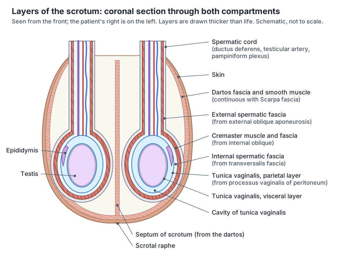
From superficial to deep, the layers of the scrotal wall are the skin, the dartos fascia with its smooth muscle, the external spermatic fascia, the cremaster muscle, the internal spermatic fascia and the tunica vaginalis. These layers are continuations of the layers of the abdominal wall. (Patel AP. Anatomy and physiology of chronic scrotal pain. Transl Androl Urol. 2017.)
Radiopaedia lists the same order: pigmented skin with rugal folds; superficial fascia and dartos muscle; external spermatic fascia, a continuation of the external oblique aponeurosis; cremasteric fascia, a continuation of the internal oblique aponeurosis; internal spermatic fascia, a continuation of the transversalis fascia; the tunica vaginalis; and the tunica albuginea covering the testis. (Radiopaedia: Scrotum.)
Strictly, Kenhub counts only two layers as the scrotum itself, the skin and the dartos fascia, because the scrotum develops separately from the testes. The deeper layers are coverings of the testis brought down from the abdominal wall as the testis descended. (Kenhub: The scrotum.)
The scrotal skin is thin and corrugated (wrinkled) and rich in sebaceous glands. Under it lies a unique subcutaneous layer filled with smooth muscle fibres and with no fat. (Lin et al. Anatomical considerations, testicular, and scrotal anatomy of nonobstructive azoospermia patients. Asian J Androl. 2025.)
The dartos is a sheet of smooth muscle immediately under the skin. When it contracts it wrinkles the skin, which reduces the surface area and so reduces heat loss. (TeachMeAnatomy: The Scrotum.)
The dartos fascia is continuous with Scarpa fascia, the membranous layer of superficial fascia on the abdominal wall, and with the superficial fascia of the perineum (Colles fascia). (Radiopaedia: Scarpa fascia.)
The three spermatic fasciae wrap each testis and spermatic cord. From outermost to innermost: the external spermatic fascia comes from the aponeurosis of external oblique; the cremaster muscle and its fascia come from internal oblique; and the internal spermatic fascia comes from the transversalis fascia. (TeachMeAnatomy: The Spermatic Cord.)
Radiopaedia gives the same three coverings and explains them by descent: the testes drag the layers of the anterior abdominal wall with them as they move down into the scrotum. (Radiopaedia: Spermatic cord.)
The innermost layer, the tunica vaginalis, lies inside the internal spermatic fascia. It has a parietal and a visceral layer, normally separated by only a little fluid. (Lam Shin Cheung et al. A sound approach to stay on the ball: a review of scrotal pathologies on ultrasound imaging. Br J Radiol. 2026.)
The tunica vaginalis is what remains of the processus vaginalis, a pouch of peritoneum that pushed down through the abdominal wall during development. Its upper part closes off, and the lower part that envelops the testis becomes the tunica vaginalis. (Radiopaedia: Testicular development and descent.)
External oblique, internal oblique, transversus abdominis and the inguinal canal. Abdominal muscles.
Layers of the scrotum and the abdominal wall layers they continue
| Scrotal layer (outside to inside) | Continuous with or derived from | Notes |
|---|---|---|
| Skin | Skin of the abdominal wall and perineum | Thin, pigmented, wrinkled, rich in sebaceous glands |
| Dartos fascia and smooth muscle | Membranous superficial fascia: Scarpa fascia (abdomen), Colles fascia (perineum) | No fat; forms the septum; wrinkles the skin |
| External spermatic fascia | External oblique aponeurosis | Outer covering of the testis and spermatic cord |
| Cremaster muscle and fascia | Internal oblique muscle and aponeurosis | Raises the testis; genital branch of the genitofemoral nerve |
| Internal spermatic fascia | Transversalis fascia | Inner covering of the spermatic cord |
| Tunica vaginalis (parietal and visceral layers) | Processus vaginalis of the peritoneum | Encloses a thin film of fluid around the testis |
From Patel (2017), Radiopaedia (scrotum, spermatic cord, Scarpa fascia, testicular descent), TeachMeAnatomy (spermatic cord) and Lin et al. (2025).
The septum and the scrotal raphe
The dartos muscle forms the subcutaneous muscle layer of the scrotum and continues inwards as the scrotal septum, a wall that divides the scrotum into two compartments, each holding one testis. (OpenStax Anatomy and Physiology 2e, 27.1: Anatomy and Physiology of the Testicular Reproductive System.)
Kenhub likewise describes the dartos fascia dividing the scrotum internally into right and left compartments through an internal fascial septum, one compartment for each testis. (Kenhub: The scrotum.)
Lin and colleagues add that the fat-free subcutaneous layer, together with the fascia of the spermatic cord, extends backwards to form the septum. (Lin et al. Anatomical considerations, testicular, and scrotal anatomy of nonobstructive azoospermia patients. Asian J Androl. 2025.)
On the outside, a ridge of skin called the scrotal raphe runs down the midline. It marks the line where the paired labioscrotal swellings fused in the embryo. (Kenhub: The scrotum.)
The midline partition between the two compartments, in the 3D model. Septum of scrotum.
What is inside the scrotum?
The scrotum contains the testes, the epididymides, the testicular and epididymal appendages and the lower part of each spermatic cord. (Radiopaedia: Scrotum.)
Each testis is the site of sperm production; the epididymis stores sperm; and the spermatic cord is a collection of muscle fibres, vessels, nerves and ducts running to and from the testis. (TeachMeAnatomy: The Scrotum.)
Normal adult testes are ovoid, about 3 to 5 cm long and 2 to 3 cm across, with a volume of 15 to 25 mL. (Radiopaedia: Testis.)
A 2025 review gives the same sizes for healthy adult men: 3 to 5 cm long, 2 to 3 cm wide and 2 to 3 cm thick, with a volume of 15 to 25 mL. (Lin et al. Anatomical considerations, testicular, and scrotal anatomy of nonobstructive azoospermia patients. Asian J Androl. 2025.)
Apart from being attached to the bottom of the scrotum by the gubernaculum, which helps prevent twisting, each testis can move within its compartment. In most men the right testis rests a little higher than the left. (Patel AP. Anatomy and physiology of chronic scrotal pain. Transl Androl Urol. 2017.)
Lam Shin Cheung and colleagues also note that one testis, typically the left, may normally hang slightly lower than the other. (Lam Shin Cheung et al. A sound approach to stay on the ball: a review of scrotal pathologies on ultrasound imaging. Br J Radiol. 2026.)
The spermatic cord contains the ductus deferens, the testicular artery from the aorta, the cremasteric artery, the artery to the ductus deferens, the pampiniform plexus of veins, the genital branch of the genitofemoral nerve, autonomic nerves, lymphatic vessels and the closed remnant of the processus vaginalis. (TeachMeAnatomy: The Spermatic Cord.)
The testis in the 3D model: structure, blood supply and function. Testis.
Blood supply and venous drainage
The scrotal wall is supplied by three sets of arteries: the posterior scrotal branches of the perineal artery, the anterior scrotal branches of the external pudendal artery, and the cremasteric artery, a branch of the inferior epigastric artery. (Kenhub: The scrotum.)
TeachMeAnatomy gives the anterior scrotal artery as a branch of the external pudendal artery and the posterior scrotal artery as a branch of the internal pudendal artery. The scrotal veins follow the major arteries; TeachMeAnatomy names the external pudendal veins as their drainage. (TeachMeAnatomy: The Scrotum.)
The testis itself has a different supply. Each testis is fed by its testicular artery, which arises from the aorta just below the renal arteries. The pampiniform plexus drains the testis and ascends as the testicular vein: the right testicular vein drains directly into the inferior vena cava and the left into the left renal vein. (Radiopaedia: Testis.)
Patel describes the supply of the scrotum and its contents as rich and interconnected, with anastomoses between branches of the internal and external iliac arteries. The superficial veins of the scrotum mostly follow the arteries: the front drains through the external pudendal branches and the back through the internal pudendal branches to the internal iliac vein. He also describes the small veins of the pampiniform plexus joining to form the gonadal (testicular) vein, which empties into the left renal vein on the left and the inferior vena cava on the right. (Patel AP. Anatomy and physiology of chronic scrotal pain. Transl Androl Urol. 2017.)
Lymph drainage: scrotum vs testis
Lymph from the scrotum drains to the nearby superficial inguinal lymph nodes in the groin. (TeachMeAnatomy: The Scrotum.)
Radiopaedia gives the same route for scrotal lymph, to the superficial inguinal nodes. (Radiopaedia: Scrotum.)
Lymph from the testis takes a completely different path. Its lymphatic vessels run up with the testicular arteries to the para-aortic lymph nodes on the back wall of the abdomen. (Radiopaedia: Testis.)
TeachMeAnatomy gives the same destination for the lymph vessels of the spermatic cord: the para-aortic nodes in the lumbar region. (TeachMeAnatomy: The Spermatic Cord.)
Patel explains the difference: lymph from the skin, the scrotal layers and the tunica vaginalis drains to the superficial and then deep inguinal nodes, while lymph from the testis and epididymis drains into the retroperitoneum along the route the testis took when it migrated down during development. (Patel AP. Anatomy and physiology of chronic scrotal pain. Transl Androl Urol. 2017.)
The inguinal region, inguinal canal and superficial inguinal lymph nodes. Groin.
Supply of the scrotal wall compared with the testis
| Scrotal wall (skin and dartos) | Testis | |
|---|---|---|
| Arteries | Anterior scrotal (external pudendal), posterior scrotal (perineal, from internal pudendal); cremasteric artery | Testicular artery from the aorta, below the renal arteries |
| Veins | Scrotal veins following the arteries: front to the external pudendal veins, back to the internal pudendal veins | Pampiniform plexus to the testicular vein: right to the inferior vena cava, left to the left renal vein |
| Lymph | Superficial inguinal nodes | Para-aortic (lumbar) nodes |
| Nerves | Ilioinguinal and genitofemoral (front), pudendal and posterior femoral cutaneous (back and below) | Autonomic nerves travelling with the vessels and ductus deferens |
From Kenhub, TeachMeAnatomy, Radiopaedia and Patel (2017).
Nerves of the scrotum
The front and side of the scrotum are supplied by the genital branch of the genitofemoral nerve (L1, L2) and the anterior scrotal nerves, branches of the ilioinguinal nerve (L1). The back is supplied by the posterior scrotal nerves from the perineal branch of the pudendal nerve (S2 to S4), and the lower part by perineal branches of the posterior cutaneous nerve of the thigh. (Kenhub: The scrotum.)
Patel gives the same map: the genital branch of the genitofemoral nerve supplies the anterolateral surface, the anterior scrotal nerves from the ilioinguinal nerve the anterior surface, the posterior scrotal nerves from the pudendal nerve the posterior surface, and the long scrotal branches of the posterior femoral cutaneous nerve the inferior surface. Overall the somatic supply of the scrotum and testes comes from the L1 to L2 and S2 to S4 roots. (Patel AP. Anatomy and physiology of chronic scrotal pain. Transl Androl Urol. 2017.)
The genital branch of the genitofemoral nerve also supplies the cremaster muscle. Stroking the skin of the upper inner thigh makes the cremaster on the same side contract and lift the testis: in this cremasteric reflex the ilioinguinal nerve is the sensory limb and the genital branch of the genitofemoral nerve the motor limb. (Radiopaedia: Cremaster muscle.)
TeachMeAnatomy describes the same reflex, with the ilioinguinal nerve carrying the sensory signal into the spinal cord at L1 and the genital branch of the genitofemoral nerve carrying the motor signal. (TeachMeAnatomy: The Spermatic Cord.)
Development: labioscrotal swellings and the descent of the testes
The scrotum develops from a pair of ridges, the labioscrotal swellings, which later fuse in the midline to form one continuous sac. The scrotal raphe marks the line of fusion. (Kenhub: The scrotum.)
TeachMeAnatomy also describes the scrotum forming from paired genital swellings that fuse in the midline, and notes that it is the biological homologue of the labia majora in females. (TeachMeAnatomy: The Scrotum.)
The testes develop from the gonadal ridge on the back wall of the abdomen. Each is attached to the gubernaculum, a band of mesenchyme that grows down towards the scrotal swellings, and a pouch of peritoneum, the processus vaginalis, pushes through the abdominal wall; together they form the inguinal canal. The testis lies within the deep inguinal ring by the 7th month and then descends into the scrotum over days to weeks. (Radiopaedia: Testicular development and descent.)
OpenStax places the descent through the abdominal musculature into the scrotum in the seventh month of fetal development. (OpenStax Anatomy and Physiology 2e, 27.1: Anatomy and Physiology of the Testicular Reproductive System.)
Ivanova and colleagues describe the gubernaculum as the mesenchymal tissue extending from the fetal testis and epididymis to the developing scrotum, and note that in humans testicular descent is complete at birth, at about the 35th week of gestation. (Ivanova et al. Gubernaculum and Epididymo-Testicular Descent: Review of the Literature. Acta Med Litu. 2022.)
Kenhub gives about 32 weeks for both testes to complete their descent from the back wall of the abdomen through the inguinal canal, so sources differ by a few weeks on the exact timing. If the testes do not descend into the scrotum but remain inside the body, the condition is called cryptorchidism. (Kenhub: The scrotum.)
How the scrotum keeps the testes cool
Sperm production works best when the testes are kept 2 to 4 °C below core body temperature. The dartos and cremaster muscles contract together to raise the testes towards the body in cold weather, and relax to let them hang lower when it is warm. (OpenStax Anatomy and Physiology 2e, 27.1: Anatomy and Physiology of the Testicular Reproductive System.)
A review of scrotal imaging puts the scrotal temperature about 2 °C lower than the rest of the body. (Studniarek M et al. Scrotal imaging. J Ultrason. 2015.)
Inside the spermatic cord, the pampiniform plexus of veins acts as a heat exchanger, cooling the arterial blood before it reaches the testis. (TeachMeAnatomy: The Spermatic Cord.)
Radiopaedia describes the same heat-exchange role of the pampiniform veins, and explains that a varicocele disrupts it so that the testis warms towards core body temperature. (Radiopaedia: Varicocele.)
Hydrocele, varicocele, torsion and inguinal hernia
A hydrocele is a collection of serous fluid between the layers of the tunica vaginalis around a testis or spermatic cord. A congenital (communicating) hydrocele collects around a processus vaginalis that failed to close; acquired hydroceles can follow trauma, epididymitis, torsion, a tumour or infarction of the testis. (Radiopaedia: Hydrocele.)
Lam Shin Cheung and colleagues define a hydrocele as excess fluid within the tunica vaginalis; most cases have no known cause and show as painless scrotal swelling. (Lam Shin Cheung et al. A sound approach to stay on the ball: a review of scrotal pathologies on ultrasound imaging. Br J Radiol. 2026.)
A varicocele is a dilatation of the pampiniform plexus of veins. Radiopaedia estimates it in about 15% of the general male population and about 40% of subfertile and infertile men. Clinically significant varicoceles are about 85% left-sided, partly because the left testicular vein is longer and joins the left renal vein at a right angle. (Radiopaedia: Varicocele.)
A 2026 ultrasound review gives the same figures, varicocele in about 15% of males and up to 40% of infertile men, and explains that the vertical course of the left testicular vein predisposes to left-sided varicoceles. (Lam Shin Cheung et al. A sound approach to stay on the ball: a review of scrotal pathologies on ultrasound imaging. Br J Radiol. 2026.)
A 2015 review of scrotal imaging likewise places 85% of varicoceles on the left spermatic cord; isolated right-sided varicocele is rare. (Studniarek M et al. Scrotal imaging. J Ultrason. 2015.)
Testicular torsion is twisting of the testis on its spermatic cord, which cuts off its blood supply. The common intravaginal type, seen in adolescents and young adults, is linked to the bell clapper deformity, an abnormally high attachment of the tunica vaginalis to the cord. The cremasteric reflex is typically absent. (Radiopaedia: Testicular torsion.)
Lam Shin Cheung and colleagues describe torsion as a surgical emergency; the bell clapper deformity lets the cord move freely, and neonates and males around puberty are at highest risk. (Lam Shin Cheung et al. A sound approach to stay on the ball: a review of scrotal pathologies on ultrasound imaging. Br J Radiol. 2026.)
An indirect inguinal hernia, the most common type of abdominal hernia, enters the inguinal canal through the deep inguinal ring and in males may extend through the superficial ring into the scrotum. Radiopaedia links its male predominance to persistence of the processus vaginalis during testicular descent. (Radiopaedia: Indirect inguinal hernia.)
Radiopaedia's article on testicular descent adds that failure of the processus vaginalis to close, more common on the right, predisposes to an indirect inguinal hernia that occupies it, and that persistence of part of the processus can produce a hydrocele of the cord or testis. (Radiopaedia: Testicular development and descent.)
Structures next to the scrotum
The nearest named structures in the 3D model, measured from the centre of each part:
- Skin · skin
- Testis · reproductive
- Septum of scrotum · reproductive
- Epididymis · reproductive
- Deferent duct · reproductive
- Femoral vein · cardiovascular
- Adductor longus · muscular
- Testicular artery · cardiovascular
- Great saphenous vein · cardiovascular
- Testicular vein · cardiovascular
Frequently asked questions
What are the layers of the scrotum?
From outside in: skin, the dartos fascia and smooth muscle, the external spermatic fascia, the cremaster muscle and fascia, the internal spermatic fascia and the tunica vaginalis. Each continues a layer of the abdominal wall: Scarpa fascia, the external oblique aponeurosis, internal oblique, the transversalis fascia and the peritoneum. Strictly, only the skin and dartos belong to the scrotum itself.
What is the function of the scrotum?
The scrotum holds and protects the testes outside the abdominal cavity, where they stay about 2 to 4 °C cooler than the body core, the temperature at which sperm production works best. Its dartos and cremaster muscles raise the testes towards the body in the cold and let them hang lower when it is warm.
What divides the scrotum into two halves?
The septum of the scrotum. The dartos layer under the skin continues inwards as a midline wall that separates the left and right compartments, each holding one testis, its epididymis and the lower spermatic cord. On the outside the scrotal raphe, a ridge of skin, marks the midline where the two labioscrotal swellings fused in the embryo.
What nerves supply the scrotum?
The front and sides are supplied by the anterior scrotal branches of the ilioinguinal nerve and the genital branch of the genitofemoral nerve, from the L1 and L2 segments. The back is supplied by the posterior scrotal nerves from the pudendal nerve (S2 to S4), and the lower part by branches of the posterior cutaneous nerve of the thigh.
Where does lymph from the scrotum drain?
Lymph from the scrotal skin and wall drains to the superficial inguinal lymph nodes in the groin. Lymph from the testis does not: it runs up with the testicular vessels to the para-aortic nodes on the back wall of the abdomen, because the testis developed there and descended later.
Is it normal for one testicle to hang lower?
Yes. One testis, typically the left, may normally hang slightly lower than the other, and Patel's review states that the right testis rests higher in most men. The two sides also drain differently: the left testicular vein joins the left renal vein while the right drains into the inferior vena cava, which helps explain why varicoceles are commoner on the left.
What is the difference between the scrotum and the testicles?
The scrotum is the sac of skin and dartos muscle; the testicles (testes) are the paired glands inside it that make sperm and testosterone. Each testis, about 3 to 5 cm long, sits in its own compartment of the scrotum with its epididymis, wrapped in the spermatic fasciae, cremaster and tunica vaginalis.
References
- OpenStax. Anatomy and Physiology 2e, §27.1 Anatomy and Physiology of the Testicular Reproductive System. Rice University, 2022.
- Jones O. The Scrotum. TeachMeAnatomy.
- Mathews N. The Spermatic Cord. TeachMeAnatomy.
- Kenhub. The scrotum.
- Jha P, Knipe H, et al. Scrotum. Radiopaedia reference article.
- Jha P, Hacking C, et al. Spermatic cord. Radiopaedia reference article.
- Hacking C, et al. Cremaster muscle. Radiopaedia reference article.
- Manickam A, et al. Scarpa fascia. Radiopaedia reference article.
- Jha P, Hacking C, Walizai T, et al. Testis. Radiopaedia reference article.
- Manickam A, Hacking C, Silverstone L, et al. Testicular development and descent. Radiopaedia reference article.
- St-Amant M, Weerakkody Y, et al. Hydrocele. Radiopaedia reference article.
- D'Souza D, Silverstone L, Knipe H, et al. Varicocele. Radiopaedia reference article.
- Gaillard F, Silverstone L, Le L, et al. Testicular torsion. Radiopaedia reference article.
- Gaillard F, Zambrano A, Weerakkody Y. Indirect inguinal hernia. Radiopaedia reference article.
- Patel AP. Anatomy and physiology of chronic scrotal pain. Transl Androl Urol. 2017;6(Suppl 1):S51–S56.
- Lin et al. Anatomical considerations, testicular, and scrotal anatomy of nonobstructive azoospermia patients. Asian J Androl. 2025.
- Lam Shin Cheung et al. A sound approach to stay on the ball: a review of scrotal pathologies on ultrasound imaging. Br J Radiol. 2026.
- Studniarek M, Skrobisz-Balandowska K, Modzelewska E. Scrotal imaging. J Ultrason. 2015.
- Ivanova et al. Gubernaculum and epididymo-testicular descent: review of the literature. Acta Med Litu. 2022.
- Foundational Model of Anatomy: Scrotum (FMA18252). EMBL-EBI Ontology Lookup Service.
- Mitsuhashi N. et al. (2009). BodyParts3D: 3D structure database for anatomical concepts. Nucleic Acids Research 37, D782–D785. Data © The Database Center for Life Science, CC BY 4.0.
Related anatomy
- Male reproductive systemMale reproductive anatomy with labelled diagrams: testes, epididymis, ductus deferens, seminal gland, the accessory…
- TestisTestis: The testis is the male gonad that produces sperm and testosterone. See where it is in an interactive 3D human…
- EpididymisEpididymis: The epididymis is a coiled tube on the posterior testis where sperm mature and are stored. See where it is…
- Septum of scrotumSeptum of scrotum: The scrotal septum is the midline partition that divides the scrotum into a left and a right…
- GroinGroin anatomy with labelled diagrams and a 3D model: inguinal ligament and canal, femoral triangle, groin muscles…
- Male anatomyMale anatomy diagram and 3D model: male body organs, urethra and bladder, pelvis, Adam's apple, muscle, pelvic floor…
Explore the whole body in the interactive 3D human anatomy model, or browse the A–Z list of human structures.