

What is the vulva?
The vulva, also called the pudendum, is the external female genitalia. It has three jobs: it is sensory tissue during intercourse, it directs the flow of urine, and it helps protect the internal reproductive tract from infection. (TeachMeAnatomy: The Vulva.)
OpenStax uses vulva as the collective name for all the external female reproductive structures. Within it, the vaginal opening lies between the opening of the urethra in front and the anus behind. (OpenStax Anatomy and Physiology 2e, 27.2: Anatomy and Physiology of the Ovarian Reproductive System.)
The vulva is the external genital area, while the vagina is a flexible, muscular canal inside the body; the vaginal opening is one part of the vulva. Many people use the word vagina for the whole area, but the correct anatomical name for the outside is the vulva. (Cleveland Clinic: Vulva.)
The internal canal from the vaginal opening to the cervix, in 3D. Vagina anatomy.
Parts of the vulva
Kenhub lists the external female genitalia as the mons pubis, the labia majora, the labia minora, the clitoris, the vestibule, the vestibular bulbs and the greater and lesser vestibular glands, with the paraurethral (Skene) glands opening near the urethra. (Kenhub: External female genitalia.)
Cleveland Clinic adds the urethral opening below the clitoris, the vaginal opening below that, the hymen at the vaginal opening and the perineum, the skin between the vaginal opening and the anus. (Cleveland Clinic: Vulva.)
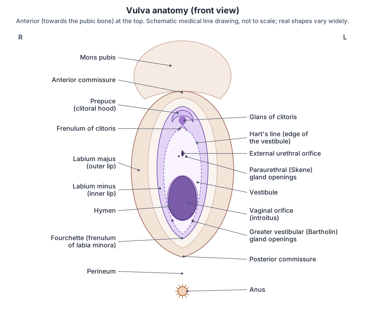
Parts of the vulva
| Part | What and where it is | Male counterpart or origin |
|---|---|---|
| Mons pubis | Fat pad in front of the pubic symphysis, with pubic hair | - |
| Labia majora (outer lips) | Folds of skin and fat from the mons to the posterior commissure; hair on the outer surface | Scrotum |
| Labia minora (inner lips) | Thin hairless folds around the vestibule; form the clitoral hood, frenulum and fourchette | Develop from the urethral folds |
| Clitoris | Erectile organ: external glans, internal body and two crura | Penis |
| Prepuce (clitoral hood) | Fold of the labia minora over the glans | Foreskin |
| Vestibule | Area between the labia minora, from Hart's line to the hymen | - |
| External urethral orifice | In the vestibule, about 2 to 3 cm behind the clitoris | - |
| Vaginal orifice and hymen | Behind the urethral opening; hymen partly covers it | - |
| Vestibular bulbs | Erectile tissue on each side of the vestibule, under bulbospongiosus | Bulb and corpus spongiosum of the penis |
| Greater vestibular (Bartholin) glands | Behind the bulbs; ducts open beside the vaginal orifice | Bulbourethral (Cowper) glands |
| Paraurethral (Skene) glands | In the wall of the lower urethra; ducts open beside the urethral orifice | Prostate |
From Kenhub, TeachMeAnatomy, Radiopaedia and Goldstein 2006. Counterparts follow the embryonic origin: genital tubercle, urethral folds and labioscrotal swellings.
Mons pubis and labia majora (outer lips)
The mons pubis is a pad of fat in front of the pubic symphysis that carries most of the pubic hair. Below it, the labia majora are thick folds of skin and fat separated by the pudendal cleft. After puberty their outer surfaces grow pubic hair while their inner surfaces stay hairless. They join in front at the anterior commissure and behind at the posterior commissure, and the round ligament of the uterus ends in them. (Kenhub: External female genitalia.)
The labia majora run from the mons pubis back to the posterior commissure, which lies over the perineal body. They develop from the labioscrotal swellings of the embryo. (TeachMeAnatomy: The Vulva.)
Pubic hair begins to grow on the mons and outer lips at puberty. After menopause, falling oestrogen can thin the pubic hair and reduce the fat in the vulva. (Cleveland Clinic: Vulva.)
Labia minora (inner lips) and the clitoral hood
The labia minora are thin, hairless folds without fat that lie inside the labia majora and surround the vestibule. At the front each one splits into two layers: the upper layers join over the clitoris to form its hood (prepuce), and the lower layers join beneath it to form the frenulum of the clitoris. (Kenhub: External female genitalia.)
At the back the labia minora merge to form the fourchette (frenulum of the labia minora). They develop from the urethral folds of the embryo. (TeachMeAnatomy: The Vulva.)
The clitoral hood may cover all, some or none of the glans. (Cleveland Clinic: Clitoris.)
On the inner surface of each labium minus a visible boundary, Hart's line, marks where keratinised skin meets the moist mucosa of the vestibule. (Goldstein A. Surgery for vulvar vestibulitis syndrome. J Sex Med. 2006.)
Clitoris anatomy: glans, body, crura and bulbs
The clitoris is an erectile organ at the front of the vulva, below the mons pubis where the labia minora meet, and the urethra opens separately just behind it. Its two crura attach to the ischiopubic rami and converge to form the paired corpora cavernosa, which make up the body and are wrapped in a tunica albuginea. The glans sits on the end of the body. Only the glans is external: about four-fifths of the clitoris lies inside the body. (Kenhub: Clitoris.)
The two corpora cavernosa are separated by a thin fibrous septum, and the glans lies at the lower end, angled backwards. Older texts did not count the bulbs of the vestibule as part of the clitoris, but newer work suggests that view is outdated. (Radiopaedia: Clitoris.)
Longhurst and colleagues describe the body as shaped like a boomerang that folds back on itself, with an ascending and a descending segment; its highest point is anchored to the pubic symphysis by the suspensory ligament. The crura follow the lower border of the ischiopubic rami deep to the ischiocavernosus muscles, and the bulbs run parallel to the crura deep to the bulbospongiosus muscles, with the greater vestibular glands at their back edge. A fully labelled image of the whole clitoris was not published in Gray's Anatomy until its 40th edition. (Longhurst GJ, Beni R, Jeong SR, et al. Beyond the tip of the iceberg: a meta-analysis of the anatomy of the clitoris. Clin Anat. 2023.)
Cleveland Clinic compares the whole organ to an upside-down wishbone. Everything except the glans is erectile tissue, and during arousal the crura and bulbs fill with blood; the bulbs can double in size. The glans has about 10,000 nerve endings. (Cleveland Clinic: Clitoris.)
Two dorsal arteries of the clitoris, branches of the internal pudendal artery, each give a deep artery to the corpus cavernosum on their side. Paired deep dorsal veins and a single superficial dorsal vein drain it. The dorsal nerve of the clitoris, the end branch of the pudendal nerve, runs along the body to the glans, which is its most densely innervated part. (Kenhub: Clitoris.)
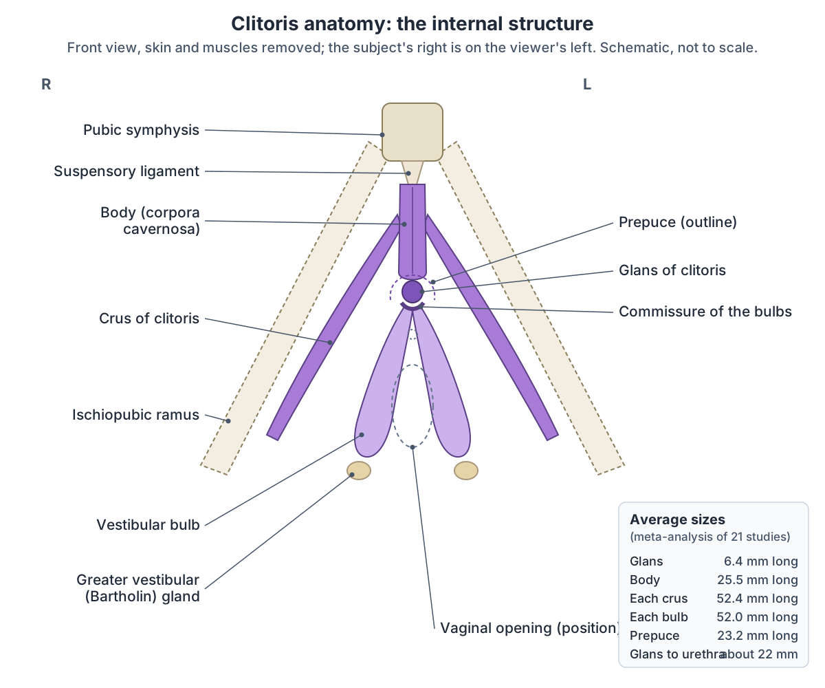
How big is the clitoris? Measurements and variation
A 2023 meta-analysis pooled measurements from 21 MRI, ultrasound, cadaver and clinical studies. The glans averaged 6.4 mm long (range 1 to 21 mm) and 5.1 mm wide, the body 25.5 mm long and 9 mm wide, each crus 52.4 mm long (range 23 to 90 mm) and 8.7 mm wide, each vestibular bulb 52 mm long (range 13 to 70 mm) and 10.3 mm wide, and the prepuce 23.2 mm. The authors conclude that every part of the clitoris varies substantially. (Longhurst GJ, Beni R, Jeong SR, et al. Beyond the tip of the iceberg: a meta-analysis of the anatomy of the clitoris. Clin Anat. 2023.)
The same review found that the glans lies on average about 22 mm in front of the urethral opening (range 7 to 52 mm), about 43 mm from the vagina and about 76 mm from the anus. Textbooks usually give the distance to the urethra as 2.5 cm and rarely mention the range. (Longhurst GJ, Beni R, Jeong SR, et al. Beyond the tip of the iceberg: a meta-analysis of the anatomy of the clitoris. Clin Anat. 2023.)
TeachMeAnatomy gives the same landmark as 2 to 3 cm: the external urethral orifice lies in front of the vaginal opening and 2 to 3 cm behind the clitoris. (TeachMeAnatomy: The Urethra.)
Simpler sources give round figures. Kenhub gives the glans as 5 to 8 mm long. Cleveland Clinic describes it as roughly pea-sized, about half an inch wide, and the whole clitoris from glans to the ends of the crura as about 3.5 to 4.25 inches long and about 2.5 inches wide. (Cleveland Clinic: Clitoris.)
Kenhub gives each vestibular bulb a length of about 3 cm, shorter than the 52 mm average of the meta-analysis, so bulb size depends on the study and method. (Kenhub: Bulb of vestibule.)
Clitoris measurements: averages and ranges
| Structure | Average length | Range of length | Average width |
|---|---|---|---|
| Glans | 6.4 mm | 1 to 21 mm | 5.1 mm (range 2 to 32 mm) |
| Body (corpora cavernosa) | 25.5 mm | 5 to 59 mm | 9.0 mm (range 5 to 20 mm) |
| Each crus | 52.4 mm | 23 to 90 mm | 8.7 mm (range 2 to 13 mm) |
| Each vestibular bulb | 52.0 mm | 13 to 70 mm | 10.3 mm (range 3 to 29 mm) |
| Prepuce (hood) | 23.2 mm | 5 to 40 mm | Not reported |
| Distance from clitoris to urethral opening | About 22 mm | 7 to 52 mm | Not applicable |
From the meta-analysis of 21 studies by Longhurst et al. (Clinical Anatomy, 2023). The paper gives the clitoris-to-urethra distance as 22.27 mm in its abstract and 22.72 mm in its results.
The vestibule: urethral and vaginal openings
The vestibule is the area enclosed by the labia minora. It contains the urethral and vaginal openings and the openings of the greater and lesser vestibular glands, and the prepuce of the clitoris lies at its front margin. (Kenhub: External female genitalia.)
Surgically, the vulvar vestibule lies between the hymenal ring on the inside and Hart's line on the inner surface of the labia minora on the outside. The minor vestibular glands lie within it. (Goldstein A. Surgery for vulvar vestibulitis syndrome. J Sex Med. 2006.)
The external urethral orifice opens in the vestibule in front of the vaginal opening, and two paraurethral (Skene) glands lie on either side of the lower urethra. The urethra is only about 4 cm long. (TeachMeAnatomy: The Urethra.)
The vaginal opening (introitus) is a midline opening whose long axis lies behind the urethral opening. (Radiopaedia: Hymen.)
The 4 cm urethra from the bladder neck to its opening in the vestibule, in 3D. Female urethra.
Hymen anatomy
The hymen is a thin fold of mucous membrane across the vaginal opening that is part of the vulva. It usually has an opening that lets menstrual blood out, and that opening can be single or multiple, central, off-centre or to one side. (Radiopaedia: Hymen.)
The hymen forms from leftover tissue during fetal development and has no known essential function. Most newborns have a ring-shaped (annular) hymen, which usually becomes crescent-shaped by early school age. It is thicker at birth, is flexible and stretches, and tends to wear gradually with hormones, exercise, tampon use, pelvic examinations and sex rather than tearing at once. Once worn away it does not regrow in adults. (Cleveland Clinic: Hymen.)
Radiopaedia lists annular, crescentic, cribriform (many small openings), septate (an opening divided by a band), bifenestrated, circular and denticular forms. In an imperforate hymen there is no opening, and fluid collects in the vagina and uterus. Small tags of tissue around the opening, the hymenal caruncles, are remnants of the hymen in adults. (Radiopaedia: Hymen.)
The appearance of the hymen is not a reliable sign of whether someone has had vaginal intercourse, and no known examination can prove it. The World Health Organization states that so-called virginity testing has no scientific merit or clinical indication. (WHO: Eliminating virginity testing, an interagency statement (2018).)
Hymen shapes
| Type | Description |
|---|---|
| Annular | Ring around the vaginal opening; usual at birth |
| Crescentic | Crescent at the back of the opening; the most common shape after early childhood |
| Cribriform | Many small openings |
| Septate | A band of tissue divides the opening in two |
| Microperforate | Covers the opening except for a tiny hole |
| Imperforate | No opening; menstrual fluid collects behind it and it is opened surgically |
From Radiopaedia and Cleveland Clinic. Shape varies between people and changes with age; it does not show sexual history.
Glands of the vulva: Bartholin, Skene and lesser vestibular glands
The greater vestibular (Bartholin) glands are paired, pea-sized glands less than 1 cm across, one on each side of the vaginal opening behind the back end of each vestibular bulb. Each drains through a duct about 2 cm long that opens at the back and side of the vaginal opening, between the hymen and the labium minus. They secrete lubricating fluid, especially during arousal, and are the counterparts of the male bulbourethral (Cowper) glands. (Radiopaedia: Bartholin gland.)
The Skene (paraurethral) glands lie in the wall of the lower urethra, and each drains through a paraurethral duct beside the urethral opening. They secrete mucus during sexual activity and are the counterparts of the prostate. A blocked duct can form a Skene duct cyst. They are named after the gynaecologist Alexander Skene (1838 to 1900). (Radiopaedia: Skene glands.)
OpenStax describes both the Bartholin glands and the lesser vestibular glands, located near the clitoris, as secreting mucus that keeps the vestibule moist. (OpenStax Anatomy and Physiology 2e, 27.2: Anatomy and Physiology of the Ovarian Reproductive System.)
If a Bartholin duct is blocked the gland swells into a fluid-filled cyst, which can become infected (bartholinitis), most often with Staphylococcus or E. coli. (TeachMeAnatomy: The Perineum.)
Vestibular bulbs, perineal muscles and the perineum
Each vestibular bulb is an elongated mass of erectile tissue on one side of the vestibule, deep to the labium minus and lying on the perineal membrane just beside the vaginal opening. Its rounded back end touches the greater vestibular gland, and the narrow front ends of the two bulbs join in a commissure that runs forwards to the clitoris. Bulbospongiosus covers it. During arousal the bulbs engorge, which is thought to help squeeze secretions from the greater vestibular glands. (Kenhub: Bulb of vestibule.)
The vulva lies in the urogenital triangle of the diamond-shaped perineum, whose corners are the pubic symphysis, the tip of the coccyx and the two ischial tuberosities. The superficial perineal pouch, below the perineal membrane, holds the erectile tissue of the clitoris, the greater vestibular glands and the ischiocavernosus, bulbospongiosus and superficial transverse perineal muscles. The perineal body, between the vagina and the external anal sphincter, supports the back wall of the vagina. (TeachMeAnatomy: The Perineum.)
Levator ani, the perineal membrane and the perineal body beneath the vulva. Pelvic floor and perineum.
Blood supply, nerves and lymph of the vulva
The vulva is supplied by the paired internal and external pudendal arteries, branches of the internal iliac and femoral arteries, and drained by the pudendal veins. Its front is supplied by the ilioinguinal nerve and the genital branch of the genitofemoral nerve, and its back by the pudendal nerve and the posterior cutaneous nerve of the thigh. Parasympathetic cavernous nerves from the uterovaginal plexus supply the clitoris and vestibule. Lymph drains to the superficial inguinal nodes. (TeachMeAnatomy: The Vulva.)
Kenhub adds that the mons pubis and front of the labia are supplied by the anterior labial nerves from the lumbar plexus and the back of the vulva by the posterior labial branches of the pudendal nerve, and that lymph may also drain to the deep inguinal nodes, or from the clitoris, vestibular bulbs and front of the labia minora directly to the internal iliac nodes. (Kenhub: External female genitalia.)
Nerve supply of the vulva
| Area | Nerve | Origin |
|---|---|---|
| Mons pubis and front of the labia | Ilioinguinal nerve and genital branch of the genitofemoral nerve (anterior labial nerves) | Lumbar plexus |
| Back of the labia and vestibule | Posterior labial branches of the pudendal nerve; posterior cutaneous nerve of the thigh | Pudendal nerve from S2 to S4 |
| Clitoris (sensation) | Dorsal nerve of the clitoris | Pudendal nerve |
| Clitoris and vestibule (erectile tissue) | Cavernous nerves (parasympathetic) | Uterovaginal plexus |
From TeachMeAnatomy (vulva, perineum) and Kenhub (external female genitalia, clitoris).
Development of the vulva and male homologues
The external genitalia of both sexes develop from the same embryonic structures. The genital tubercle forms the clitoris, the urethral folds form the labia minora and the labioscrotal swellings form the labia majora. (TeachMeAnatomy: The Vulva.)
Without testosterone, the cells that would form the glans of the penis form the clitoris. (OpenStax Anatomy and Physiology 2e, 27.3: Development of the Male and Female Reproductive Systems.)
The clitoris is the homologue of the penis, the vestibular bulbs of the bulb and corpus spongiosum of the penis, the greater vestibular glands of the bulbourethral glands, and the paraurethral glands of the prostate. (Kenhub: External female genitalia.)
The labia majora are homologous to the scrotum. (Kenhub: Female reproductive organs.)
How the female body differs from the male body, with a labelled organ diagram. Female anatomy.
Is my vulva normal? Variation in size, shape and colour
All vulvas have the same parts, but no two look alike. The labia come in many shapes and sizes, the inner lips may be symmetrical or asymmetrical, and they may sit inside the outer lips or extend beyond them. (Cleveland Clinic: Vulva.)
A study of photographs of 400 Chinese women found that the length, width and colour of the labia minora vary widely and that there is no standard for an ideal shape. The point of greatest width (maximum protrusion) of the labia minora was in the middle third in 47% of women, the front third in 34% and the back third in 19%. (Che K, Wang K, Yuan Y, Li F, Li Q. Quantitative analysis of the labia minora morphology in 400 Chinese women. Front Surg. 2023.)
The clitoral meta-analysis found that textbook figures do not reflect how much clitoral size varies. Its authors note that the American College of Obstetricians and Gynecologists advises clinicians to reassure patients that the size, shape and colour of the external genitalia vary considerably. (Longhurst GJ, Beni R, Jeong SR, et al. Beyond the tip of the iceberg: a meta-analysis of the anatomy of the clitoris. Clin Anat. 2023.)
A clitoris that is much larger than usual (clitoromegaly) can be present from birth or develop later, most often because of exposure to androgen hormones. (Cleveland Clinic: Clitoris.)
Clinical anatomy of the vulva
Conditions of the vulva include Bartholin cysts, labial adhesions, an imperforate hymen, skin conditions such as lichen sclerosus, infections, pain without a clear cause (vulvodynia) and vulvar cancer, which is usually linked to lichen sclerosus or HPV. (Cleveland Clinic: Vulva.)
Genital warts on the vulva are benign growths caused by HPV types 6 and 11. The high-risk types 16 and 18 can cause precancerous changes (dysplasia) of the cervix, vagina and anus. (TeachMeAnatomy: The Vulva.)
Female genital mutilation (FGM) is any procedure that partly or totally removes the external female genitalia, or otherwise injures them, for non-medical reasons. More than 230 million girls and women alive today have undergone it. Type 1 removes part or all of the visible clitoral glans and/or its hood, type 2 also removes the labia minora, with or without the labia majora, type 3 (infibulation) narrows the vaginal opening with a seal made from the cut labia, and type 4 covers other harmful procedures. FGM has no health benefits. (WHO fact sheet: Female genital mutilation.)
Frequently asked questions
What are the parts of the vulva?
The vulva is made up of the mons pubis, the labia majora (outer lips), the labia minora (inner lips), the clitoris with its hood, and the vestibule between the inner lips. The vestibule contains the urethral opening, the vaginal opening with the hymen, and the openings of the Bartholin and Skene glands. The vestibular bulbs lie under the surface on each side.
Is the vulva the same as the vagina?
No. The vulva is the external genital area that you can see. The vagina is the internal muscular canal that starts at the vaginal opening in the vestibule and runs up to the cervix. The two are often confused because people commonly use the word vagina for the whole area.
How big is the clitoris?
Most of the clitoris is internal. A 2023 meta-analysis of 21 studies found an average glans length of 6.4 mm, a body of about 25 mm, and crura and vestibular bulbs of about 52 mm each, with wide ranges for every part. Cleveland Clinic describes the whole structure as about 3.5 to 4.25 inches long from glans to crura.
Where is the urethral opening in females?
The external urethral orifice opens in the vestibule, between the clitoris in front and the vaginal opening behind. Textbooks place it 2 to 3 cm behind the clitoris; measurements in real people average about 22 mm but range from 7 to 52 mm. The Skene glands open on either side of it.
What are Bartholin glands?
Bartholin glands, or greater vestibular glands, are two pea-sized glands less than 1 cm across, one on each side of the vaginal opening behind the vestibular bulbs. Each opens through a duct about 2 cm long between the hymen and the labium minus and releases lubricating fluid, especially during arousal. A blocked duct can form a Bartholin cyst.
Is it normal for the labia minora to be uneven or longer than the labia majora?
Yes. The inner lips vary widely in length, width, shape and colour, they are often asymmetrical, and they may extend beyond the outer lips. A study of 400 women found no standard ideal shape, and gynaecologists advise that the size, shape and colour of the external genitalia vary considerably.
Does the hymen show whether someone has had sex?
No. The hymen varies in shape from birth, usually changes from ring-shaped to crescent-shaped in childhood, and stretches or wears gradually with hormones, exercise, tampons and examinations as well as sex. The World Health Organization states that its appearance is not a reliable sign of intercourse and that no examination can prove sexual history.
References
- Thompson L. The Vulva. TeachMeAnatomy, updated 2025.
- TeachMeAnatomy. The Perineum.
- TeachMeAnatomy. The Urethra.
- External female genitalia. Kenhub anatomy library, reviewed 2022.
- Clitoris. Kenhub anatomy library.
- Bulb of vestibule. Kenhub anatomy library, reviewed 2024.
- Sendić G. Female reproductive organs. Kenhub, reviewed 2023.
- Clitoris. Radiopaedia reference article.
- Hymen. Radiopaedia reference article.
- Bartholin gland. Radiopaedia reference article.
- Skene glands. Radiopaedia reference article.
- OpenStax. Anatomy and Physiology 2e, §27.2 Anatomy and Physiology of the Ovarian Reproductive System. Rice University, 2022.
- OpenStax. Anatomy and Physiology 2e, §27.3 Development of the Male and Female Reproductive Systems. Rice University, 2022.
- Cleveland Clinic. Vulva: Location, Anatomy, Function, Conditions and Care.
- Cleveland Clinic. Clitoris: Anatomy, Location, Purpose and Conditions.
- Cleveland Clinic. Hymen: Overview, Function and Anatomy.
- Longhurst GJ, Beni R, Jeong SR, Pianta M, Soper AL, Leitch P, De Witte G, Fisher L. Beyond the tip of the iceberg: a meta-analysis of the anatomy of the clitoris. Clin Anat. 2023. doi:10.1002/ca.24113 (open access, CC BY).
- Che K, Wang K, Yuan Y, Li F, Li Q. Quantitative analysis of the labia minora morphology in 400 Chinese women: a new method for assessing the shape of the labia minora. Front Surg. 2023;9:961247.
- Goldstein A. Surgery for vulvar vestibulitis syndrome. J Sex Med. 2006;3:559-562.
- World Health Organization. Eliminating virginity testing: an interagency statement. WHO/RHR/18.15, 2018.
- World Health Organization. Female genital mutilation: fact sheet.
Related anatomy
- VaginaVagina anatomy with labelled diagrams and a 3D model: vaginal canal length, walls, fornices, rugae, wall layers…
- Female anatomyFemale anatomy explained with a labelled body diagram and 3D model: reproductive organs, vulva, breasts, pelvis…
- Female reproductive systemLabelled female reproductive system diagrams: uterus anatomy and layers, uterine tube parts, ovaries, ligaments…
- Pelvic floor and perineumPerineum and pelvic floor anatomy with labelled diagrams and a 3D model: levator ani, coccygeus, perineal body, both…
- Female urethraFemale urethra: The female urethra is a short tube, about 4 cm long, that carries urine from the bladder to the…
Explore the whole body in the interactive 3D human anatomy model, or browse the A–Z list of human structures.